

TP53-Activated lncRNA GHRLOS Regulates Cell Proliferation, Invasion, and Apoptosis of Non-Small Cell Lung Cancer by Modulating the miR-346/APC Axis
- PMID: 33968785
- PMCID: PMC8097184
- DOI: 10.3389/fonc.2021.676202
TP53-Activated lncRNA GHRLOS Regulates Cell Proliferation, Invasion, and Apoptosis of Non-Small Cell Lung Cancer by Modulating the miR-346/APC Axis
Abstract
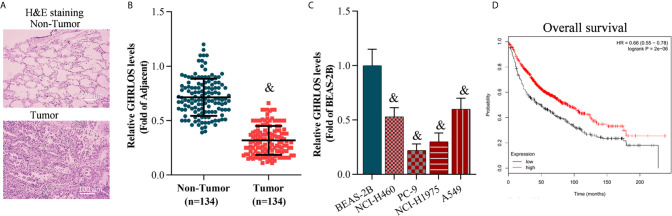
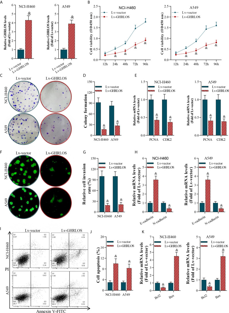
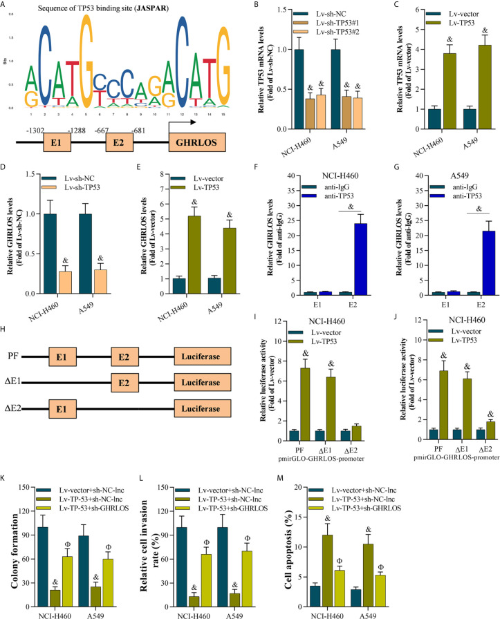
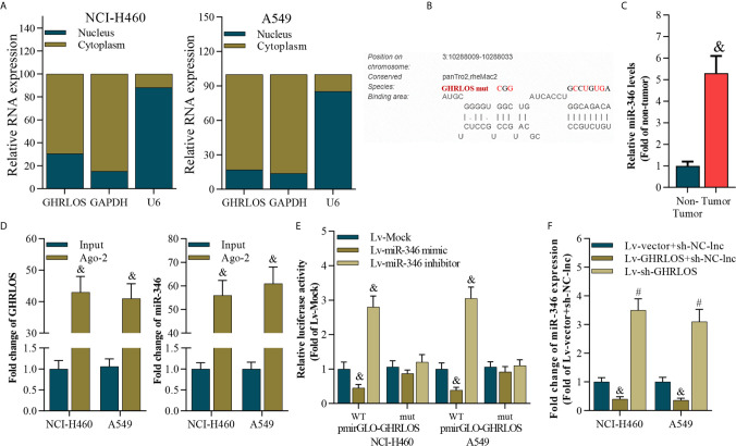
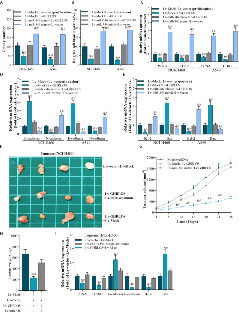
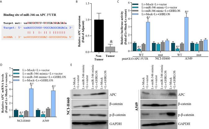
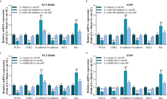
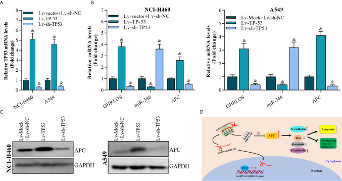
Non-small cell lung cancer (NSCLC) is the main type of lung cancer with high mortality worldwide. To improve NSCLC therapy, the exploration of molecular mechanisms involved in NSCLC progression and identification of their potential therapy targeting is important. Long noncoding RNAs (lncRNAs) have shown important roles in regulating various tumors progression, including NSCLC. We found lncRNA GHRLOS was decreased in NSCLC cell lines and tissues which correlated with poor prognosis of NSCLC patients. However, the role and underlying mechanisms of lncRNA GHRLOS in NSCLC progression remains elusive. The expression of lncRNA GHRLOS was examined in NSCLC cell lines and biopsy specimens of patients with NSCLC by quantitative real time polymerase chain reaction (qRT-PCR). The effects of GHRLOS on proliferation, invasion and apoptosis of NSCLC cells were determined by both in vitro and in vivo experiments. The interaction between GHRLOS and TP53 was determined by dual-luciferase reporter assay and chromatin immunoprecipitation (ChIP) combined with qRT-PCR analysis. RNA immunoprecipitation (RIP) was conducted to validate the binding between GHRLOS and microRNA-346 (miR-346). Dual-luciferase reporter assays were also carried out to reveal the interaction between miR-346 and the 3' untranslated region (3'UTR) of adenomatous polyposis coli (APC) mRNA.Our data demonstrated that overexpression of lncRNA GHRLOS suppressed cancer cell proliferation and invasion as well as promoted cell apoptosis by regulating the expression of CDK2, PCNA, E-cadherin, N-cadherin, Bax, and Bcl-2 in NSCLC cells. Moreover, lncRNA GHRLOS was upregulated by the binding of TP53 to the GHRLOS promoter. The binding target of lncRNA GHRLOS was identified to be miR-346. Impressively, overexpression of miR-346 promoted cell proliferation and invasion, as well as inhibited cell apoptosis, however, these effects can be blocked by overexpression of lncRNA GHRLOS both in vitro and in vivo. In summary, this study reveals lncRNA GHRLOS, upregulated by TP53, acts as a molecule sponge of miR-346 to cooperatively modulates expression of APC, a miR-346 target, and potentially inhibits NSCLC progression via TP53/lncRNA GHRLOS/miR-346/APC axis, which represents a novel pathway that could be useful in targeted therapy against NSCLC.
Keywords: APC; NSCLC; TP53; lncRNA GHRLOS; miR-346.
Copyright © 2021 Ren, Sun, Liu, Yang, Li, Wang, Deng, Hou, Qiu and Zhao.
Conflict of interest statement
The authors declare that the research was conducted in the absence of any commercial or financial relationships that could be construed as a potential conflict of interest.
Figures
Similar articles
-
Downregulation of long non-coding RNA XIST inhibits cell proliferation, migration, invasion and EMT by regulating miR-212-3p/CBLL1 axis in non-small cell lung cancer cells.Eur Rev Med Pharmacol Sci. 2019 Oct;23(19):8391-8402. doi: 10.26355/eurrev_201910_19150. Eur Rev Med Pharmacol Sci. 2019. PMID: 31646569
-
The long non-coding RNA PVT1/miR-145-5p/ITGB8 axis regulates cell proliferation, apoptosis, migration and invasion in non-small cell lung cancer cells.Neoplasma. 2020 Jul;67(4):802-812. doi: 10.4149/neo_2020_190723N657. Epub 2020 Mar 24. Neoplasma. 2020. PMID: 32202906
-
Long noncoding RNA Linc00210 promotes non-small cell lung cancer progression via sponging miR-16-5p/PTK2 axis.Eur Rev Med Pharmacol Sci. 2020 Sep;24(18):9438-9452. doi: 10.26355/eurrev_202009_23029. Eur Rev Med Pharmacol Sci. 2020. PMID: 33015786
-
LncRNAs in non-small cell lung cancer: novel diagnostic and prognostic biomarkers.Front Mol Biosci. 2023 Dec 13;10:1297198. doi: 10.3389/fmolb.2023.1297198. eCollection 2023. Front Mol Biosci. 2023. PMID: 38152110 Free PMC article. Review.
-
LncRNA in tumorigenesis of non-small-cell lung cancer: From bench to bedside.Cell Death Discov. 2022 Aug 13;8(1):359. doi: 10.1038/s41420-022-01157-4. Cell Death Discov. 2022. PMID: 35963868 Free PMC article. Review.
Cited by
-
Various LncRNA Mechanisms in Gene Regulation Involving miRNAs or RNA-Binding Proteins in Non-Small-Cell Lung Cancer: Main Signaling Pathways and Networks.Int J Mol Sci. 2023 Sep 3;24(17):13617. doi: 10.3390/ijms241713617. Int J Mol Sci. 2023. PMID: 37686426 Free PMC article. Review.
-
Identification and Evaluation of Hub Long Non-Coding RNAs and mRNAs in PM2.5-Induced Lung Cell Injury.Int J Mol Sci. 2025 Jan 22;26(3):911. doi: 10.3390/ijms26030911. Int J Mol Sci. 2025. PMID: 39940682 Free PMC article.
-
Integrating single-cell and multi-omic approaches reveals Euphorbiae Humifusae Herba-dependent mitochondrial dysfunction in non-small-cell lung cancer.J Cell Mol Med. 2024 May;28(10):e18317. doi: 10.1111/jcmm.18317. J Cell Mol Med. 2024. PMID: 38801409 Free PMC article.
-
Comprehensive analysis of circulating cell-free RNAs in blood for diagnosing non-small cell lung cancer.Comput Struct Biotechnol J. 2023 Aug 28;21:4238-4251. doi: 10.1016/j.csbj.2023.08.029. eCollection 2023. Comput Struct Biotechnol J. 2023. PMID: 37692082 Free PMC article.
-
Roles of hypoxic environment and M2 macrophage-derived extracellular vesicles on the progression of non-small cell lung cancer.BMC Pulm Med. 2023 Jul 3;23(1):239. doi: 10.1186/s12890-023-02468-7. BMC Pulm Med. 2023. PMID: 37400770 Free PMC article.
References
LinkOut - more resources
Full Text Sources
Other Literature Sources
Research Materials
Miscellaneous