

Axial Spondyloarthritis: Mimics and Pitfalls of Imaging Assessment
- PMID: 33968964
- PMCID: PMC8100693
- DOI: 10.3389/fmed.2021.658538
Axial Spondyloarthritis: Mimics and Pitfalls of Imaging Assessment
Abstract
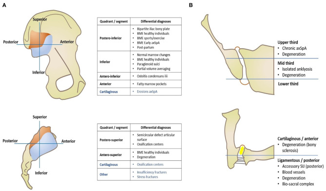
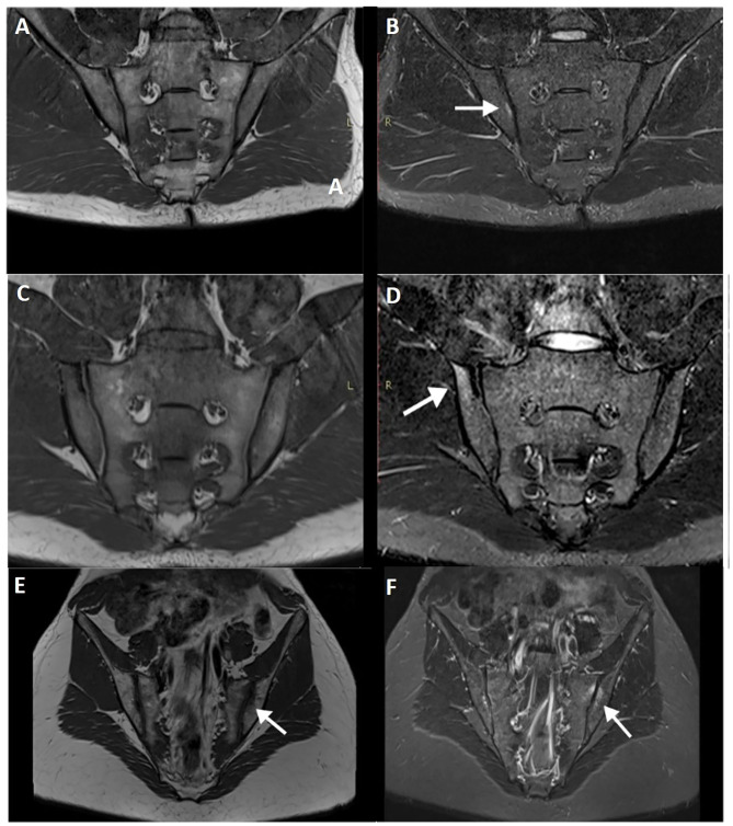
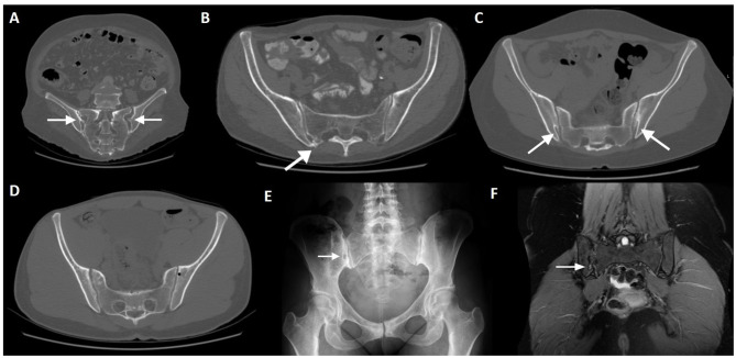
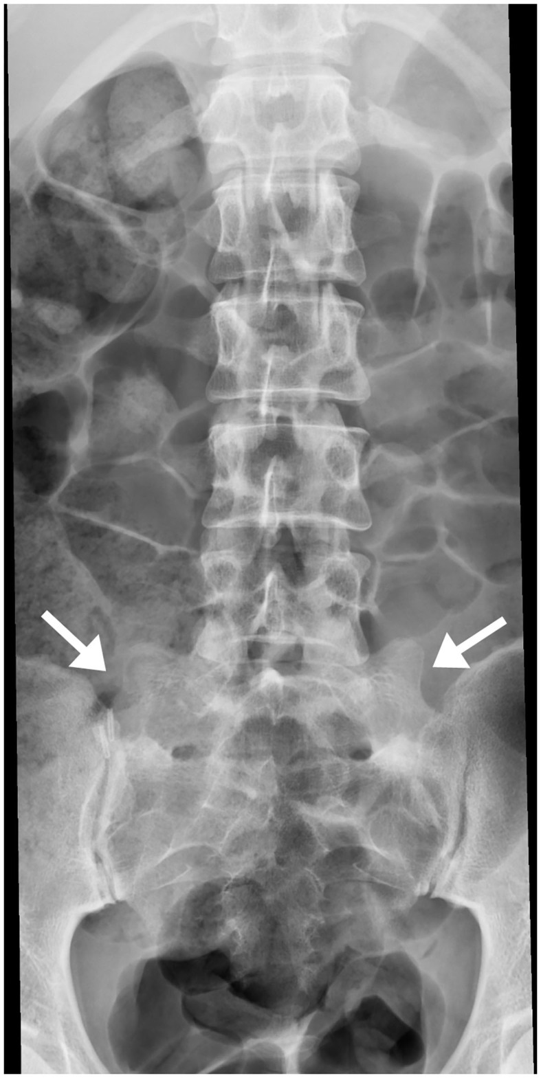
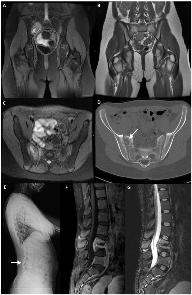
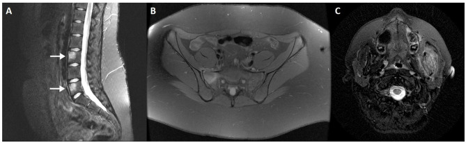
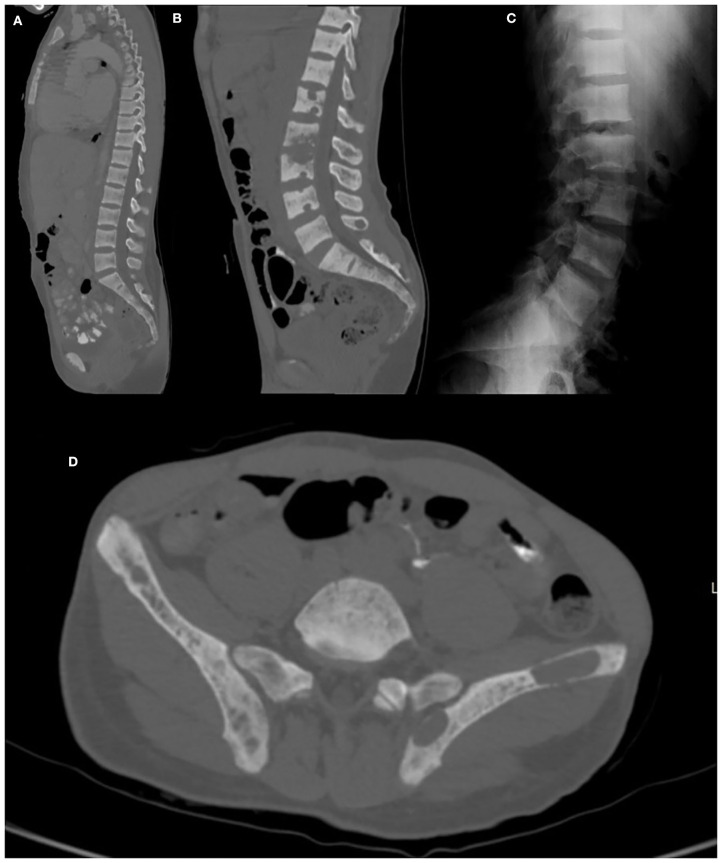
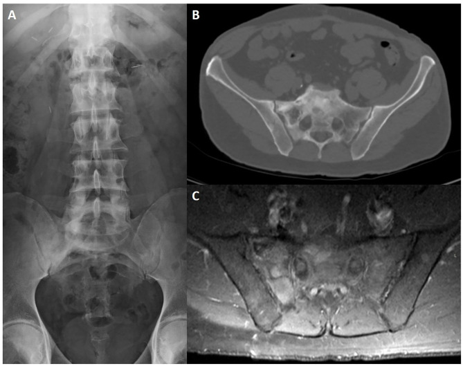
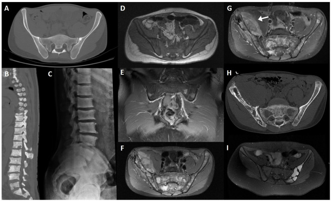
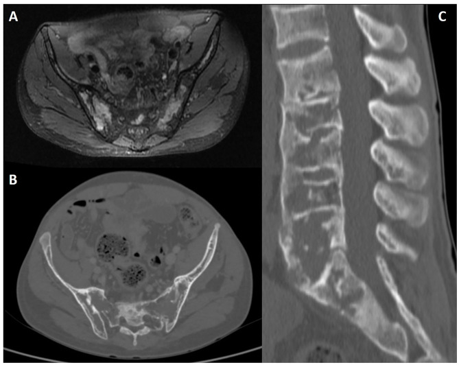
Axial spondyloarthritis (axSpA) is a chronic inflammatory disorder that predominantly involves the axial skeleton. Imaging findings of axSpA can be divided into active changes, which include bone marrow edema, synovitis, enthesitis, capsulitis, and intra-articular effusion, and structural changes, which include erosions, sclerosis, bone fatty infiltration, fat deposition in an erosion cavity, and bone bridging or ankylosis. The ability to distinguish between imaging lesions suggestive of axSpA and artifacts or lesions suggestive of other disorders is critical for the accurate diagnosis of axSpA. Diagnosis may be challenging, particularly in early-stage disease and magnetic resonance imaging (MRI) plays a key role in the detection of subtle or inflammatory changes. MRI also allows the detection of structural changes in the subchondral bone marrow that are not visible on conventional radiography and is of prognostic and monitoring value. However, bone structural changes are more accurately depicted using computed tomography. Conventional radiography, on the other hand, has limitations, but it is easily accessible and may provide insight on gross changes as well as rule out other pathological features of the axial skeleton. This review outlines the imaging evaluation of axSpA with a focus on imaging mimics and potential pitfalls when assessing the axial skeleton.
Keywords: axial spondyloarthritis; computed tomography; differential diagnosis; magnetic resonance imaging; mimic; normal variant; pitfall; radiography.
Copyright © 2021 Caetano, Mascarenhas and Machado.
Conflict of interest statement
PM has received consulting/speaker's fees from AbbVie, BMS, Celgene, Eli Lilly, Janssen, MSD, Novartis, Pfizer, Roche, and UCB all unrelated to this manuscript. The remaining authors declare that the research was conducted in the absence of any commercial or financial relationships that could be construed as a potential conflict of interest.
Figures
References
-
- Maksymowych WP, Lambert RG, Ostergaard M, Pedersen SJ, Machado PM, Weber U, et al. . MRI lesions in the sacroiliac joints of patients with spondyloarthritis: an update of definitions and validation by the ASAS MRI working group. Ann Rheum Dis. (2019) 78:1550–8. 10.1136/annrheumdis-2019-215589 - DOI - PubMed
-
- Maksymowych WP, Eshed I, Machado PM, Pedersen SJ, Weber U, de Hooge M, et al. . Consensus definitions for MRI lesions in the spine of patients with acial spondyloarthritis: first analysis from the assessments in spondyloarthritis international society classification cohort. Ann Rheum Dis. (2020) 79:749–50. 10.1136/annrheumdis-2020-eular.6304 - DOI
-
- Hermann KG, Baraliakos X, van der Heijde DM, Jurik AG, Landewe R, Marzo-Ortega H, et al. . Descriptions of spinal MRI lesions and definition of a positive MRI of the spine in axial spondyloarthritis: a consensual approach by the ASAS/OMERACT MRI study group. Ann Rheum Dis. (2012) 71:1278–88. 10.1136/ard.2011.150680 - DOI - PubMed
Publication types
LinkOut - more resources
Full Text Sources