

Sclerosing polycystic adenosis of the submandibular gland: Two case reports
- PMID: 33969079
- PMCID: PMC8058660
- DOI: 10.12998/wjcc.v9.i12.2930
Sclerosing polycystic adenosis of the submandibular gland: Two case reports
Abstract
Background: Sclerosing polycystic adenosis (SPA) is a rare disease of salivary glands, similar to fibrocystic disease of the breast. It occurs over a wide age range and exhibits a slight female preference. Most SPA cases have occurred in the parotid gland. The exact nature of SPA is unclear, but its tumor nature has recently been proposed. Although SPA has a good prognosis after adequate surgery, atypical lesions might occur, ranging from mild dysplasia to carcinoma in situ in some cases. To the best of our knowledge, only five cases of SPA in the submandibular gland have been reported to date. Here, we present two new cases of SPA involving the submandibular gland.
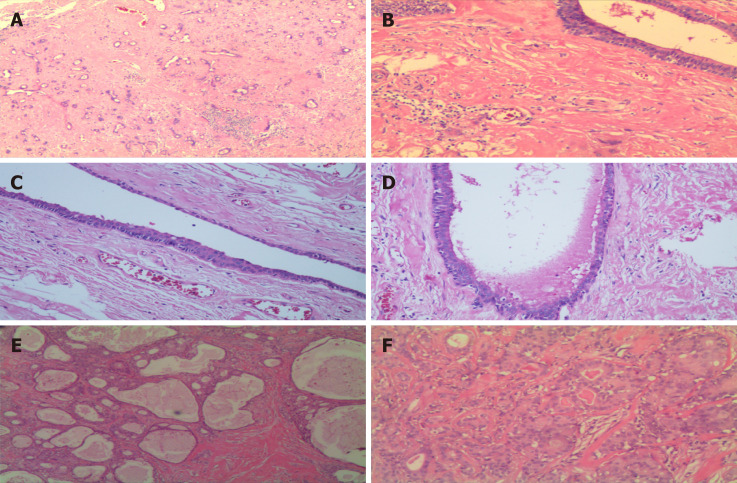
Case summary: A 50-year-old woman and a 52-year-old woman were referred to Tongji Hospital in Wuhan, China, with complaints of moderate pain, recurrent swelling, and a mass in the submandibular area. After admission, the two cases of the submandibular mass were examined physically. The boundary of the submandibular tumor was clear, and the range of motion was good. After preoperative examinations, surgery was performed on a selective basis. Postoperative histopathological examination revealed a well-defined mass with acinar structures, ducts, or cystic dilated glands of various sizes scattered in a large number of proliferative sclerosing stroma. There were flat and cuboidal cells, and eosinophils in the duct epithelium. There was also a eosinophilic substance in the lumen of dilated cysts. No atypical epithelial hyperplasia, invasive growth, or carcinoma in situ was found. Based on the above findings, the mass was diagnosed as SPA. Both patients have remained asymptomatic and no recurrence or distant metastasis had occurred by the 7-mo and 5-year follow-up, respectively.
Conclusion: SPA is a rare disease of the salivary gland. Even though it has a good prognosis after adequate surgery, atypical lesions may occur from mild dysplasia to carcinoma in situ. However, no recurrence, distant metastasis, or mortality has been reported for submandibular gland SPA. Clinicians and pathologists should be familiar with the characteristics of SPA in the submandibular gland to avoid misdiagnosis and overtreatment.
Keywords: Case report; Diagnosis; Histopathology; Sclerosing polycystic adenosis; Submandibular gland; Treatment.
©The Author(s) 2021. Published by Baishideng Publishing Group Inc. All rights reserved.
Conflict of interest statement
Conflict-of-interest statement: The authors declare that they have no conflict of interest.
Figures

References
-
- Smith BC, Ellis GL, Slater LJ, Foss RD. Sclerosing polycystic adenosis of major salivary glands. A clinicopathologic analysis of nine cases. Am J Surg Pathol. 1996;20:161–170. - PubMed
-
- Gnepp DR, Wang LJ, Brandwein-Gensler M, Slootweg P, Gill M, Hille J. Sclerosing polycystic adenosis of the salivary gland: a report of 16 cases. Am J Surg Pathol. 2006;30:154–164. - PubMed
-
- Noonan VL, Kalmar JR, Allen CM, Gallagher GT, Kabani S. Sclerosing polycystic adenosis of minor salivary glands: report of three cases and review of the literature. Oral Surg Oral Med Oral Pathol Oral Radiol Endod. 2007;104:516–520. - PubMed
Publication types
LinkOut - more resources
Full Text Sources
Other Literature Sources
