

An electrophysiological evaluation method for the ovine facial nerve
- PMID: 33969162
- PMCID: PMC8060511
- DOI: 10.1016/j.reth.2021.03.008
An electrophysiological evaluation method for the ovine facial nerve
Abstract
Introduction: Large-animal models such as sheep for facial nerve regeneration research have not yet been established because of the lack of methods for assessing the electrophysiological function of regenerated nerves. In this study, we developed a percutaneous measurement method for the evoked compound muscle action potential (CMAP) of the facial nerve in sheep.
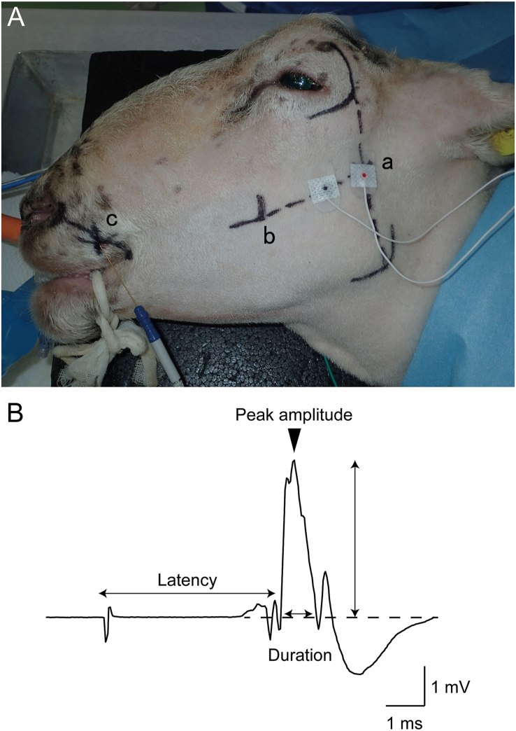
Methods: Six 3-year-old castrated male Corriedale sheep were used in this study. Under general anesthesia, an anatomical exploration was performed to identify the course of the buccal branch of the facial nerve and its innervating muscles on one side, followed by the application of surface stimulating electrodes to the contralateral side of the face along the course of the buccal branch of the facial nerve to obtain CMAP measurements of the nasolabial levator muscle.
Results: Percutaneous CMAP measurements of the nasolabial levator muscle could be obtained in all animals by placing stimulating electrodes 1 cm apart on the line coinciding with the course of the buccal branch of the facial nerve revealed by the preceding anatomical exploration. Mean values for electrophysiological parameters were amplitude 4.7 ± 0.7 mV, duration 2.1 ± 0.6 ms, and latency 3.6 ± 0.4 ms.
Conclusion: We have established a percutaneous measurement method for CMAP of the buccal branch of the facial nerve in sheep. This method is expected to be very useful in future studies of facial nerve regeneration for long nerve defects in sheep.
Keywords: ADSC, adipose-derived stem cell; CFNG, cross-facial nerve grafting; CMAP, compound muscle action potential; Compound muscle action potential; Facial nerve; Nasolabial levator muscle; Paralysis.
© 2021 The Japanese Society for Regenerative Medicine. Production and hosting by Elsevier B.V.
Conflict of interest statement
The authors declare no conflict of interest.
Figures


References
-
- Sampath P., Holliday M.J., Brem H., Niparko J.K., Long D.M. Facial nerve injury in acoustic neuroma (vestibular schwannoma) surgery: etiology and prevention. J Neurosurg. 1997;87(1):60–66. - PubMed
-
- Matsumine H., Kamei W., Fujii K., Shimizu M., Osada A., Sakurai H. One-stage reconstruction by dual-innervated double muscle flap transplantation with the neural interconnection between the ipsilateral masseter and contralateral facial nerve for reanimating established facial paralysis: a report of 2 cases. Microsurgery. 2019;39(5):457–462. - PubMed
-
- Matsumine H., Sasaki R., Yamato M., Okano T., Sakurai H. A polylactic acid non-woven nerve conduit for facial nerve regeneration in rats. J Tissue Eng Regen Med. 2014;8:454–462. - PubMed
-
- Matsumine H., Sasaki R., Tabata Y., Matsui M., Yamato M., Okano T. Facial nerve regeneration using basic fibroblast growth factor-impregnated gelatin microspheres in a rat model. J Tissue Eng Regen Med. 2016;10:E559–E567. - PubMed
LinkOut - more resources
Full Text Sources
Other Literature Sources
