. 2021 Apr 26;6(1):55-74.
doi: 10.20411/pai.v6i1.408. eCollection 2021.

Alpha 1 Antitrypsin is an Inhibitor of the SARS-CoV-2-Priming Protease TMPRSS2

Affiliations

Alpha 1 Antitrypsin is an Inhibitor of the SARS-CoV-2-Priming Protease TMPRSS2

Nurit P Azouz et al. Pathog Immun. .

Abstract

Background: Host proteases have been suggested to be crucial for dissemination of MERS, SARS-CoV, and SARS-CoV-2 coronaviruses, but the relative contribution of membrane versus intracellular proteases remains controversial. Transmembrane serine protease 2 (TMPRSS2) is regarded as one of the main proteases implicated in the coronavirus S protein priming, an important step for binding of the S protein to the angiotensin-converting enzyme 2 (ACE2) receptor before cell entry.

Methods: We developed a cell-based assay to identify TMPRSS2 inhibitors. Inhibitory activity was established in SARS-CoV-2 viral load systems.

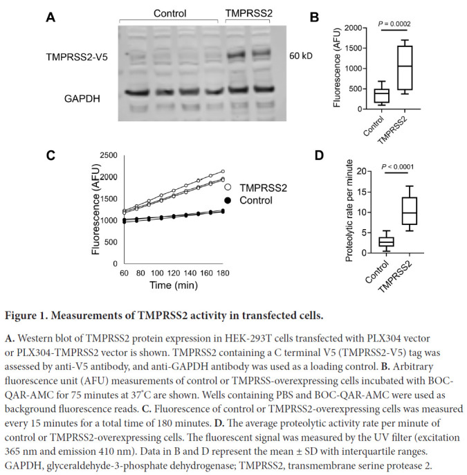
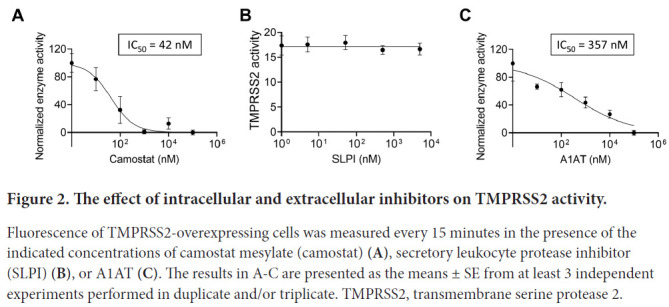
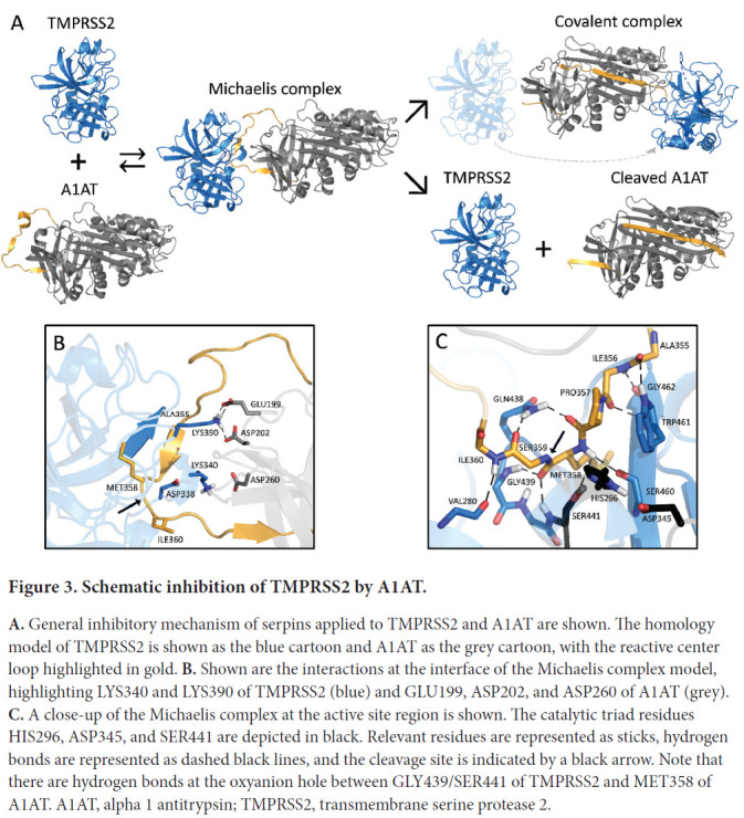
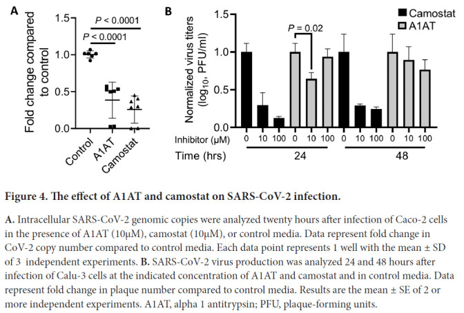
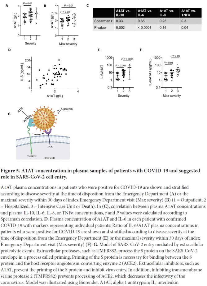
Results: We identified the human extracellular serine protease inhibitor (serpin) alpha 1 anti-trypsin (A1AT) as a novel TMPRSS2 inhibitor. Structural modeling revealed that A1AT docked to an extracellular domain of TMPRSS2 in a conformation that is suitable for catalysis, resembling similar serine protease inhibitor complexes. Inhibitory activity of A1AT was established in a SARS-CoV-2 viral load system. Notably, plasma A1AT levels were associated with COVID-19 disease severity.

Conclusions: Our data support the key role of extracellular serine proteases in SARS CoV-2 infections and indicate that treatment with serpins, particularly the FDA-approved drug A1AT, may be effective in limiting SARS-CoV-2 dissemination by affecting the surface of the host cells.

Keywords: COVID; TMPRSS2; alpha 1 antitrypsin; camostat mesylate; coronavirus; protease.


Conflict of interest statement

M.E.R. is a consultant for Pulm One, Spoon Guru, ClostraBio, Serpin Pharma, Celgene, Astra Zeneca, Allakos, Arena Pharmaceuticals, Guidepoint, and Suvretta Capital Management and has an equity interest in the first 4 listed and royalties from reslizumab (Teva Pharmaceuticals), PEESSv2 (Mapi Research Trust), and UpToDate. M.E.R. is an inventor of patents owned by Cincinnati Children's Hospital. M.E.R. and N.P.A. are inventors of a patent owned by Cincinnati Children's Hospital with the provisional number of 63/017,027.

Figures

Figure 1.
Figure 1.
Measurements of TMPRSS2 activity in transfected cells. A. Western blot of TMPRSS2 protein expression in HEK-293T cells transfected with PLX304 vector or PLX304-TMPRSS2 vector is shown. TMPRSS2 containing a C terminal V5 (TMPRSS2-V5) tag was assessed by anti-V5 antibody, and anti-GAPDH antibody was used as a loading control. B. Arbitrary fluorescence unit (AFU) measurements of control or TMPRSS-overexpressing cells incubated with BOCQAR-AMC for 75 minutes at 37°C are shown. Wells containing PBS and BOC-QAR-AMC were used as background fluorescence reads. C. Fluorescence of control or TMPRSS2-overexpressing cells was measured every 15 minutes for a total time of 180 minutes. D. The average proteolytic activity rate per minute of control or TMPRSS2-overexpressing cells. The fluorescent signal was measured by the UV filter (excitation 365 nm and emission 410 nm). Data in B and D represent the mean ± SD with interquartile ranges. GAPDH, glyceraldehyde-3-phosphate dehydrogenase; TMPRSS2, transmembrane serine protease 2.
Figure 2.
Figure 2.
The effect of intracellular and extracellular inhibitors on TMPRSS2 activity. Fluorescence of TMPRSS2-overexpressing cells was measured every 15 minutes in the presence of the indicated concentrations of camostat mesylate (camostat) (A), secretory leukocyte protease inhibitor (SLPI) (B), or A1AT (C). The results in A-C are presented as the means ± SE from at least 3 independent experiments performed in duplicate and/or triplicate. TMPRSS2, transmembrane serine protease 2.
Figure 3.
Figure 3.
Schematic inhibition of TMPRSS2 by A1AT. A. General inhibitory mechanism of serpins applied to TMPRSS2 and A1AT are shown. The homology model of TMPRSS2 is shown as the blue cartoon and A1AT as the grey cartoon, with the reactive center loop highlighted in gold. B. Shown are the interactions at the interface of the Michaelis complex model, highlighting LYS340 and LYS390 of TMPRSS2 (blue) and GLU199, ASP202, and ASP260 of A1AT (grey). C. A close-up of the Michaelis complex at the active site region is shown. The catalytic triad residues HIS296, ASP345, and SER441 are depicted in black. Relevant residues are represented as sticks, hydrogen bonds are represented as dashed black lines, and the cleavage site is indicated by a black arrow. Note that there are hydrogen bonds at the oxyanion hole between GLY439/SER441 of TMPRSS2 and MET358 of A1AT. A1AT, alpha 1 antitrypsin; TMPRSS2, transmembrane serine protease 2.
Figure 4.
Figure 4.
The effect of A1AT and camostat on SARS-CoV-2 infection. A. Intracellular SARS-CoV-2 genomic copies were analyzed twenty hours after infection of Caco-2 cells in the presence of A1AT (10µM), camostat (10µM), or control media. Data represent fold change in CoV-2 copy number compared to control media. Each data point represents 1 well with the mean ± SD of 3 independent experiments. B. SARS-CoV-2 virus production was analyzed 24 and 48 hours after infection of Calu-3 cells at the indicated concentration of A1AT and camostat and in control media. Data represent fold change in plaque number compared to control media. Results are the mean ± SE of 2 or more independent experiments. A1AT, alpha 1 antitrypsin; PFU, plaque-forming units.
Figure 5.
Figure 5.
A1AT concentration in plasma samples of patients with COVID-19 and suggested role in SARS-CoV-2 cell entry. A1AT plasma concentrations in patients who were positive for COVID-19 are shown and stratified according to disease severity at the time of disposition from the Emergency Department (A) or the maximal severity within 30 days of index Emergency Department visit (Max severity) (B) (1 = Outpatient, 2 = Hospitalized, 3 = Intensive Care Unit or Death). In (C), correlation between plasma A1AT concentrations and plasma IL-10, IL-6, IL-8, or TNFα concentrations, r and P values were calculated according to Spearman correlation. D. Plasma concentration of A1AT and IL-6 in each patient with confirmed COVID-19 with markers representing individual patients. Ratio of IL-6/A1AT plasma concentrations in patients who were positive for COVID-19 are shown and stratified according to disease severity at the time of disposition from the Emergency Department (E) or the maximal severity within 30 days of index Emergency Department visit (Max severity) (F). G. Model of SARS-CoV-2 entry mediated by extracellular proteolytic events. Extracellular proteases, such as TMPRSS2, process the S protein on the SARS-CoV-2 envelope in a process called priming. Priming of the S protein is necessary for binding between the S protein and the host receptor angiotensin-converting enzyme 2 (ACE2). Extracellular inhibitors, such as A1AT, prevent the priming of the S protein and inhibit virus entry. In addition, inhibiting transmembrane serine protease 2 (TMPRSS2) prevents processing of ACE2, which decreases the infectivity of the coronavirus. Model was illustrated using Biorender. A1AT, alpha 1 antitrypsin; IL, interleukin

Update of

References

    1. Hoffmann M, Kleine-Weber H, Schroeder S, Kruger N, Herrler T, Erichsen S, Schiergens TS, Herrler G, Wu NH, Nitsche A, Muller MA, Drosten C, Pohlmann S. SARS-CoV-2 cell entry depends on ACE2 and TMPRSS2 and is blocked by a clinically proven protease inhibitor. Cell. 2020;181(2):271–80 e8. doi: 10.1016/j.cell.2020.02.052. PubMed PMID: ; PMCID: . - DOI - PMC - PubMed
    1. Qing E, Hantak MP, Galpalli GG, Gallagher T. Evaluating MERS-CoV entry pathways. Methods Mol Biol. 2020;2099:9–20. doi: 10.1007/978-1-0716-0211-9_2. PubMed PMID: ; PMCID: . - DOI - PMC - PubMed
    1. Heurich A, Hofmann-Winkler H, Gierer S, Liepold T, Jahn O, Pohlmann S. TMPRSS2 and ADAM17 cleave ACE2 differentially and only proteolysis by TMPRSS2 augments entry driven by the severe acute respiratory syndrome coronavirus spike protein. J Virol. 2014;88(2):1293–307. doi: 10.1128/JVI.02202-13. PubMed PMID: ; PMCID: . - DOI - PMC - PubMed
    1. Iwata-Yoshikawa N, Okamura T, Shimizu Y, Hasegawa H, Takeda M, Nagata N. TMPRSS2 Contributes to virus spread and immunopathology in the airways of murine models after coronavirus infection. J Virol. 2019;93(6). doi: 10.1128/JVI.01815-18. PubMed PMID: ; PMCID: . - DOI - PMC - PubMed
    1. Matsuyama S, Nao N, Shirato K, Kawase M, Saito S, Takayama I, Nagata N, Sekizuka T, Katoh H, Kato F, Sakata M, Tahara M, Kutsuna S, Ohmagari N, Kuroda M, Suzuki T, Kageyama T, Takeda M. Enhanced isolation of SARS-CoV-2 by TMPRSS2-expressing cells. Proc Natl Acad Sci U S A. 2020;117(13):7001–3. doi: 10.1073/pnas.2002589117. PubMed PMID: ; PMCID: . - DOI - PMC - PubMed

LinkOut - more resources