. 2021 May 14;2(5):591-610.e10.
doi: 10.1016/j.medj.2021.04.016. Epub 2021 Apr 30.

Maternal respiratory SARS-CoV-2 infection in pregnancy is associated with a robust inflammatory response at the maternal-fetal interface

Affiliations

Maternal respiratory SARS-CoV-2 infection in pregnancy is associated with a robust inflammatory response at the maternal-fetal interface

Alice Lu-Culligan et al. Med. .

Abstract

Background: Pregnant women are at increased risk for severe outcomes from coronavirus disease 2019 (COVID-19), but the pathophysiology underlying this increased morbidity and its potential effect on the developing fetus is not well understood.

Methods: We assessed placental histology, ACE2 expression, and viral and immune dynamics at the term placenta in pregnant women with and without respiratory severe acute respiratory syndrome coronavirus 2 (SARS-CoV-2) infection.

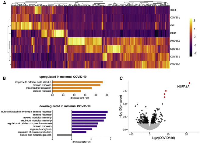
Findings: The majority (13 of 15) of placentas analyzed had no detectable viral RNA. ACE2 was detected by immunohistochemistry in syncytiotrophoblast cells of the normal placenta during early pregnancy but was rarely seen in healthy placentas at full term, suggesting that low ACE2 expression may protect the term placenta from viral infection. Using immortalized cell lines and primary isolated placental cells, we found that cytotrophoblasts, the trophoblast stem cells and precursors to syncytiotrophoblasts, rather than syncytiotrophoblasts or Hofbauer cells, are most vulnerable to SARS-CoV-2 infection in vitro. To better understand potential immune mechanisms shielding placental cells from infection in vivo, we performed bulk and single-cell transcriptomics analyses and found that the maternal-fetal interface of SARS-CoV-2-infected women exhibited robust immune responses, including increased activation of natural killer (NK) and T cells, increased expression of interferon-related genes, as well as markers associated with pregnancy complications such as preeclampsia.

Conclusions: SARS-CoV-2 infection in late pregnancy is associated with immune activation at the maternal-fetal interface even in the absence of detectable local viral invasion.

Funding: NIH (T32GM007205, F30HD093350, K23MH118999, R01AI157488, U01DA040588) and Fast Grant funding support from Emergent Ventures at the Mercatus Center.

Keywords: COVID-19; SARS-CoV-2; placenta; pregnancy.


Conflict of interest statement

A.I. is a scientific advisor for 4BIO and is on the advisory board of Med. The laboratory of A.I. received sponsored research funding from Spring Discovery.

Figures

None
Graphical abstract
Figure 1
Figure 1
Histopathology of representative COVID-19 and matched control placentas (A) COVID-19 placenta at low magnification revealed extensive intervillous fibrin deposition with only occasional areas of open (I) spaces. (A1) High magnification of the edge of a blood-filled I space and the earliest fibrin deposition (asterisks). Trapped chorionic villi (V) have become avascular and fibrotic. Initial fibrillar fibrin (arrowheads) can be seen at the blood-fibrin interface. (A2) Older area of I fibrin (asterisks) and trapped villi (V) revealing migration of trophoblasts (arrowheads) into the fibrin matrix. (A3) The oldest area of I fibrin became calcified (green asterisks), encasing villous remnants (V). (B) In sharp contrast, the control placenta revealed virtually no fibrin in the I space. (B1 and B2) Representative magnified areas revealed normal villi (V) and open, maternal blood containing I space, with only occasional foci of fibrin formation (arrowheads). Scale bars represents 200 μM for (A) and (B) and 50 μM for (A1)–(B2).
Figure 2
Figure 2
ACE2 protein expression in the placenta varies with gestational age (A) Human kidney used as a positive control revealed strong apical staining of the proximal tubules (P). The distal tubules (D) and glomerulus (G) were negative. The inset shows a serial section of the same kidney stained with non-immune rabbit serum, resulting in no staining. (B–D) Placentas derived from normal pregnancies between 7 and 15 weeks of gestation demonstrated strong, uniform, apical microvillus syncytiotrophoblast staining (arrowheads) and patchy strong basolateral staining at the cytotrophoblast-syncytiotrophoblast contact zone (arrows). V, villous core (E) A normal 21-week placenta still exhibited syncytiotrophoblast surface staining (arrowhead) but to a lesser extent than the earlier samples. Cytotrophoblast-syncytiotrophoblast contact zone staining was still prominent (arrow). (F) A representative normal placenta at 39 weeks revealed almost no ACE2 staining. Occasionally, staining at the cytotrophoblast-syncytiotrophoblast contact zone was noted (arrow). (G) Normal extravillous invasive trophoblasts from a 39-week placenta demonstrated strong surface expression of ACE2 with variable cytoplasmic staining. (H) Representative image of ACE2 expression in a 38-week placenta derived from an individual with symptomatic maternal COVID-19. Reappearance of strong apical microvillus syncytiotrophoblast (arrowheads) and cytotrophoblast-syncytiotrophoblast contact zone staining (arrows) was observed. All sections were cut at 5 μM, except (E), which was cut at 10 μM. (A–H) Scale bars represent 50 μM. (I) ACE2 H-score demonstrated steady loss of placental ACE2 with increasing gestational age in healthy pregnancies (p < 0.001). Linear regression (blue line) was fit to data from healthy controls (circles). 95% confidence interval is shown with dashed lines. Placentas derived from individuals with COVID-19 are depicted as red squares. (J) ACE2 H-score was increased significantly in term placentas from individuals with COVID-19 (squares) compared with uninfected, matched control individuals (circles).
Figure 3
Figure 3
SARS-CoV-2 infection of placental cells in vitro (A) Representative images of icSARS-CoV-2-mNG infection of primary placental cells, immortalized placental cell lines, and Vero E6 cells as measured by mNG expression and immunofluorescence staining of SARS-CoV-2 nucleocapsid (N). Images are displayed as maximum intensity projections of z stacks, and grayscale bars indicate measured fluorescence intensity in arbitrary digital units. (B) Fold change quantification of SARS-CoV-2 N1 by qRT-PCR 24 h after infection. Cells were infected at an MOI of 5 for 1 h and washed three times with PBS before addition of fresh medium. Cells were washed and collected 24 h after infection. Data presented are representative results from one of three replicates.
Figure 4
Figure 4
HSPA1A is significantly upregulated in maternal COVID-19 (A) Hierarchal clustering and heatmap of differentially expressed genes (p < 0.05). Bulk RNA sequencing (RNA-seq) was performed on placental villi isolated from control and maternal COVID cases. (B) Gene Ontology of differentially expressed genes (p < 0.05) in bulk RNA-seq. (C) Volcano plot indicating differentially expressed genes between control and maternal COVID-19 groups from bulk RNA-seq. Significant hits are depicted in red (padj < 0.05) and black (p < 0.05). Non-significant genes are shown in gray.
Figure 5
Figure 5
Single-cell RNA-seq of placental cells demonstrates gene expression changes in placental immune cells during COVID-19 (A) UMAP projection of 83,378 single placenta cells from COVID-19 cases (n = 2 decidual and n = 2 villous samples) and uninfected controls (n = 2 decidual and n = 3 villous samples). Cell type annotations are based on correlation with reference datasets,, , followed by manual examination of marker genes. (B) Dotplot of the top 5 genes that are upregulated between COVID-19 and uninfected control samples for each annotated cell type based on fold difference. The size of the dots represents the percentage of cells in each cluster expressing the gene of interest; the intensity of the color reflects average scaled expression. Significantly altered expression between COVID-19 cases and controls (Bonferroni-adjusted, two-tailed Wilcoxon rank-sum test, p < 0.05) is marked by a solid black line. (C) Interferome analysis demonstrating the fraction of differentially expressed genes in each cell type that are IFN responsive in COVID-19 cases compared with controls; p values for enrichment (observed over the expected fraction) were calculated using hypergeometric distribution. (D) Clustered heatmap showing the top enriched functional terms according to Metascape among differentially expressed genes between COVID-19 and control samples in the annotated placental cell types. Bars are colored to encode p values of increasing statistical significance. (E) Heatmap depicting the log-transformed ratio (COVID-19 cases over controls) of number of ligand-receptor interactions between all placental cell type pairs, inferred using the CellphoneDB repository of ligands, receptors, and their interactions. Red indicates cell type pairs with more interactions in COVID-19 cases compared with controls; blue indicates the opposite. (F) Violin plots of HSPA1A expression at the placental villi and maternal decidua obtained by single-cell RNA-seq (scRNA-seq).

Update of

References

    1. Zambrano L.D., Ellington S., Strid P., Galang R.R., Oduyebo T., Tong V.T., Woodworth K.R., Nahabedian J.F., 3rd, Azziz-Baumgartner E., Gilboa S.M., Meaney-Delman D., CDC COVID-19 Response Pregnancy and Infant Linked Outcomes Team Update: Characteristics of Symptomatic Women of Reproductive Age with Laboratory-Confirmed SARS-CoV-2 Infection by Pregnancy Status - United States, January 22-October 3, 2020. MMWR Morb. Mortal. Wkly. Rep. 2020;69:1641–1647. - PMC - PubMed
    1. Ahlberg M., Neovius M., Saltvedt S., Söderling J., Pettersson K., Brandkvist C., Stephansson O. Association of SARS-CoV-2 Test Status and Pregnancy Outcomes. JAMA. 2020;324:1782–1785. - PMC - PubMed
    1. Abasse S., Essabar L., Costin T., Mahisatra V., Kaci M., Braconnier A., Serhal R., Collet L., Fayssoil A. Neonatal COVID-19 Pneumonia: Report of the First Case in a Preterm Neonate in Mayotte, an Overseas Department of France. Children (Basel) 2020;7:E87. - PMC - PubMed
    1. Zimmermann P., Curtis N. COVID-19 in Children, Pregnancy and Neonates: A Review of Epidemiologic and Clinical Features. Pediatr. Infect. Dis. J. 2020;39:469–477. - PMC - PubMed
    1. Sherer M.L., Lei J., Creisher P., Jang M., Reddy R., Voegtline K., Olson S., Littlefield K., Park H.-S., Ursin R.L., et al. 2020. Dysregulated immunity in SARS-CoV-2 infected pregnant women. medRxiv. - DOI

Publication types

Substances