. 2021 May 28;41(6):BSR20204250.
doi: 10.1042/BSR20204250.

Reduction of TMAO level enhances the stability of carotid atherosclerotic plaque through promoting macrophage M2 polarization and efferocytosis

Affiliations

Reduction of TMAO level enhances the stability of carotid atherosclerotic plaque through promoting macrophage M2 polarization and efferocytosis

Weihao Shi et al. Biosci Rep. .

Abstract

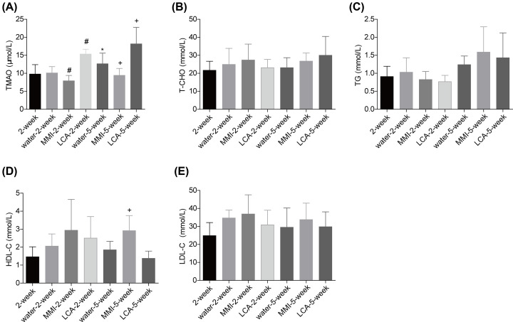
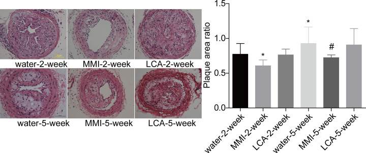
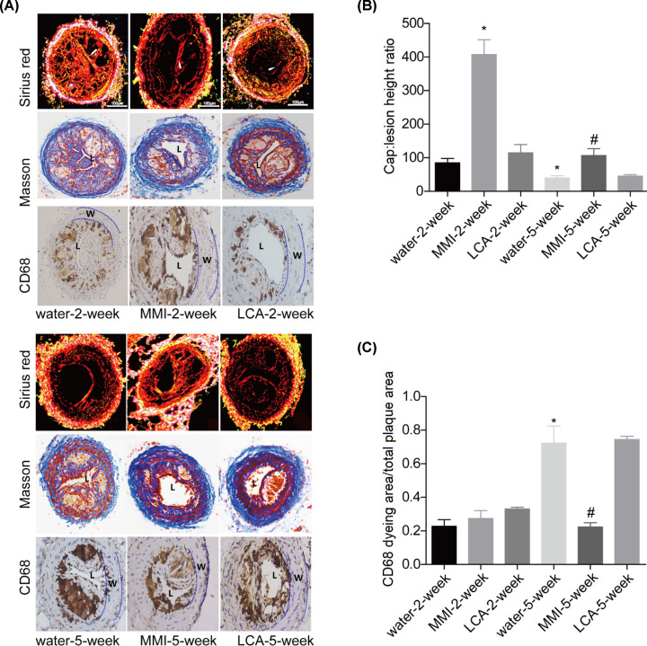
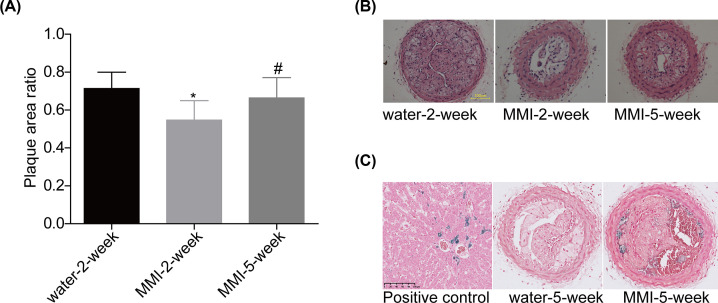
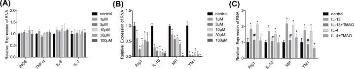
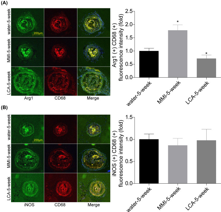
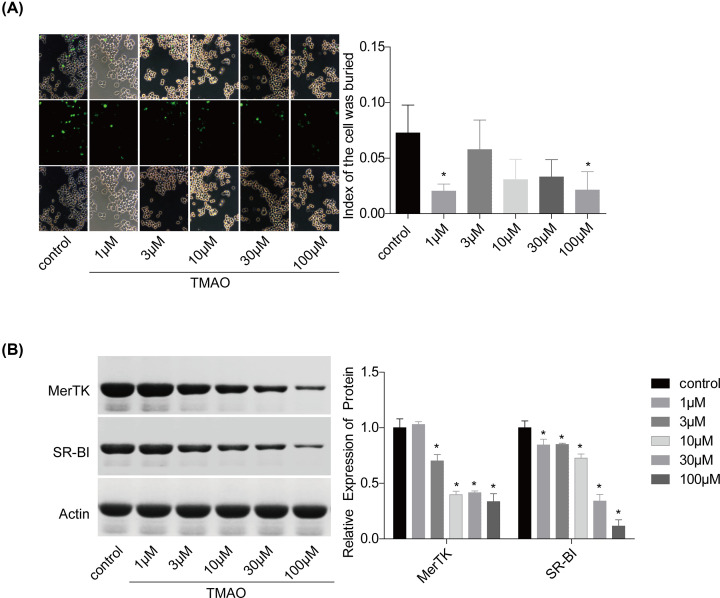
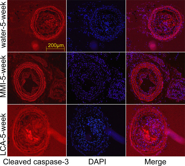
It has been demonstrated that trimethylamine N-oxide (TMAO) serves as a driver of atherosclerosis, suggesting that reduction of TMAO level might be a potent method to prevent the progression of atherosclerosis. Herein, we explored the role of TMAO in the stability of carotid atherosclerotic plaques and disclosed the underlying mechanisms. The unstable carotid artery plaque models were established in C57/BL6 mice. L-carnitine (LCA) and methimazole (MMI) administration were applied to increase and reduce TMAO levels. Hematoxylin and eosin (H&E) staining, Sirius red, Perl's staining, Masson trichrome staining and immunohistochemical staining with CD68 staining were used for histopathology analysis of the carotid artery plaque. M1 and M2 macrophagocyte markers were assessed by RT-PCR to determine the polarization of RAW264.7 cells. MMI administration for 2 weeks significantly decreased the plaque area, increased the thickness of the fibrous cap and reduced the size of the necrotic lipid cores, whereas 5-week of administration of MMI induced intraplate hemorrhage. LCA treatment further deteriorated the carotid atherosclerotic plaque but with no significant difference. In mechanism, we found that TMAO treatment impaired the M2 polarization and efferocytosis of RAW264.7 cells with no obvious effect on the M1 polarization. In conclusion, the present study demonstrated that TMAO reduction enhanced the stability of carotid atherosclerotic plaque through promoting macrophage M2 polarization and efferocytosis.

Keywords: efferocytosis; methimazole; plaque stability; polarization; trimethylamine N-oxide.


Conflict of interest statement

The authors declare that there are no competing interests associated with the manuscript.

Figures

Figure 1
Figure 1. Effect of MMI and LCA on the serum lipid of mice with unstable carotid artery plaques
After being anaesthetized, mice common carotid artery and the bifurcation of the carotid artery were separated and ligated to establish the unstable carotid artery plaque models. Then, the mice were divided into 2-week group (n=6), water-2-week group (n=10), MMI-2-week group (n=10), LCA-2-week group (n=10), water-5-week group (n=10), MMI-5-week group (n=10) and LCA-5-week group (n=10) and the plasma samples were collected for the following detection. (A) Plasma level of TMAO was measured by using the ultrahigh performance liquid chromatography tandem mass spectrometry (UHPLC-MS). (B–E) The serum levels of total cholesterol (T-CHO), triacylglycerol (TG), high-density lipoprotein cholesterol (HDL-C) and low-density lipoprotein cholesterol (LDL-C) were measured by using the enzymatic reagent kits (*P<0.05, compared with the 2-week group; #P<0.05, compared with the water-2-week group; +P<0.05, compared with the water-5-week group).
Figure 2
Figure 2. MMI and LCA roles in the plaque area in the unstable carotid artery plaque models
Carotid artery plaques were collected from mice in water-2-week group (n=10), MMI-2-week group (n=10), LCA-2-week group (n=10), water-5-week group (n=10), MMI-5-week group (n=10) and LCA-5-week group (n=10), and HE staining was then used to assess the plaque size (*P<0.05, compared with the water-2-week group; #P<0.05, compared with water-5-week group).
Figure 3
Figure 3. Effects of MMI and LCA on plaque composition in the unstable carotid artery plaque models
Carotid artery plaques in mice from the 2-week group (n=6), water-2-week group (n=10), MMI-2-week group (n=10), LCA-2-week group (n=10), water-5-week group (n=10), MMI-5-week group (n=10) and LCA-5-week group (n=10) were collected to the following assessments. (A) Sirius red staining, Masson trichrome staining and immunohistochemical staining of CD68 were used to assess plaque composition of the right common carotid artery in the unstable carotid artery plaque models (The lumen of the vessel has been indicated as “L”, and the vessel wall has been marked as “W”). (B) Bar graph of the Sirius red staining. (C) Bar graph of the CD68 staining (*P<0.05, compared with the water-2-week group; #P<0.05, compared with water-5-week group).
Figure 4
Figure 4. Long-term management of MMI induced intracellular hemorrhage in the unstable carotid artery plaque models
(A and B) HE staining was used to assess the plaque size in different groups. (C) Sporadic hemosiderin-laden macrophages were stained by Perl’s staining for iron (stained blue; *P<0.05, compared with the water-2-week group; #P<0.05, compared with the MMI-2-week group).
Figure 5
Figure 5. Effects of TMAO on the M1 and M2 polarization of RAW264.7 cells
(A and B) The expression levels of M1 (iNOS, TNF-α, IL-6 and IL-7) and M2 (Arg1, IL-10, MR and YM1) macrophagocyte markers in RAW264.7 cells treated with TMAO (0, 1, 3, 10, 30, 100 μM of 24 h) were determined by using RT-PCR. (C) The mRNA levels of Arg1, IL-10, MR and YM1 were detected by RT-PCR in RAW264.7 cells treated with IL-4 (20 ng/ml; 12 h) or IL-13 (10 ng/ml; 72 h) and TMAO (100 μM; 24 h; n=3, *P<0.05, compared with the control group; #P<0.05, compared with the IL-13 group; +P<0.05, compared with the IL-4 group).
Figure 6
Figure 6. MMI administration promoted M2 polarization and inhibited efferocytosis in vivo
(A and B) Immunofluorescence staining was used to evaluate the expression and location of Arg1, CD68 and iNOS in carotid arteries samples of mice in water-5-week, MMI-5-week and LCA-5-week groups. The co-locations of CD68 and Arg1/iNOS were quantitated (*P<0.05, compared with the water-5-week group).
Figure 7
Figure 7. Effects of TMAO on the efferocytosis of RAW264.7 cells
(A) The efferocytosis of RAW264.7 cells with different treatments (0, 1, 3, 10, 30, 100 μM of TMAO; 24 h) were assessed. (B) The expression levels of MerTK and SR-BI were measured by Western blotting (n=3, *P<0.05, compared with the control group).
Figure 8
Figure 8. MMI administration inhibited efferocytosis in vivo
Immunofluorescence staining was used to evaluate the expression of cleaved caspase-3 in carotid arteries samples of mice in water-5-week, MMI-5-week and LCA-5-week groups.

References

    1. Lloyd-Jones D., Adams R.J., Brown T.M., Carnethon M., Dai S., De Simone G.et al. . (2010) Heart disease and stroke statistics–2010 update: a report from the American Heart Association. Circulation 121, e46–e215, Epub 2009/12/19 - PubMed
    1. Wezel A., Welten S.M., Razawy W., Lagraauw H.M., de Vries M.R., Goossens E.A.et al. . (2015) Inhibition of MicroRNA-494 reduces carotid artery atherosclerotic lesion development and increases plaque stability. Ann. Surg. 262, 841–847, discussion 7-8. Epub 2015/11/20 10.1097/SLA.0000000000001466 - DOI - PubMed
    1. Chaturvedi S., Bruno A., Feasby T., Holloway R., Benavente O., Cohen S.N.et al. . (2005) Carotid endarterectomy–an evidence-based review: report of the Therapeutics and Technology Assessment Subcommittee of the American Academy of Neurology. Neurology 65, 794–801, Epub 2005/09/28 10.1212/01.wnl.0000176036.07558.82 - DOI - PubMed
    1. Shah P.K. (2003) Mechanisms of plaque vulnerability and rupture. J. Am. Coll. Cardiol. 41, 15S–22S, Epub 2003/03/20 10.1016/S0735-1097(02)02834-6 - DOI - PubMed
    1. Huang X.Z., Wang Z.Y., Dai X.H., Yun Z. and Zhang M. (2013) Velocity vector imaging of longitudinal mechanical properties of upstream and downstream shoulders and fibrous cap tops of human carotid atherosclerotic plaque. Echocardiography 30, 211–218, Epub 2012/10/26 10.1111/echo.12005 - DOI - PubMed

Publication types

MeSH terms