. 2021 May 28;478(10):1943-1958.
doi: 10.1042/BCJ20210203.

Histone H3 N-terminal mimicry drives a novel network of methyl-effector interactions

Affiliations

Histone H3 N-terminal mimicry drives a novel network of methyl-effector interactions

Jianji Chen et al. Biochem J. .

Abstract

The reader ability of PHD fingers is largely limited to the recognition of the histone H3 N-terminal tail. Distinct subsets of PHDs bind either H3K4me3 (a transcriptional activator mark) or H3K4me0 (a transcriptional repressor state). Structural studies have identified common features among the different H3K4me3 effector PHDs, including (1) removal of the initiator methionine residue of H3 to prevent steric interference, (2) a groove where arginine-2 binds, and (3) an aromatic cage that engages methylated lysine-4. We hypothesize that some PHDs might have the ability to engage with non-histone ligands, as long as they adhere to these three rules. A search of the human proteome revealed an enrichment of chromatin-binding proteins that met these criteria, which we termed H3 N-terminal mimicry proteins (H3TMs). Seven H3TMs were selected, and used to screen a protein domain microarray for potential effector domains, and they all had the ability to bind H3K4me3-interacting effector domains. Furthermore, the binding affinity between the VRK1 peptide and the PHD domain of PHF2 is ∼3-fold stronger than that of PHF2 and H3K4me3 interaction. The crystal structure of PHF2 PHD finger bound with VRK1 K4me3 peptide provides a molecular basis for stronger binding of VRK1 peptide. In addition, a number of the H3TMs peptides, in their unmethylated form, interact with NuRD transcriptional repressor complex. Our findings provide in vitro evidence that methylation of H3TMs can promote interactions with PHD and Tudor domain-containing proteins and potentially block interactions with the NuRD complex. We propose that these interactions can occur in vivo as well.

Keywords: NuRD complex; PHD domain; PHF2; Tudor domain; VRK1.


Conflict of interest statement

M.T.B. is a co-founder of EpiCypher.

Figures

Figure 1.
Figure 1.. Identification of H3 N-terminal mimicry proteins.
PANTHER Protein Class annotation of 20996 proteins in the human proteome (A), 209 internal motif-containing proteins (B), and 48 N-terminal motif-containing proteins (excluding histone H3 and variants) (C). (D) Phylogenetic analysis of the N-terminal sequences of 48 H3TMs and histone H3. (E) Red asterisks indicate seven H3TMs candidates for follow-up study and their N-terminal sequences and functions are summarized in Figure S1.
Figure 2.
Figure 2.. Identification of novel methylation-dependent interactions between H3TMs and PHD fingers.
N-terminal peptide (1–12 amino acids) were generated with K4me3 modified, for seven H3TMs and histone H3, and used to probe our methyl-reader microarray that contains 108 PHD fingers, 40 TUDOR and 31 Chromo domains. Duplicates of each protein domain were used to facilitate identification and reproducibility. The histone H3K4me3 peptide was used as positive control to visualize H3K4me3-interacting methyl reader domains.
Figure 3.
Figure 3.. Validation of novel interactions using in vitro interaction assays.
(A) Peptides pull down GST-tagged PHD fingers. Unmodified and K4me3 peptide probes for VRK1, BCL11B, TSHZ1, and H3 were used to pull down GST-tagged PHF2 PHD, ING2 PHD, ING3 PHD, and SPIN1 TUDOR. (B) Peptides pull down endogenous PHD fingers and NuRD complex from 293T whole cell lysates. Unmodified and K4me3 peptide probes for VRK1, BCL11B, TSHZ1, and H3 were used to pull down PHF2, ING2, ING3, SPIN1 and NuRD complex components including MTA1 and RBAP46/48. Immunoblot using streptavidin-HRP was used to ensure equal loading of all biotin-tagged peptide probes. Molecular Weight markers (kDa) are shown to the right of each panel.
Figure 4.
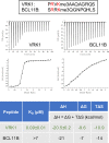
Figure 4.. Investigating the strength of the interaction between PHF2 and the N-termini of VRK1 and BCL11B.
Isothermal titration calorimetry measurements were performed with recombinant PHF2 (residues 1–451) and indicated peptides of VRK1 and BCL11B. The top panel is the raw data showing the heat released and measured by the sensitive calorimeter during gradual titration of the peptide into the sample cell containing PHF2 until the binding reaction has reached equilibrium. The bottom panel shows each peak in the raw data is integrated and plotted versus the molar ratio of peptide to protein. The resulting isotherm can be fitted to a binding model from which the affinity (KD) is derived. The Y-axis measures enthalpy change (ΔH) using the relationship ΔH = ΔG + TΔS where ΔG is the Gibbs free energy, ΔS is the entropy change and T is the absolute temperature.
Figure 5.
Figure 5.. Structure of PHD domain of PHF2 (residues 1–70) in complex with VRK1 peptide.
(A) A surface representation of the PHD domain displayed in red for negative, blue for positive and white for neutral. The bound VRK1 peptide is shown as a stick model in yellow. The bottom panel shows Fo-Fc omit electron density for the peptide, contoured at 5s above the mean. (B) The PHD domain in green is displayed in a ribbon model with the bound VRK1 peptide in yellow. The two zinc ions are coordinated with Cys4 and His-Cys3, respectively. The bottom panel shows antiparallel arrangement between VRK1 peptide (in yellow) and the PHF2 strand (in green). The 2Fo-Fc electron density is contoured at 2s above the mean. (C) Superimposition of PHF2 with bound VRK1 peptide (yellow) and histone H3 peptide (grey). (D) Interaction with VRK1 P1 residue. (E) Interaction with histone H3 A1 residue (PDB 3KQI). (F) Interaction with VRK1 R2 residue. (G) Interaction with VRK1 V3 residue. (H) Aromatic cage for binding K4me3. (I) The bound K4me3 from the viewpoint of Tyr7 of PHF2. (J and K) Interactions with VRK1 A5 and A6 residues. (L and M) Interactions with VRK1 Q7.
Figure 6.
Figure 6.. Three H3TMs can be methylated at Lys4 by H3K4 Methyltransferases.
Unmodified and K4me3 peptide probes for VRK1, BCL11B, TSHZ1, and H3 were incubated with three H3K4-specific methyltransferases, Set7/9 (upper), MLL1 (middle) and PRDM9 (lower). Enzymes and 3H-labeled Sam were used as a negative control (most right lane). Immunoblot using streptavidin-HRP was used to ensure equal loading of all biotin-tagged peptide probes. Molecular Weight markers (kDa) are shown to the right of each panel.
Figure 7.
Figure 7.. The Model of H3TM interactions.
In their methylated state, H3TMs can bind PHD and Tudor domains that are implicated in maintaining an open chromatin state, and are generally involved in promoting transcription. If the H3TMs are unmethylated they have the potential of interacting with the NuRD transcriptional repressor complex.

Similar articles

Cited by

References

    1. Jenuwein, T. and Allis, C.D. (2001) Translating the histone code. Science 293, 1074–1080 10.1126/science.1063127 - DOI - PubMed
    1. Kouzarides, T. (2007) Chromatin modifications and their function. Cell 128, 693–705 10.1016/j.cell.2007.02.005 - DOI - PubMed
    1. Ruthenburg, A.J., Li, H., Patel, D.J. and Allis, C.D. (2007) Multivalent engagement of chromatin modifications by linked binding modules. Nat. Rev. Mol. Cell Biol. 8, 983–994 10.1038/nrm2298 - DOI - PMC - PubMed
    1. Taverna, S.D., Li, H., Ruthenburg, A.J., Allis, C.D. and Patel, D.J. (2007) How chromatin-binding modules interpret histone modifications: lessons from professional pocket pickers. Nat. Struct. Mol. Biol. 14, 1025–1040 10.1038/nsmb1338 - DOI - PMC - PubMed
    1. Strahl, B.D. and Allis, C.D. (2000) The language of covalent histone modifications. Nature 403, 41–45 10.1038/47412 - DOI - PubMed

Publication types

MeSH terms