Comparative Study
. 2021 May 10:10:e59525.
doi: 10.7554/eLife.59525.

Recovery of consciousness and cognition after general anesthesia in humans

Affiliations
Comparative Study

Recovery of consciousness and cognition after general anesthesia in humans

George A Mashour et al. Elife. .

Abstract

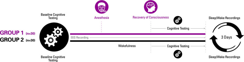
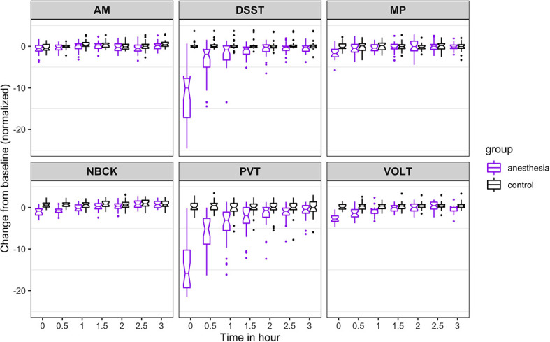
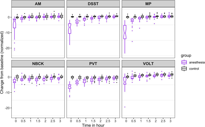
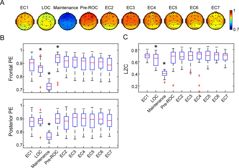
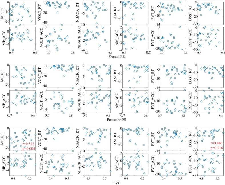
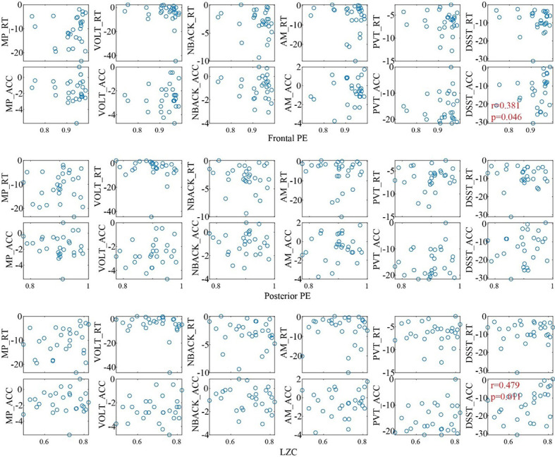
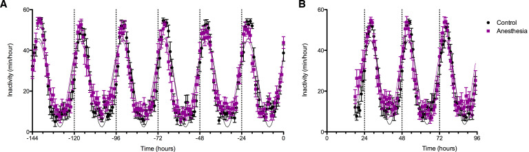
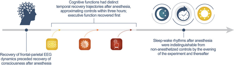
Understanding how the brain recovers from unconsciousness can inform neurobiological theories of consciousness and guide clinical investigation. To address this question, we conducted a multicenter study of 60 healthy humans, half of whom received general anesthesia for 3 hr and half of whom served as awake controls. We administered a battery of neurocognitive tests and recorded electroencephalography to assess cortical dynamics. We hypothesized that recovery of consciousness and cognition is an extended process, with differential recovery of cognitive functions that would commence with return of responsiveness and end with return of executive function, mediated by prefrontal cortex. We found that, just prior to the recovery of consciousness, frontal-parietal dynamics returned to baseline. Consistent with our hypothesis, cognitive reconstitution after anesthesia evolved over time. Contrary to our hypothesis, executive function returned first. Early engagement of prefrontal cortex in recovery of consciousness and cognition is consistent with global neuronal workspace theory.

Trial registration: ClinicalTrials.gov NCT01911195.

Keywords: anesthesia; cognition; consciousness; frontal cortex; human; medicine; neuroscience.

Plain language summary

Anesthesia is a state of reversable, controlled unconsciousness. It has enabled countless medical procedures. But it also serves as a tool for scientists to study how the brain regains consciousness after disruptions such as sleep, coma or medical procedures requiring general anesthesia. It is still unclear how exactly the brain regains consciousness, and less so, why some patients do not recover normally after general anesthesia or fail to recover from brain injury. To find out more, Mashour et al. studied the patterns of reemerging consciousness and cognitive function in 30 healthy adults who underwent general anesthesia for three hours. While the volunteers were under anesthesia, their brain activity was measured with an EEG; and their sleep-wake activity was measured before and after the experiment. Each participant took part in a series of cognitive tests designed to measure the reaction speed, memory and other functions before receiving anesthesia, right after the return of consciousness, and then every 30 minutes thereafter. Thirty healthy volunteers who did not have anesthesia also completed the scans and tests as a comparison group. The experiments showed that certain normal EEG patterns resumed just before a person wakes up from anesthesia. The return of thinking abilities was an extended, multistep process, but volunteers recovered their cognitive abilities to nearly the same level as the volunteers within three hours of being deeply anesthetized. Mashour et al. also unexpectedly found that abstract problem-solving resumes early in the process, while other functions such as reaction time and attention took longer to recover. This makes sense from an evolutionary perspective. Sleep leaves individuals vulnerable. Quick evaluation and decision-making skills would be key to respond to a threat upon waking. The experiments confirm that the front of the brain, which handles thinking and decision-making, was especially active around the time of recovery. This suggests that therapies targeting this part of the brain may help people who experience loss of consciousness after a brain injury or have difficulties waking up after anesthesia. Moreover, disorders of cognition, such as delirium, in the days following surgery may be caused by factors other than the lingering effects of anesthetic drugs on the brain.


Conflict of interest statement

GM, BP, MB, DL, WW, SB, NL, KM, MM, VT, GV, EO, RH, MS, HM, RH, EJ, GG, PP, AM, MA, MK No competing interests declared

Figures

Figure 1.
Figure 1.. Experimental design.
Participants were randomized to one of two groups for investigating recovery of consciousness and cognition after general anesthesia. Sleep-wake actigraphy data were acquired in the week leading up to the day of the experiment, which started with baseline cognitive testing followed by either a period of general anesthesia (1.3 age-adjusted minimum alveolar concentration of isoflurane) or wakefulness. Upon recovery of consciousness (or similar time point for controls), recurrent cognitive testing was performed for 3 hr. Actigraphy resumed for 3 days after the experiment.
Figure 2.
Figure 2.. Time course for recovery of (normalized) accuracy in cognitive task performance after general anesthesia (time 0 is just after recovery of consciousness in the group that was anesthetized).
AM, Abstract Matching; DSST, Digit Symbol Substitution Test; MP, Motor Praxis; NBCK, Fractal 2-Back; PVT, Psychomotor Vigilance Test; VOLT, Visual Object Learning Test. The six cognitive tests are all represented.
Figure 3.
Figure 3.. Time course for recovery of (normalized) speed of cognitive task performance after general anesthesia (time 0 is just after recovery of consciousness in the group that was anesthetized).
AM, Abstract Matching; DSST, Digit Symbol Substitution Test; MP, Motor Praxis; NBCK, Fractal 2-Back; PVT, Psychomotor Vigilance Test; VOLT, Visual Object Learning Test. The six cognitive tests are all represented.
Figure 4.
Figure 4.. Cortical dynamics before, during, and after general anesthesia.
(A) Scalp topographic maps of the group-level permutation entropy (PE; median average across N = 30 participants) at the ten studied epochs. (B) The box plots of average PE values in frontal (Fp1, Fp2, Fpz, F3, F4, and Fz) and posterior channels (P3, P4, Pz, O1, O2, and Oz) for the studied epochs. On each box, the central mark is the median, the edges are the 25th and 75th percentiles, the whiskers extend to the most extreme data points determined by the MATLAB algorithm to be non-outliers, and the points deemed by the algorithm to be outliers are plotted individually (red cross). (C) The box plots of LZC values for the studied epochs. EC = eyes closed resting state (EC1 is baseline consciousness, EC2-7 are post-emergence just prior to cognitive testing), LOC = loss of consciousness, Pre-ROC = 2 min epoch just before recovery of consciousness. *indicates significant difference relative to EC1, using linear mixed model analysis (Bonferroni-corrected p<0.05).
Figure 4—figure supplement 1.
Figure 4—figure supplement 1.. Confirmatory results of permutation entropy (PE) with different settings of embedding dimension (dE) and time delay (τ).
The description of the figure and statistical results are the same as those in Figure 4A-B.
Figure 4—figure supplement 2.
Figure 4—figure supplement 2.. Confirmatory results of Lempel-Ziv Complexity (LZC).
(A) Sensitivity of the LZC measure to the selection of threshold for binarization. (B) Confirmatory results of LZC by using spatial shuffling (top) and phase randomization (bottom) in the generation of surrogate data for normalization. The spatial shuffling method permutates the spatial order (at each time point) of the spatiotemporal matrix. The phase randomization method preserves the spectral profile of the signals and reflects the complexity changes beyond the power spectrogram. The description of the figure and the statistical results are same with those in Figure 4C.
Figure 4—figure supplement 3.
Figure 4—figure supplement 3.. Cortical dynamics as assessed by permutation (PE) and Lempel-Ziv complexity (LZC) for the non-anesthetized control group.
(A) Scalp topographic maps of the group-level PE at the seven resting-state eyes-closed (EC) epochs. (B) The box plots of average PE values in frontal and posterior channels for the studied epochs. (C) The box plots of LZC values for the studied epochs.
Figure 4—figure supplement 4.
Figure 4—figure supplement 4.. Associations between EEG measures during pre-anesthetic baseline (EC1) with the impairment of cognitive functions at emergence (just after recovery of consciousness).
The EEG measures do not appear to be related to the degree of performance impairment via Spearman’s rank correlation analysis. AM, Abstract Matching; DSST, Digit Symbol Substitution Test; MP, Motor Praxis; NBCK, Fractal 2-Back; PVT, Psychomotor Vigilance Test; VOLT, Visual Object Learning Test; RT, Response time; ACC, Accuracy.
Figure 4—figure supplement 5.
Figure 4—figure supplement 5.. Associations between EEG measures during maintenance with the impairment of cognitive functions at emergence.
Only significant correlations were indicated (Spearman’s rank correlation, p<0.05).
Figure 4—figure supplement 6.
Figure 4—figure supplement 6.. Associations between EEG measures during pre-ROC with the impairment of cognitive functions at emergence.
Only significant correlations are indicated (Spearman’s rank correlation, p<0.05).
Figure 5.
Figure 5.. Effects of anesthetic exposure on rest-activity rhythms.
(A) Rest activity plots are displayed in the week prior to the study day for volunteers that were subsequently randomized to anesthetized (purple) or control (black) conditions. (B) Rest-activity rhythms in the same participants are displayed on the evening of the study day and for the ensuing days. Time = 0 corresponds to midnight on the evening of the study day.
Figure 6.
Figure 6.. Summary of the study findings.
Author response image 1.
Author response image 2.

Similar articles

Cited by

References

    1. Abásolo D, Simons S, Morgado da Silva R, Tononi G, Vyazovskiy VV. Lempel-Ziv complexity of cortical activity during sleep and waking in rats. Journal of Neurophysiology. 2015;113:2742–2752. doi: 10.1152/jn.00575.2014. - DOI - PMC - PubMed
    1. Avidan MS, Evers AS. The Fallacy of Persistent Postoperative Cognitive Decline. Anesthesiology. 2016;124:255–258. doi: 10.1097/ALN.0000000000000958. - DOI - PMC - PubMed
    1. Bandt C, Pompe B. Permutation entropy: a natural complexity measure for time series. Physical Review Letters. 2002;88:174102. doi: 10.1103/PhysRevLett.88.174102. - DOI - PubMed
    1. Banks MI, Krause BM, Endemann CM, Campbell DI, Kovach CK, Dyken ME, Kawasaki H, Nourski KV. Cortical functional connectivity indexes arousal state during sleep and anesthesia. NeuroImage. 2020;211:116627. doi: 10.1016/j.neuroimage.2020.116627. - DOI - PMC - PubMed
    1. Basner M, Mollicone D, Dinges DF. Validity and Sensitivity of a Brief Psychomotor Vigilance Test (PVT-B) to Total and Partial Sleep Deprivation. Acta Astronautica. 2011;69:949–959. doi: 10.1016/j.actaastro.2011.07.015. - DOI - PMC - PubMed

Publication types

Associated data