. 2021 Jun 22;144(5):1422-1434.
doi: 10.1093/brain/awab041.

Biallelic variants in HPDL cause pure and complicated hereditary spastic paraplegia

Manuela Wiessner  1 Reza Maroofian  2 Meng-Yuan Ni  3 Andrea Pedroni  4 Juliane S Müller  5 Rolf Stucka  1 Christian Beetz  6 Stephanie Efthymiou  2 Filippo M Santorelli  7 Ahmed A Alfares  8 Changlian Zhu  9   10   11 Anna Uhrova Meszarosova  12 Elham Alehabib  13 Somayeh Bakhtiari  14 Andreas R Janecke  15   16 Maria Gabriela Otero  17 Jin Yun Helen Chen  18 James T Peterson  19 Tim M Strom  20 Peter De Jonghe  21   22   23 Tine Deconinck  24 Willem De Ridder  21   22   23 Jonathan De Winter  21   22   23 Rossella Pasquariello  7 Ivana Ricca  7 Majid Alfadhel  25 Bart P van de Warrenburg  26 Ruben Portier  27 Carsten Bergmann  28   29 Saghar Ghasemi Firouzabadi  30 Sheng Chih Jin  31 Kaya Bilguvar  32   33 Sherifa Hamed  34 Mohammed Abdelhameed  34 Nourelhoda A Haridy  2   34 Shazia Maqbool  35 Fatima Rahman  35 Najwa Anwar  35 Jenny Carmichael  36 Alistair Pagnamenta  37 Nick W Wood  2   38 Frederic Tran Mau-Them  39 Tobias Haack  40 Genomics England Research Consortium, PREPARE networkMaja Di Rocco  41 Isabella Ceccherini  42 Michele Iacomino  43 Federico Zara  43   44 Vincenzo Salpietro  44   45 Marcello Scala  44   45 Marta Rusmini  42 Yiran Xu  9 Yinghong Wang  46 Yasuhiro Suzuki  47 Kishin Koh  48 Haitian Nan  48 Hiroyuki Ishiura  49 Shoji Tsuji  50 Laëtitia Lambert  51 Emmanuelle Schmitt  52 Elodie Lacaze  53 Hanna Küpper  54 David Dredge  18 Cara Skraban  55   56 Amy Goldstein  19   56 Mary J H Willis  57 Katheryn Grand  58 John M Graham  58 Richard A Lewis  59 Francisca Millan  60 Özgür Duman  61 Nihal Dündar  62 Gökhan Uyanik  63   64 Ludger Schöls  65   66 Peter Nürnberg  67 Gudrun Nürnberg  67 Andrea Catala Bordes  12 Pavel Seeman  12 Martin Kuchar  68 Hossein Darvish  69 Adriana Rebelo  70 Filipa Bouçanova  4   71 Jean-Jacques Medard  4   71 Roman Chrast  4   71 Michaela Auer-Grumbach  72 Fowzan S Alkuraya  73 Hanan Shamseldin  73 Saeed Al Tala  74 Jamileh Rezazadeh Varaghchi  75 Maryam Najafi  6   76 Selina Deschner  77 Dieter Gläser  77 Wolfgang Hüttel  78 Michael C Kruer  14 Erik-Jan Kamsteeg  76 Yoshihisa Takiyama  48 Stephan Züchner  70 Jonathan Baets  21   22   23 Matthis Synofzik  65   66 Rebecca Schüle  65   66 Rita Horvath  5 Henry Houlden  2 Luca Bartesaghi  4   71 Hwei-Jen Lee  3 Konstantinos Ampatzis  4 Tyler Mark Pierson  17   59   79   80 Jan Senderek  1
Affiliations

Biallelic variants in HPDL cause pure and complicated hereditary spastic paraplegia

Manuela Wiessner et al. Brain. .

Erratum in

  • Erratum to: Biallelic variants in HPDL cause pure and complicated hereditary spastic paraplegia.
    Wiessner M, Maroofian R, Ni MY, Pedroni A, Müller JS, Stucka R, Beetz C, Efthymiou S, Santorelli FM, Alfares AA, Zhu C, Uhrova Meszarosova A, Alehabib E, Bakhtiari S, Janecke AR, Otero MG, Chen JYH, Peterson JT, Strom TM, De Jonghe P, Deconinck T, De Ridder W, De Winter J, Pasquariello R, Ricca I, Alfadhel M, van de Warrenburg BP, Portier R, Bergmann C, Ghasemi Firouzabadi S, Jin SC, Bilguvar K, Hamed S, Abdelhameed M, Haridy NA, Maqbool S, Rahman F, Anwar N, Carmichael J, Pagnamenta AT, Wood NW, Tran Mau-Them F, Haack T; Genomics England Research Consortium, PREPARE network; Di Rocco M, Ceccherini I, Iacomino M, Zara F, Salpietro V, Scala M, Rusmini M, Xu Y, Wang Y, Suzuki Y, Koh K, Nan H, Ishiura H, Tsuji S, Lambert L, Schmitt E, Lacaze E, Küpper H, Dredge D, Skraban C, Goldstein A, Willis MJH, Grand K, Graham JM, Lewis RA, Millan F, Duman Ö, Olgac Dundar N, Uyanik G, Schöls L, Nürnberg P, Nürnberg G, Català-Bordes A, Seeman P, Kuchar M, Darvish H, Rebelo A, Bouçanova F, Medard JJ, Chrast R, Auer-Grumbach M, Alkuraya FS, Shamseldin H, Al Tala S, Rezazadeh Varaghchi J, Najafi M, Deschner S, Gläser D, Hüttel W, Kruer MC, Kamsteeg EJ, Takiyama Y, Züchner S, Baets J, Synofzik M, Sch… See abstract for full author list ➔ Wiessner M, et al. Brain. 2021 Sep 4;144(8):e70. doi: 10.1093/brain/awab193. Brain. 2021. PMID: 34480796 Free PMC article. No abstract available.

Abstract

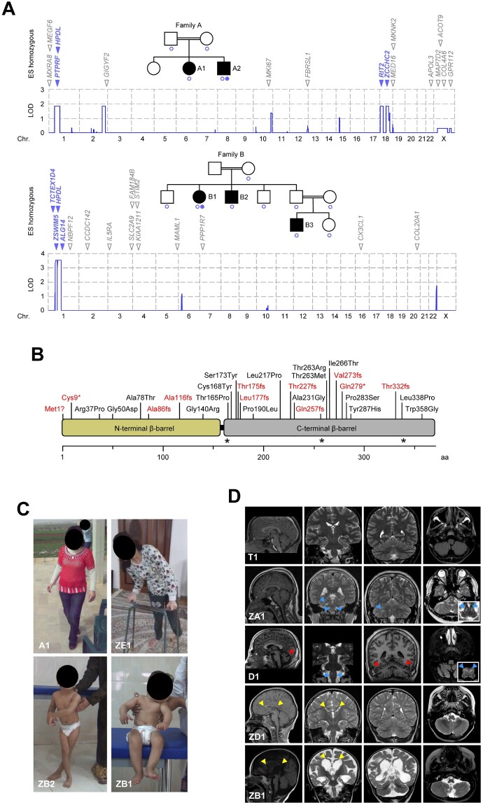
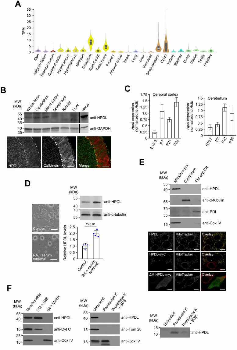
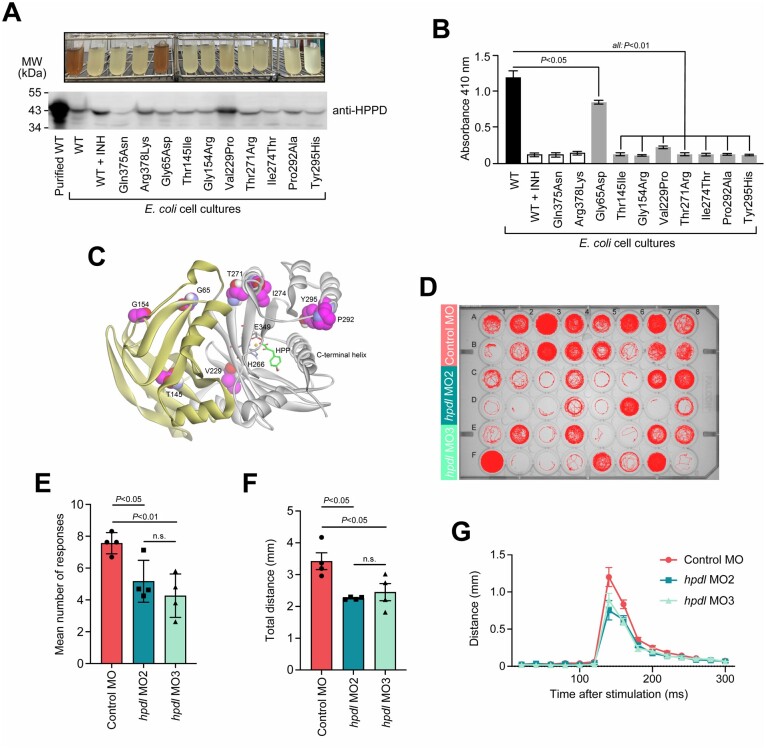
Human 4-hydroxyphenylpyruvate dioxygenase-like (HPDL) is a putative iron-containing non-heme oxygenase of unknown specificity and biological significance. We report 25 families containing 34 individuals with neurological disease associated with biallelic HPDL variants. Phenotypes ranged from juvenile-onset pure hereditary spastic paraplegia to infantile-onset spasticity and global developmental delays, sometimes complicated by episodes of neurological and respiratory decompensation. Variants included bona fide pathogenic truncating changes, although most were missense substitutions. Functionality of variants could not be determined directly as the enzymatic specificity of HPDL is unknown; however, when HPDL missense substitutions were introduced into 4-hydroxyphenylpyruvate dioxygenase (HPPD, an HPDL orthologue), they impaired the ability of HPPD to convert 4-hydroxyphenylpyruvate into homogentisate. Moreover, three additional sets of experiments provided evidence for a role of HPDL in the nervous system and further supported its link to neurological disease: (i) HPDL was expressed in the nervous system and expression increased during neural differentiation; (ii) knockdown of zebrafish hpdl led to abnormal motor behaviour, replicating aspects of the human disease; and (iii) HPDL localized to mitochondria, consistent with mitochondrial disease that is often associated with neurological manifestations. Our findings suggest that biallelic HPDL variants cause a syndrome varying from juvenile-onset pure hereditary spastic paraplegia to infantile-onset spastic tetraplegia associated with global developmental delays.

Keywords: HPDL; HSP; autosomal recessive; hereditary spastic paraplegia; mitochondrial disorder.


Figures

Figure 1
Figure 1
Biallelic HPDL variants and associated phenotypes. (A) Genome-wide linkage analysis and exome sequencing in two consanguineous HSP families. SNP-based linkage analysis pinpointed four chromosomal regions where the maximum theoretical LOD score was obtained in Family A (LOD 1.81) and a single linkage interval reaching genome-wide significance in Family B (LOD 3.56). Blue circles indicate individuals included in linkage studies. Exome sequencing on one subject from each family identified several rare non-synonymous homozygous variants that were located in regions of interest as defined by linkage analysis (blue) or outside these regions (grey). HPDL was the only gene residing in the linked region on chromosome 1 that harboured rare homozygous non-synonymous variants in both families. Filled blue circles indicate individuals whose DNA was analysed by exome sequencing. (B) Schematic representation of the human HPDL protein and distribution of truncating (red) and missense (black) variants. By analogy to the crystallographic structures of HPPD, the structure of HPDL is predicted to contain two open β-barrels. The carboxy-terminal domain (grey) would possess the putative catalytic centre, while the amino-terminal domain (yellow) may not have a direct catalytic function but may play a role in dimer formation. Amino acid numbering is shown along the bottom. Asterisks mark the position of residues predicted to coordinate an iron ion to form the active site (similar to HPPD). (C) Variable severity of clinical presentation of individuals with HPDL variants. Subject A1 is able to walk without support but with scissoring gait (age 16 years). Subject ZE1 can only ambulate with a walker (age 15 years). Subject ZB2 needs support to stand with legs crossed at age 6 years. Subject ZB1 cannot sit independently at age 2.5 years. (D) Brain MRI findings. Subject T1 (age 17 years) had mild juvenile-onset pure HSP and normal neuroimaging. Subjects ZA1 (age 15 years) and D1 (age 21 years) had earlier-onset, intermediate disease associated with cerebellar atrophy (red arrowheads, Subject D1) and T2 hyperintensities (blue arrowheads) in the medulla oblongata (Subject ZA1 and D1) and middle cerebellar peduncle (Subject ZA1). Subjects ZD1 (age 5 years) and ZB1 (age 19 months) had severe disease associated with a hypoplastic corpus callosum (yellow arrowheads, Subjects ZD1 and ZB1), generalized reduction of white matter volume (Subjects ZD1 and ZB1) as well as ventriculomegaly, global cerebral atrophy and a simplified gyral pattern (Subject ZB1).
Figure 2
Figure 2
Expression and subcellular localization of HPDL. (A) Gene expression for HPDL across 24 human tissues. Expression values are shown as transcripts per million (TPM). Box plots represent median and 25th and 75th percentiles. Data were retrieved from the GTEx web resource [http://www.gtexportal.org/home/ (date accessed 20/06/2019)]. (B) Detection of the HPDL protein in young adult mice and rats (both P56). Top: HPDL levels in mouse neural and non-neural tissues were analysed by immunoblotting using an anti-HPDL antibody. Levels of the housekeeping protein GAPDH were determined to ensure equal loading of samples. Bottom: Immunofluorescence microscopy with an anti-HPDL antibody demonstrated presence of HPDL in rat cerebellar neurons (Purkinje cells). Calbindin was used as a marker to visualize Purkinje cells. Merge represents the overlay of calbindin and HPDL signals. Scale bar = 100 µm. (C) Hpdl expression during development of the mouse nervous system. Hpdl mRNA levels in embryonic and postnatal stages were determined by quantitative RT-PCR and normalized to Actb expression. Bars correspond to means of three biological and technical replicates and error bars represent SD. (D) HPDL expression in an in vitro model of neural differentiation. HPDL protein levels in undifferentiated (control) and retinoic acid-differentiated (RA + serum removal) Neuro2a murine neuroblastoma cells were analysed by immunoblotting. Bars represent the mean of four independent experiments and error bars indicate SD. The P-value was calculated by Student’s unpaired two-tailed t-test. Changes of cell morphology were recorded to document successful differentiation of cells. Scale bars = 50 µm. (E) Association of HPDL with mitochondria. Top: Fractionation of HeLa cells using the Qproteome Mitochondria Isolation Kit (Qiagen) showed co-isolation of HPDL with mitochondrial marker Cox IV. ER = endoplasmic reticulum; PM = plasma membrane. Bottom: Immunofluorescence microscopy of HeLa cells showed co-localization of endogenous HPDL with the mitochondrial stain MitoTracker Red. Myc-tagged HPDL showed an identical pattern of expression, while an artificial myc-tagged mutant lacking the mitochondrial targeting signal (Δ-M-HPDL-myc) displayed a diffuse cytoplasmic signal. Scale bars = 10 µm. (F) Submitochondrial localization of HPDL. Left: Upon subfractionation of HeLa mitochondria, HPDL was found within the fraction containing the outer membrane (OM) and intermembrane space (IMS) but was not detectable in the fraction consisting of inner membrane (IM) and the mitochondrial matrix. Middle: Following proteinase K digestion of intact HeLa mitochondria, signals for HPDL and OM protein Tom20 but not for IMS protein cytochrome C (Cyt C) were lost. Right: Using a high-resolution gel and short blotting time, a low molecular weight band was detected by the anti-HPDL antibody after proteinase K treatment, possibly corresponding to the region that is N-terminal of a predicted transmembrane domain and projects into the IMS. Uncropped versions of blots in B and DF are shown in Supplementary Fig. 15.
Figure 3
Figure 3
Functional analysis of HPDL. (A) Enzymatic activity of recombinant E. coli expressing HPDL missense variants on a HPPD backbone. The variants tested included artificial variants known to render HPPD functionless (Gln375Asn, Arg378Lys29) or affecting amino acid residues invariant in HPDL and HPPD amino-terminal (HPDL-Thr131 = HPPD-Thr145) and carboxy-terminal domains (HPDL-Pro284 = HPPD-Pro292) as well as six amino acid changes corresponding to disease-related HPDL variants (HPDL-Gly50 = HPPD-Gly65, HPDL-Gly140 = HPPD-Gly154, HPDL-Leu217 = HPDD-Val229, HPDL-Thr263 = HPPD-Thr271, HPDL-Ile266 = HPPD-Ile274, and HPDL-Tyr287 = HPPD-Tyr295). Catalytic activity of HPPD species in growth medium was reflected by formation of a brown pigment (top). Expression of HPPD species was confirmed by immunoblotting of total bacterial protein extracts (bottom, wild-type HPPD purified from bacteria served as a size control). An uncropped version of the blot is shown in Supplementary Fig. 15. (B) Pigment formation reflecting HPPD activity was quantified by measuring the absorbance at 410 nm. Bars represent the mean of three independent experiments and error bars indicate SD. The P-values were calculated by one-way ANOVA followed by Tukey’s post hoc correction for multiple comparisons. (C) Structural model of human HPPD (Protein Data Bank accession number 3ISQ) with bound 4-HPP. The protein folds into an amino-terminal (yellow) and a carboxy-terminal β-barrel (grey), which are represented as a ribbon backbone trace. Residues that bind to the substrate 4-HPP (green) and coordinate the iron ion within the catalytic centre are displayed in ball and stick model format (grey). Residues affected by substitutions that were tested in the enzymatic assay (A and B) are drawn as a CPK model (magenta). (D) Behavioural analysis of hpdl zebrafish morphants. MO2 and MO3 were morpholinos targeting zebrafish hpdl. The control morpholino was a random sequence not predicted to target any known gene. A single fish was placed in each well of the plate and movements were recorded automatically. The image shows representative traces (red) of the entire path the animals swam during the whole experiment (20-min acclimation phase followed by 10-min experimental phase, 1 vibratory stimulus/min). (E) Average number of zebrafish’s positive responses to stimulation. The maximum reachable number of responses was 10 responses to 10 stimuli. (F) Average distance zebrafish larvae swam in response to stimulation. Data were only included for measurements where fish responded to stimulation. (G) Representation of the dislocation of fish over time. The graph shows the Euclidian distance between the positions of fish in consecutive frames (50 frames/s). In all three experimental groups, maximum motor response was recorded at ∼140 ms (time point 0 ms corresponds to the time of stimulation). Data were only included for measurements where fishes responded to stimulation. Data-points in EG correspond to means from four independent experiments each involving 16 embryos per condition. Error bars represent SEM. The P-values were calculated by one-way ANOVA. INH = inhibitor sulcotrione; WT = wild-type.

References

    1. Harding AE. Classification of the hereditary ataxias and paraplegias. Lancet. 1983;321:1151–1155. - PubMed
    1. Hedera P. Hereditary spastic paraplegia overview. In: Adam MP, Ardinger HH, Pagon RA, et al., editors. GeneReviews ®. Seattle (WA): University of Washington, Seattle; 2000. - PubMed
    1. Trummer B, Haubenberger D, Blackstone C.. Clinical trial designs and measures in hereditary spastic paraplegias. Front Neurol. 2018;9:1017. - PMC - PubMed
    1. Shribman S, Reid E, Crosby AH, Houlden H, Warner TT.. Hereditary spastic paraplegia: From diagnosis to emerging therapeutic approaches. Lancet Neurol. 2019;18:1136–1146. - PubMed
    1. Casari G, De Fusco M, Ciarmatori S, et al.Spastic paraplegia and OXPHOS impairment caused by mutations in paraplegin, a nuclear-encoded mitochondrial metalloprotease. Cell. 1998;93:973–983. - PubMed

Publication types