

Quantitative magnetic resonance imaging towards clinical application in multiple sclerosis
- PMID: 33970206
- PMCID: PMC8219362
- DOI: 10.1093/brain/awab029
Quantitative magnetic resonance imaging towards clinical application in multiple sclerosis
Abstract
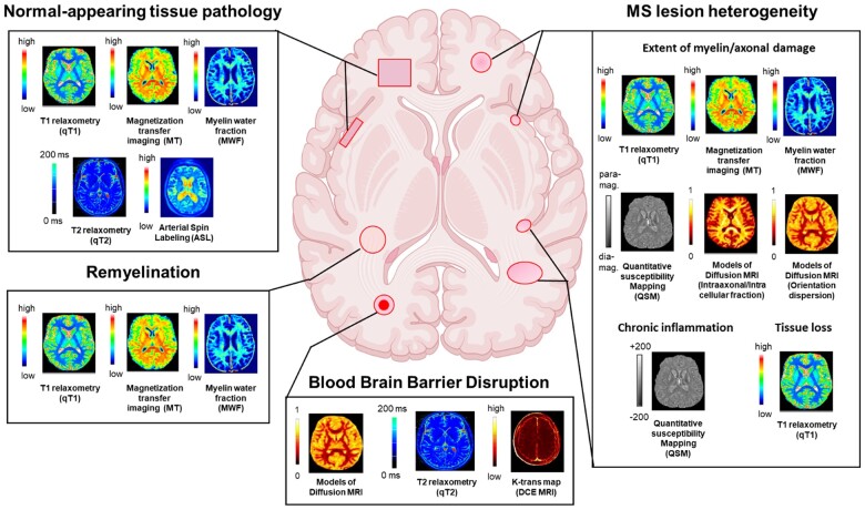
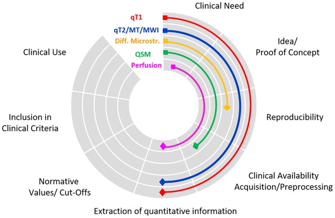
Quantitative MRI provides biophysical measures of the microstructural integrity of the CNS, which can be compared across CNS regions, patients, and centres. In patients with multiple sclerosis, quantitative MRI techniques such as relaxometry, myelin imaging, magnetization transfer, diffusion MRI, quantitative susceptibility mapping, and perfusion MRI, complement conventional MRI techniques by providing insight into disease mechanisms. These include: (i) presence and extent of diffuse damage in CNS tissue outside lesions (normal-appearing tissue); (ii) heterogeneity of damage and repair in focal lesions; and (iii) specific damage to CNS tissue components. This review summarizes recent technical advances in quantitative MRI, existing pathological validation of quantitative MRI techniques, and emerging applications of quantitative MRI to patients with multiple sclerosis in both research and clinical settings. The current level of clinical maturity of each quantitative MRI technique, especially regarding its integration into clinical routine, is discussed. We aim to provide a better understanding of how quantitative MRI may help clinical practice by improving stratification of patients with multiple sclerosis, and assessment of disease progression, and evaluation of treatment response.
Keywords: imaging; multiple sclerosis; quantitative MRI.
© The Author(s) (2021). Published by Oxford University Press on behalf of the Guarantors of Brain.
Figures
References
-
- Rovira A, Wattjes MP, Tintore M, et al. Evidence-based guidelines: MAGNIMS consensus guidelines on the use of MRI in multiple sclerosis-clinical implementation in the diagnostic process. Nat Rev Neurol. 2015;11:471-482. - PubMed
-
- Wattjes MP, Rovira A, Miller D, et al. Evidence-based guidelines: MAGNIMS consensus guidelines on the use of MRI in multiple sclerosis–establishing disease prognosis and monitoring patients. Nat Rev Neurol. 2015;11:597-606. - PubMed
-
- Lucchinetti C, Brück W, Parisi J, Scheithauer B, Rodriguez M, Lassmann H.. Heterogeneity of multiple sclerosis lesions: Implications for the pathogenesis of demyelination. Ann Neurol. 2000;47:707-717. - PubMed
Publication types
MeSH terms
Grants and funding
LinkOut - more resources
Full Text Sources
Other Literature Sources
Medical
