

Loss of H3K27me3 in meningiomas
- PMID: 33970242
- PMCID: PMC8328029
- DOI: 10.1093/neuonc/noab036
Loss of H3K27me3 in meningiomas
Abstract
Background: There is a critical need for objective and reliable biomarkers of outcome in meningiomas beyond WHO classification. Loss of H3K27me3 has been reported as a prognostically unfavorable alteration in meningiomas. We sought to independently evaluate the reproducibility and prognostic value of H3K27me3 loss by immunohistochemistry (IHC) in a multicenter study.
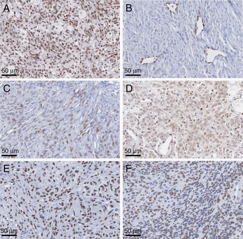
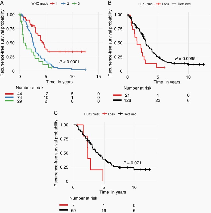
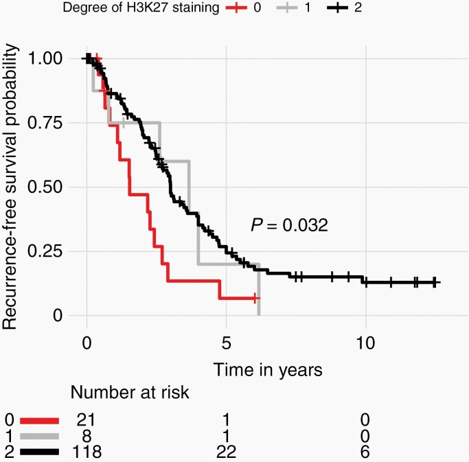
Methods: IHC staining for H3K27me3 and analyses of whole slides from 181 meningiomas across three centers was performed. Staining was analyzed by dichotomization into loss and retained immunoreactivity, and using a 3-tiered scoring system in 151 cases with clear staining. Associations of grouping with outcome were performed using Kaplan-Meier survival estimates.
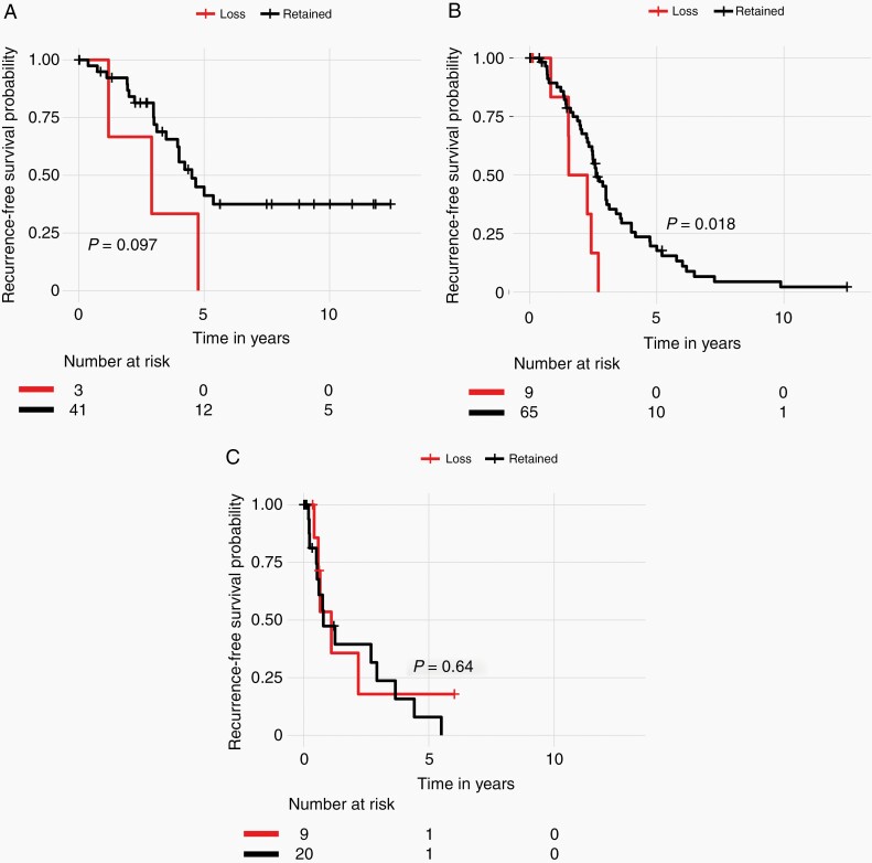
Results: A total of 21 of 151 tumors (13.9%) demonstrated complete loss of H3K27me3 staining in tumor with retained endothelial staining. Overall, loss of H3K27me3 portended a worse outcome with shorter times to recurrence in our cohort, particularly for WHO grade 2 tumors which were enriched in our study. There were no differences in recurrence-free survival (RFS) for WHO grade 3 patients with retained vs loss of H3K27me3. Scoring by a 3-tiered system did not add further insights into the prognostic value of this H3K27me3 loss. Overall, loss of H3K27me3 was not independently associated with RFS after controlling for WHO grade, extent of resection, sex, age, and recurrence status of tumor on multivariable Cox regression analysis.
Conclusions: Loss of H3K27me3 identifies a subset of WHO grade 2 and possibly WHO grade 1 meningiomas with increased recurrence risk. Pooled analyses of a larger cohort of samples with standardized reporting of clinical definitions and staining patterns are warranted.
Keywords: H3K27; immunohistochemistry; meningioma; trimethylation.
© The Author(s) 2021. Published by Oxford University Press on behalf of the Society for Neuro-Oncology. All rights reserved. For permissions, please e-mail: journals.permissions@oup.com.
Figures
Comment in
-
Prognostication for meningiomas: H3K27me3 to the rescue?Neuro Oncol. 2021 Aug 2;23(8):1218-1219. doi: 10.1093/neuonc/noab083. Neuro Oncol. 2021. PMID: 33822195 Free PMC article. No abstract available.
References
-
- Gauchotte G, Peyre M, Pouget C, et al. . Prognostic value of histopathological features and loss of H3K27me3 immunolabeling in anaplastic meningioma: a multicenter retrospective study. J Neuropathol Exp Neurol. 2020;79(7):754–762. - PubMed
-
- Katz LM, Hielscher T, Liechty B, et al. . Loss of histone H3K27me3 identifies a subset of meningiomas with increased risk of recurrence. Acta Neuropathol. 2018;135(6):955–963. - PubMed
-
- Perry A. Meningiomas. In: Practical Surgical Neuropathology: A Diagnostic Approach. Elsevier. 2018:259–298.
-
- Marosi C, Hassler M, Roessler K, et al. . Meningioma. Crit Rev Oncol Hematol. 2008;67(2):153–171. - PubMed
Publication types
MeSH terms
Substances
Grants and funding
LinkOut - more resources
Full Text Sources
Other Literature Sources
