. 2021 Feb;10(2):e1170.
doi: 10.1002/mbo3.1170.

A study of innate immune kinetics reveals a role for a chloride transporter in a virulent Francisella tularensis type B strain

Affiliations

A study of innate immune kinetics reveals a role for a chloride transporter in a virulent Francisella tularensis type B strain

Lisa M Matz et al. Microbiologyopen. 2021 Feb.

Abstract

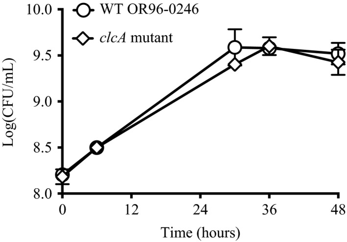
Tularemia is a zoonotic disease of global proportions. Francisella tularensis subspecies tularensis (type A) and holarctica (type B) cause disease in healthy humans, with type A infections resulting in higher mortality. Repeated passage of a type B strain in the mid-20th century generated the Live Vaccine Strain (LVS). LVS remains unlicensed, does not protect against high inhalational doses of type A, and its exact mechanisms of attenuation are poorly understood. Recent data suggest that live attenuated vaccines derived from type B may cross-protect against type A. However, there is a dearth of knowledge regarding virulent type B pathogenesis and its capacity to stimulate the host's innate immune response. We therefore sought to increase our understanding of virulent type B in vitro characteristics using strain OR96-0246 as a model. Adding to our knowledge of innate immune kinetics in macrophages following infection with virulent type B, we observed robust replication of strain OR96-0246 in murine and human macrophages, reduced expression of pro-inflammatory cytokine genes from "wild type" type B-infected macrophages compared to LVS, and delayed macrophage cell death suggesting that virulent type B may suppress macrophage activation. One disruption in LVS is in the gene encoding the chloride transporter ClcA. We investigated the role of ClcA in macrophage infection and observed a replication delay in a clcA mutant. Here, we propose its role in acid tolerance. A greater understanding of LVS attenuation may reveal new mechanisms of pathogenesis and inform strategies toward the development of an improved vaccine against tularemia.

Keywords: Francisella tularensis subsp. holarctica; Live Vaccine Strain; TargeTron™ chromosomal insertion; innate immunity; proton:chloride antiporter; tularemia.


Conflict of interest statement

None declared.

Figures

FIGURE 1
FIGURE 1
Primed human and murine macrophages are permissive for replication of WT type B, while clcA mutant shows delayed replication. Primed human THP‐1 (left) or primed murine J774A.1 (right) macrophages were grown to confluence in 24‐well plates and infected with virulent type B strain OR96‐0246 (WT), clcA mutant, or LVS as described in Materials and Methods. After phagocytosis, cells were washed and treated with gentamicin to remove extracellular bacteria. At 4‐, 18‐, and 36‐h post‐infection, macrophages were lysed to release intracellular bacteria. Bacteria were plated on MHII agar and incubated for 48 h at 37°C with 5% CO2 before enumeration. Growth is reported as log10 (CFU/ml) over time. Data represent four independent experiments for WT and two independent experiments for LVS and clcA mutant. Treatments were performed in triplicate or quadruplicate, with triplicate spot plating. Counts were averaged for each dilution. Bars represent standard error of the mean (SEM). *p < 0.05; **p < 0.01; ****p < 0.0001 as determined by two‐way ANOVA (Table 2). Comparisons to LVS in human macrophages were not performed since no CFU were recovered
FIGURE 2
FIGURE 2
Pro‐inflammatory cytokine gene expression profiles in macrophages reflect the virulence status of strains. Total RNA from infected macrophages was collected, reverse‐ transcribed, and analyzed via qRT‐PCR for human (a–c) and murine (d–h) macrophages as described in Materials and Methods. Fold change is normalized to β‐actin. Samples are calibrated to transcript levels from uninfected macrophages, represented by the dashed line at 1. Means and SEM are shown. Samples were tested in duplicate for each gene with technical triplicates. Human data represents one independent experiment, and no significant differences are reported. Murine data represents two independent experiments each with two technical replicates. *p < 0.05; **p < 0.01; ***p < 0.001; ****p < 0.0001 as determined by two‐way ANOVA
FIGURE 3
FIGURE 3
Cell death of infected macrophages. 96‐well plates were seeded with 5 × 104 J774A.1 murine (a) or THP‐1 human (b) macrophages and infected with either WT type B, attenuated type B strain LVS, or clcA mutant in quadruplicate. At the indicated time points supernatants were transferred to a new plate and cell death was determined by the lactate dehydrogenase assay as described in Materials and Methods. Absorbance values were averaged and converted into percentages. A representative experiment is shown for clcA mutant and human macrophage cell death. Two independent experiments are shown for WT‐ and LVS‐infected murine macrophages. Mean and SEM shown. No significant differences were detected
FIGURE 4
FIGURE 4
WT Francisella type B ClcA can partially restore acid tolerance in an acid‐sensitive E. coli mutant. WT E. coli strain MG1655, double mutant ΔclcAΔclcB, or double mutant complemented with pFTClcA or pWSK29 empty vector control (both induced with 2.5 mM IPTG) were plated for survival after incubation for 2 h in acid buffer with 1.5 mM glutamate at pH 2.5. Counts were normalized to PBS input controls, and survival ratios were converted into percentages. For all strains, survival in the absence of glutamate was less than 0.05%. Data represents 4 to 6 independent experiments, with averaged triplicate CFU counts. Mean with SEM shown. *p < 0.05 as determined by the Kruskal–Wallis test with Dunn's post‐test to adjust for multiple comparisons
FIGURE A1
FIGURE A1
Transmembrane prediction of ClcA. Probability of ClcA membrane insertion determined by TMHMM Server v. 2.0 (www.cbs.dtu.dk) for OR96‐0246 (top, AAX59_00075) and LVS (bottom, FTL_0102). Amino acid length is displayed on the x‐axis and probability on the y‐axis. Red regions represent helices within ClcA predicted to be inserted into the membrane
FIGURE A2
FIGURE A2
LVS growth is determined by the activation state of murine macrophages. J774A.1 murine macrophages were primed with LPS‐EB or left untreated and infected with LVS as described in Materials and Methods. An MOI of 1:150 CFU:macrophage was used. After phagocytosis, cells were washed and treated with gentamicin to remove extracellular bacteria. At 4, 18, and 36 hours post‐infection (HPI) macrophages were lysed to release intracellular bacteria. Bacteria were plated on MHII agar and incubated for 48 h at 37°C with 5% CO2 before enumeration. Growth is reported as Log10(CFU/ml) over time. Representative data are shown. Treatments were performed in triplicate, with triplicate spot plating. Counts were averaged for each dilution. Bars represent standard error of the mean (SEM)
FIGURE A3
FIGURE A3
MacVector protein alignments of Francisella (WT and LVS) and Escherichia coli and chloride transporter proteins. The consensus is shown in gray. Important conserved residues are serine (107), glutamic acid (148, 203), and tyrosine (445). LVS truncation occurs at position 106 resulting in an early stop codon, producing only 20% of the full‐length protein
FIGURE A4
FIGURE A4
Confirmation of clcA disruption. Gel 1, Top: TargeTron™ colony PCR products from electroporation of OR96‐0246 with pJFP1004 to generate clcA mutant. M, marker. Gene‐specific 0095 F/R primers: Lane 1, WT control; Lanes 2–4, transformants; Lane 5, NTC. Duplicate PCR reaction run on Lanes 6–10 to verify results. Band in Lane 5 (NTC) is likely an artifact from colony PCR/gel loading and is confirmed negative in Lane 10 (NTC). Gel 1, Bottom: Lanes 1–5, same as Lanes 1–5 above using intron‐specific F/R primers. Lanes 7–10 are products from Kan probe F/R primers to confirm plasmid curing: Lane 7, KanR positive control; Lanes 8–10, transformants restreaked on Kan MHII plates; Lane 11, NTC (partially cropped). Gel 2: Colony PCR of recovered CFU from clcA mutant‐infected wells at 36 HPI were run on 1% agarose confirming the presence of disruption. Lanes 1 and 3, supernatants from J774A.1 and THP‐1 wells, respectively; Lanes 2 and 4, colonies from plated J774A.1 and THP‐1 lysates; Lane 5, NTC (all with gene‐specific 0095 F/R primers). Lanes 6–9, same as Lanes 1–4 but with intron‐specific F/R primers; Lane 10, NTC
FIGURE A5
FIGURE A5
Growth of clcA mutant matches that of WT OR96‐0246 in broth culture. WT and clcA mutant strains were grown in modified Mueller‐Hinton broth as described in Materials and Methods. Data represent two independent experiments with standard deviation shown
FIGURE A6
FIGURE A6
Cytokine profiling shows diminished secretion of pro‐inflammatory cytokines by primed murine macrophages when infected with WT compared to LVS, and for IL‐6 in human macrophages. Primed human (A‐C) or J774A.1 murine (D‐F) macrophages were infected with WT, LVS, and clcA mutant (B and C only) as described in Materials and Methods. Supernatants were inactivated and analyzed by ELISA for the indicated inflammatory cytokines. Murine IL‐6 values in panel D are as follows: WT means at 4, 18, and 36 HPI are 179, 1064, and 3107 pg/ml, respectively; LVS means at 4, 18, and 36 HPI are 291, 1632, and 4351 pg/ml, respectively; Untreated means at 4, 18, and 36 HPI are 239, 2381, and 1688 pg/ml, respectively; Pam3CSK4 means at 4, 18, and 36 HPI are 360, 9206, and 4187 pg/ml, respectively. HS—high sensitivity. Values below the limit of detection are reported as zero. Mean with SEM shown for two independent experiments. Pam3CSK4 is a TLR2 agonist and serves as a positive control
FIGURE A7
FIGURE A7
LVS elicits cell death in a dose‐dependent manner in murine macrophages. Primed (a) or unprimed (b) murine macrophages were infected with LVS at five different MOIs. At the indicated time points, supernatants were transferred to a new plate and cell death was determined by the lactate dehydrogenase assay as described in Materials and Methods. Absorbance values were averaged and converted into percentages. Mean and SEM shown for two independent experiments

Similar articles

Cited by

  • Ex vivo infection model for Francisella using human lung tissue.
    Köppen K, Fatykhova D, Holland G, Rauch J, Tappe D, Graff M, Rydzewski K, Hocke AC, Hippenstiel S, Heuner K. Köppen K, et al. Front Cell Infect Microbiol. 2023 Jul 10;13:1224356. doi: 10.3389/fcimb.2023.1224356. eCollection 2023. Front Cell Infect Microbiol. 2023. PMID: 37492528 Free PMC article.

References

    1. Abu, K. Y. (2013). Targeting nutrient retrieval by Francisella tularensis . Frontiers in Cellular and Infection Microbiology, 3, 64. - PMC - PubMed
    1. Abu Kwaik, Y., & Bumann, D. (2013). Microbial quest for food in vivo: “Nutritional virulence” as an emerging paradigm. Cellular Microbiology, 15, 882–890. - PubMed
    1. Adcock, N. J., Morris, B. J., & Rice, E. W. (2014). Acid resistance in Francisella tularensis . Microbiologyopen, 3, 133–138. - PMC - PubMed
    1. Appelt, S., Faber, M., Köppen, K., Jacob, D., Grunow, R., & Heuner, K. (2020). Francisella tularensis subspecies holarctica and tularemia in Germany. Microorganisms, 8, 1–14. - PMC - PubMed
    1. Atianand, M. K., Duffy, E. B., Shah, A., Kar, S., Malik, M., & Harton, J. A. (2011). Francisella tularensis reveals a disparity between human and mouse NLRP3 inflammasome activation. Journal of Biological Chemistry, 286, 39033–39042. - PMC - PubMed

Publication types

MeSH terms

LinkOut - more resources