Review
. 2020 May;4(5):e1900126.
doi: 10.5435/JAAOSGlobal-D-19-00126.

Late Effects of Clubfoot Deformity in Adolescent and Young Adult Patients Whose Initial Treatment Was an Extensive Soft-tissue Release: Topic Review and Clinical Case Series

Affiliations
Review

Late Effects of Clubfoot Deformity in Adolescent and Young Adult Patients Whose Initial Treatment Was an Extensive Soft-tissue Release: Topic Review and Clinical Case Series

Jeffrey E Johnson et al. J Am Acad Orthop Surg Glob Res Rev. 2020 May.

Abstract

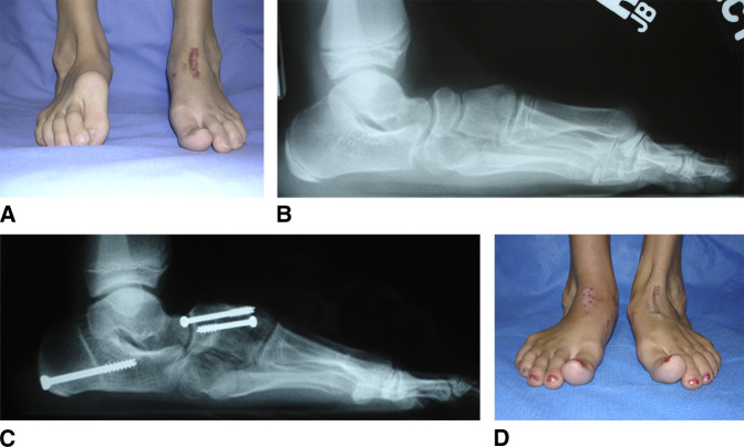
Children with congenital clubfoot often have residual deformity, pain, and limited function in adolescence and young adulthood. These patients represent a heterogeneous group that often requires an individualized management strategy. This article reviews the available literature on this topic while proposing a descriptive classification system based on a review of patients at our institution who underwent surgery for problems related to previous clubfoot deformity during the period between January 1999 and January 2012. Seventy-two patients (93 feet) underwent surgical treatment for the late effects of clubfoot deformity at an average age of 13 years (range 9 to 19 years). All patients had been treated at a young age with serial casting, and most had at least one previous surgery on the affected foot or feet. Five common patterns of pathology identified were as follows: undercorrection, overcorrection, dorsal bunion, anterior ankle impingement, and lateral hindfoot impingement. Management pathways for each group of the presenting problems is described. To our knowledge, this topic review represents the largest report of adolescent and young adult patients with residual clubfoot deformity in the literature.


Conflict of interest statement

None of the following authors or any immediate family member has received anything of value from or has stock or stock options held in a commercial company or institution related directly or indirectly to the subject of this article: Dr. Johnson, Dr. Fortney, Dr. Luk, Dr. Klein, Dr. McCormick, Dr. Dobbs, Dr. Gordon, and Schoenecker.

Figures

Figure 1
Figure 1
Overcorrection. AF, Preoperative and postoperative radiographs and photographs of a 14-year-old boy with a history of bilateral congenital clubfoot, who underwent extensive soft-tissue releases at 6 months of age. He presented with pes planovalgus deformity consistent with overcorrection. He was treated with a medial displacement calcaneal osteotomy, calcaneocuboid joint bone-block distraction fusion, midtarsal fusion, advancement of the posterior tibialis tendon, spring ligament reefing, open tendoachilles Z-lengthening, and intramuscular lengthening of peroneus longus and peroneus brevis.
Figure 2
Figure 2
Undercorrection. AH, Preoperative and postoperative radiographs and photographs of a 17-year-old boy with a history of idiopathic clubfoot and an extensive soft-tissue release at 6 months of age, presenting with recurrent (undercorrected) deformity. The deformity was centered around the midfoot. He was treated with an open plantar fascia release, biplanar midfoot osteotomy with lateral closing and medial opening wedge, dorsiflexion osteotomy of the first metatarsal, anterior tibialis tendon transfer to the third cuneiform, and a peroneus longus to peroneus brevis tendon transfer.
Figure 3
Figure 3
Dorsal bunion. AD, Preoperative and postoperative radiographs and clinical photographs of a 10-year-old girl with a history of bilateral congenital clubfoot treated with extensive soft-tissue releases at 6 months of age, who presented with bilateral dorsal bunion deformities. She was treated with a double bone-block fusion of the navicular-first cuneiform and first-second intercuneiform joints, anterior tibial tendon transfer to the second cuneiform, flexor hallucis brevis intramuscular lengthening, first metatarsophalangeal joint release and pinning, and flexor hallucis longus tendon transfer to the first metatarsal neck. The photographs illustrate the preoperative deformity on the right foot after left foot correction and postoperative correction of both feet.
Figure 4
Figure 4
Anterior Impingement. AD, Preoperative, intraoperative, and postoperative radiographs and preoperative clinical photographs of an 11-year-old boy with a history of congenital clubfoot and multiple previous surgeries. His primary report was anterior ankle pain and inability to achieve neutral dorsiflexion. Residual cavovarus deformity was also present. He was treated with a dorsiflexion-producing anterior closing-wedge tibial osteotomy with fibular osteotomy, midfoot osteotomy, plantar fascia release, and anterior tibialis tendon transfer to second cuneiform.
Figure 5
Figure 5
Lateral Impingement. A and B, Preoperative CT scan of a patient with a history of congenital clubfoot deformity and report of lateral hindfoot pain. The CT scan demonstrates subfibular impingement of the lateral calcaneus. Treatment included a lateral calcaneal wall exostectomy and débridement of the subfibular region. The black line represents an approximation of the bone removal.

References

    1. Cooper DM, Dietz FR: Treatment of idiopathic clubfoot. A thirty-year follow-up note. Bone Joint Surg Am 1995;77:1477-1489. - PubMed
    1. Dobbs MB, Gurnett CA: Update on clubfoot: Etiology and treatment. Clin Orthop Relat Res 2009;467:1146-1153. - PMC - PubMed
    1. Ryoppy S, Sairanen H: Neonatal operative treatment of club foot. A preliminary report. J Bone Joint Surg Br 1983;65:320-325. - PubMed
    1. Simons GW: Complete subtalar release in club feet. Part II. Comparison with less extensive procedures. J Bone Joint Surg Am 1985;67:1056-1065. - PubMed
    1. Turco VJ: Resistant congenital club foot--one-stage posteromedial release with internal fixation. A follow-up report of a fifteen-year experience. J Bone Joint Surg Am 1979;61:805-814. - PubMed