Review
. 2021 Sep;288(17):5021-5041.
doi: 10.1111/febs.15961. Epub 2021 May 20.

Infection-induced inflammation from specific inborn errors of immunity to COVID-19

Affiliations
Review

Infection-induced inflammation from specific inborn errors of immunity to COVID-19

Cheng-Lung Ku et al. FEBS J. 2021 Sep.

Abstract

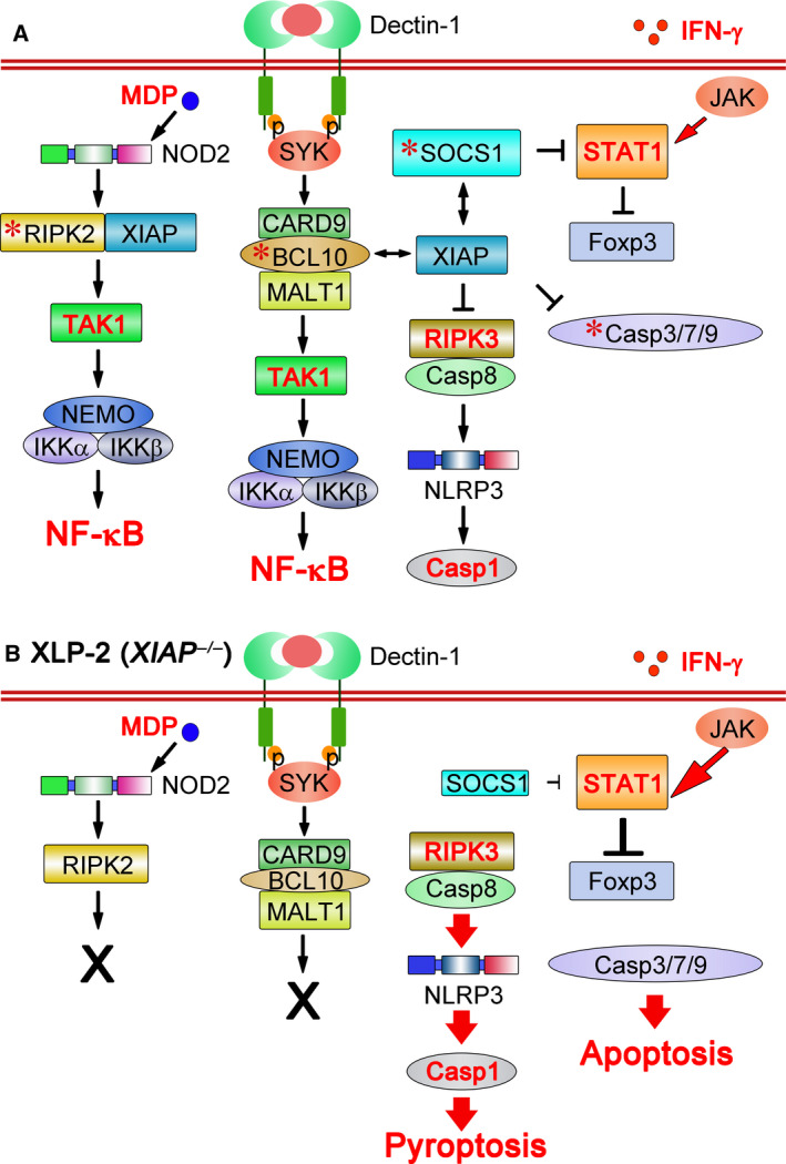
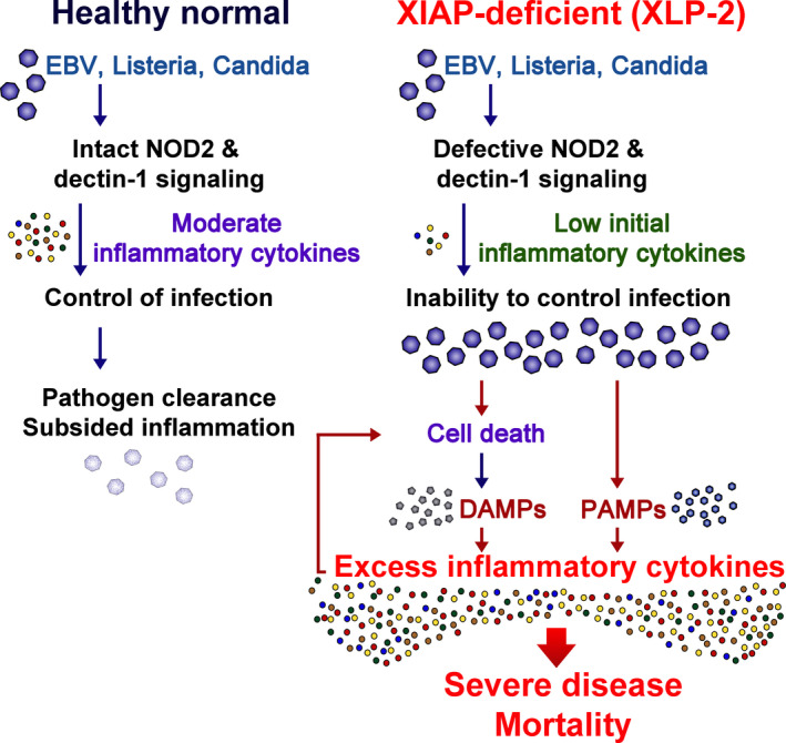
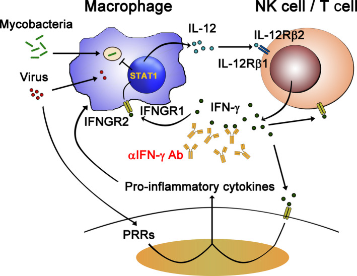
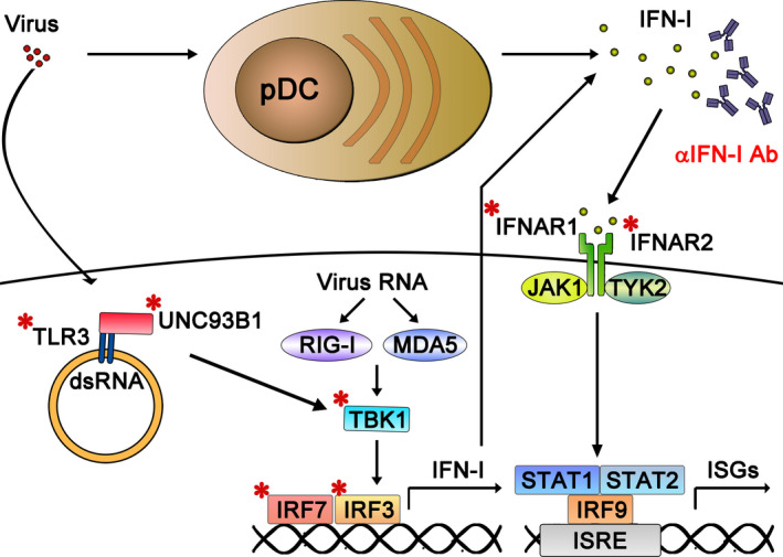
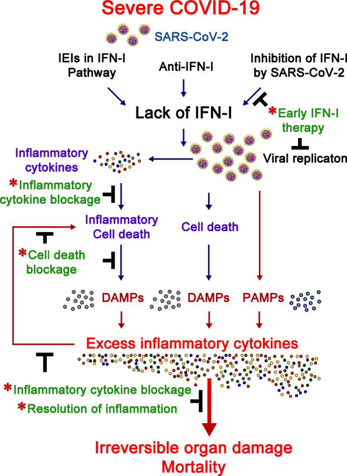
Inborn errors of immunity (IEIs) are a group of genetically defined disorders leading to defective immunity. Some IEIs have been linked to mutations of immune receptors or signaling molecules, resulting in defective signaling of respective cascades essential for combating specific pathogens. However, it remains incompletely understood why in selected IEIs, such as X-linked lymphoproliferative syndrome type 2 (XLP-2), hypo-immune response to specific pathogens results in persistent inflammation. Moreover, mechanisms underlying the generation of anticytokine autoantibodies are mostly unknown. Recently, IEIs have been associated with coronavirus disease 2019 (COVID-19), with a small proportion of patients that contract severe COVID-19 displaying loss-of-function mutations in genes associated with type I interferons (IFNs). Moreover, approximately 10% of patients with severe COVID-19 possess anti-type I IFN-neutralizing autoantibodies. Apart from IEIs that impair immune responses to severe acute respiratory syndrome coronavirus 2 (SARS-CoV-2), SARS-CoV-2 encodes several proteins that suppress early type I IFN production. One primary consequence of the lack of type I IFNs during early SARS-CoV-2 infection is the increased inflammation associated with COVID-19. In XLP-2, resolution of inflammation rescued experimental subjects from infection-induced mortality. Recent studies also indicate that targeting inflammation could alleviate COVID-19. In this review, we discuss infection-induced inflammation in IEIs, using XLP-2 and COVID-19 as examples. We suggest that resolving inflammation may represent an effective therapeutic approach to these diseases.

Keywords: COVID-19; SARS-CoV-2; XLP-2; anticytokine autoantibodies; inborn errors of immunity.


Conflict of interest statement

The authors declare no conflict of interest.

Figures

Fig. 1
Fig. 1
Signaling defects in XIAP‐deficient individuals. (A) XIAP regulates NOD2, dectin‐1, and Treg cells by binding to RIPK2, BCL10, and SOCS1. Binding of XIAP to RIPK2 polyubiquitinates RIPK2 and activates NOD2 signaling [27, 28, 29, 30]. XIAP also binds and stabilizes BCL10 to enable dectin‐1 signaling [33]. Interaction of XIAP with SOCS1 stabilizes this latter, helping to maintain regulatory T‐cell populations by suppressing IFN‐γ‐STAT1 signaling [34]. XIAP also inhibits RIPK3 and caspase‐8‐mediated NLRP3 inflammasome activation [41, 42]. Moreover, it binds and inactivates caspase‐3, caspase‐7, and caspase‐9. *, XIAP‐interacting protein. That most TLR activation is XIAP‐independent is not shown in the figure. (B) Infection by EBV, Listeria monocytogenes or Candida albicans induces signaling defects in XLP‐2 (XIAP‐deficient) cells. As a consequence of XIAP loss, the NOD2 signaling cascade is blocked and dectin‐1‐initiated signal transduction is diminished. In addition, reduced SOCS1 levels fail to antagonize STAT1‐directed Foxp3 destabilization, leading to attenuated Treg function. The absence of XIAP also facilitates caspase‐8‐dependent NLRP3 inflammasome activation, bypassing the dual signal requirement, resulting in caspase‐1 activation and pyroptosis. XIAP deficiency increases the activity of caspase‐3, caspase‐7, and caspase‐9 for apoptosis induction. Most TLR signaling activity remains intact, leading to TNF expression, which primes necroptosis.
Fig. 2
Fig. 2
How does infection trigger persistent inflammation in XLP‐2? During infection by EBV, Listeria monocytogenes, or Candida albicans in normal healthy individuals (left), intact innate immunity generates moderate amounts of inflammatory cytokines such as TNF, IL‐6, and IFN‐γ that help control pathogen replication. Consequently, the pathogens are cleared and inflammatory cytokines subside. In contrast, XIAP‐deficient individuals cannot handle the same infections due to defective NOD2 and dectin‐1 signaling pathways (right). Accordingly, inflammatory cytokine levels are low upon immediate infection. The inability to control the pathogen results in increased pathogen load and PAMPs. Moreover, XIAP‐deficient cells are more susceptible to apoptosis, pyroptosis, and necroptosis, leading to the release of DAMPs. This buildup of PAMPs and DAMPs results in persistent release of inflammatory cytokines that further induce inflammatory cell death. The excess in inflammatory cytokines leads to severe disease and mortality.
Fig. 3
Fig. 3
Role of IFN‐γ in host immunity to microbes. Intramacrophagic pathogens (such as mycobacteria) infect macrophages and strongly induce production of IL‐12, which binds IL‐12 receptors (IL‐12Rβ1 and IL‐12Rβ2 heterodimer) and activates T/NK cells to produce IFN‐γ. IFN‐γ then binds IFN‐γ receptors (IFNGR1 and IFNGR2 heterodimer), enabling STAT1 activation and expression of ISGs to eliminate the engulfed microbes. IFN‐γ also stimulates other cell types to induce ISG expression in a similar way. Anti‐IFN‐γ autoantibodies have been identified in adult patients with disseminated mycobacterial infections [18]. The direct role of IFN‐γ in antiviral responses has not been established. During viral infection, cells recognize viral invasion through PRRs and they produce pro‐inflammatory cytokines. The pro‐inflammatory cytokines further enhance IFN‐γ production. The synergistic effects of IFN‐γ and pro‐inflammatory cytokines may orchestrate the host immune system to tackle the viral invasion. However, hyperactivity of this pro‐inflammatory response may also cause cytokine release syndrome and cause host tissue damage. Thus, blocking these over‐reacting cytokine responses by means of anticytokine antibodies, such as anti‐IFN‐γ antibodies, might have a clinical benefit.
Fig. 4
Fig. 4
Virus‐induced type I IFN and TLR3 responses and their dysregulation in severe COVID‐19 pneumonia. Nonhematopoietic cells sense viral invasion through a panel of PPRs (such as TLR3, RIG‐I, MDA‐5) and produce type I IFNs by activating IRF3/7. Moreover, plasmacytoid dendritic cells (pDCs) have an extraordinary ability to detect virus and to secrete type I IFNs, particularly IFN‐α. These type I IFNs act via autocrine or paracrine modes to induce antiviral immunity in pDCs and surrounding cells through IFNARs (IFNAR1 and INFAR2 heterodimer). Recently, IEI in the genes encoding TLR3, Unc93B1, TBK1, IRF3, IRF7, IFNAR1, and IFNAR2 (marked with *) have been shown to impair induction of type I/III IFN responses to viral infection, and this scenario has been linked to severe COVID‐19 disease [9]. Moreover, the presence of neutralizing autoantibodies against IFN‐α and IFN‐ω, which can block the function of these crucial antiviral cytokines, can also cause severe COVID‐19‐related pneumonia [8].
Fig. 5
Fig. 5
Infection‐induced inflammation in COVID‐19. Type I IFNs are critical to early control of SARS‐CoV‐2 infection. A small proportion of patients with severe COVID‐19 bear IEI of type I IFN pathway genes. Other such patients carry anti‐IFN‐I antibodies, and SARS‐CoV‐2 suppresses the expression of type I IFNs and their downstream signaling molecules in other severe COVID‐19 cases. Deficiency in type I IFN cascades leads to unrestricted viral replication and enhanced inflammatory cytokine production. PAMPs arising from SARS‐CoV‐2 infection, DAMPs from cell death induced by viral replication, and DAMPs from inflammatory cell death activated by inflammatory cytokines trigger excess inflammatory cytokine production that, in turn, induce more inflammatory cell death. This scenario may lead to irreversible organ damage and patient mortality. Infection‐induced inflammation in COVID‐19 may be alleviated (marked by *) by early type I IFN administration, inflammatory cytokine blockage, inflammatory cell death blockage, or resolution of inflammation. For details of each therapeutic approach, refer to the text.

References

    1. V'Kovski P, Kratzel A, Steiner S, Stalder H & Thiel V (2021) Coronavirus biology and replication: implications for SARS‐CoV‐2. Nat Rev Microbiol 19, 155–170. - PMC - PubMed
    1. Finkel Y, Mizrahi O, Nachshon A, Weingarten‐Gabbay S, Morgenstern D, Yahalom‐Ronen Y, Tamir H, Achdout H, Stein D, Israeli O et al. (2020) The coding capacity of SARS‐CoV‐2. Nature 589, 125–130. - PubMed
    1. Morens DM & Fauci AS (2020) Emerging pandemic diseases: how we got to COVID‐19. Cell 182, 1077–1092. - PMC - PubMed
    1. Lucas C, Wong P, Klein J, Castro TBR, Silva J, Sundaram M, Ellingson MK, Mao T, Oh JE, Israelow B et al. (2020) Longitudinal analyses reveal immunological misfiring in severe COVID‐19. Nature 584, 463–469. - PMC - PubMed
    1. Schultze JL & Aschenbrenner AC (2021) COVID‐19 and the human innate immune system. Cell 184, 1671–1692. - PMC - PubMed

Publication types

Substances