. 2021 Jun;594(7864):553-559.
doi: 10.1038/s41586-021-03594-0. Epub 2021 May 10.

Neutralizing antibody vaccine for pandemic and pre-emergent coronaviruses

Affiliations

Neutralizing antibody vaccine for pandemic and pre-emergent coronaviruses

Kevin O Saunders et al. Nature. 2021 Jun.

Abstract

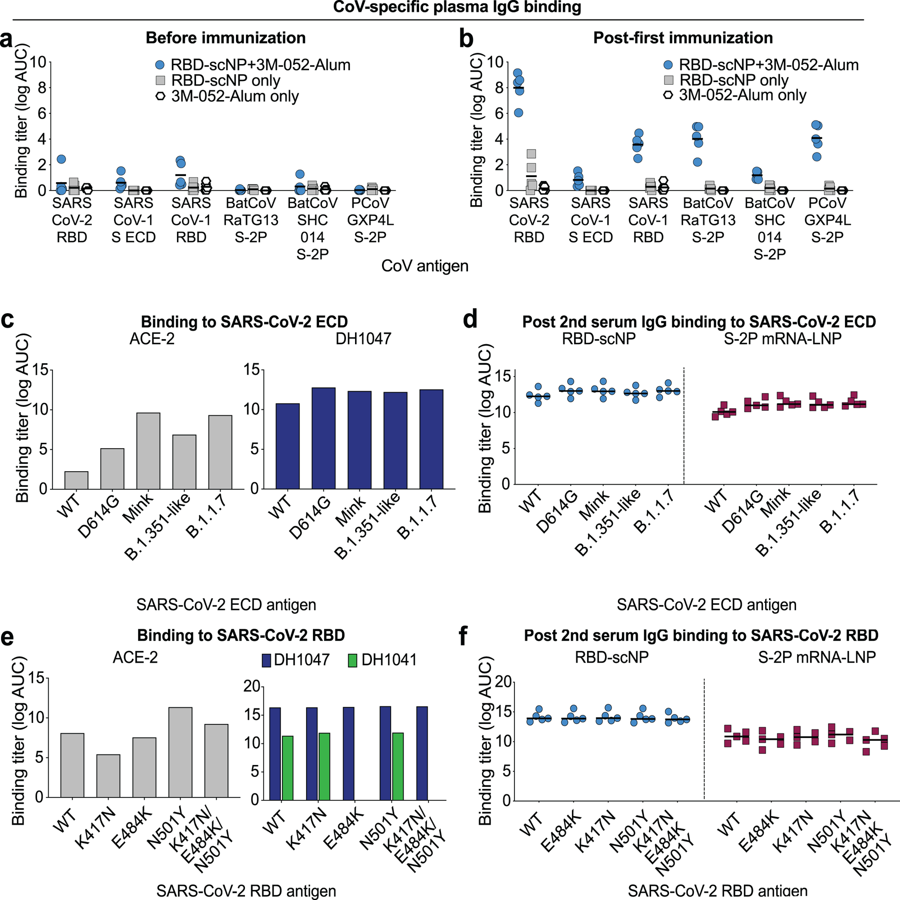
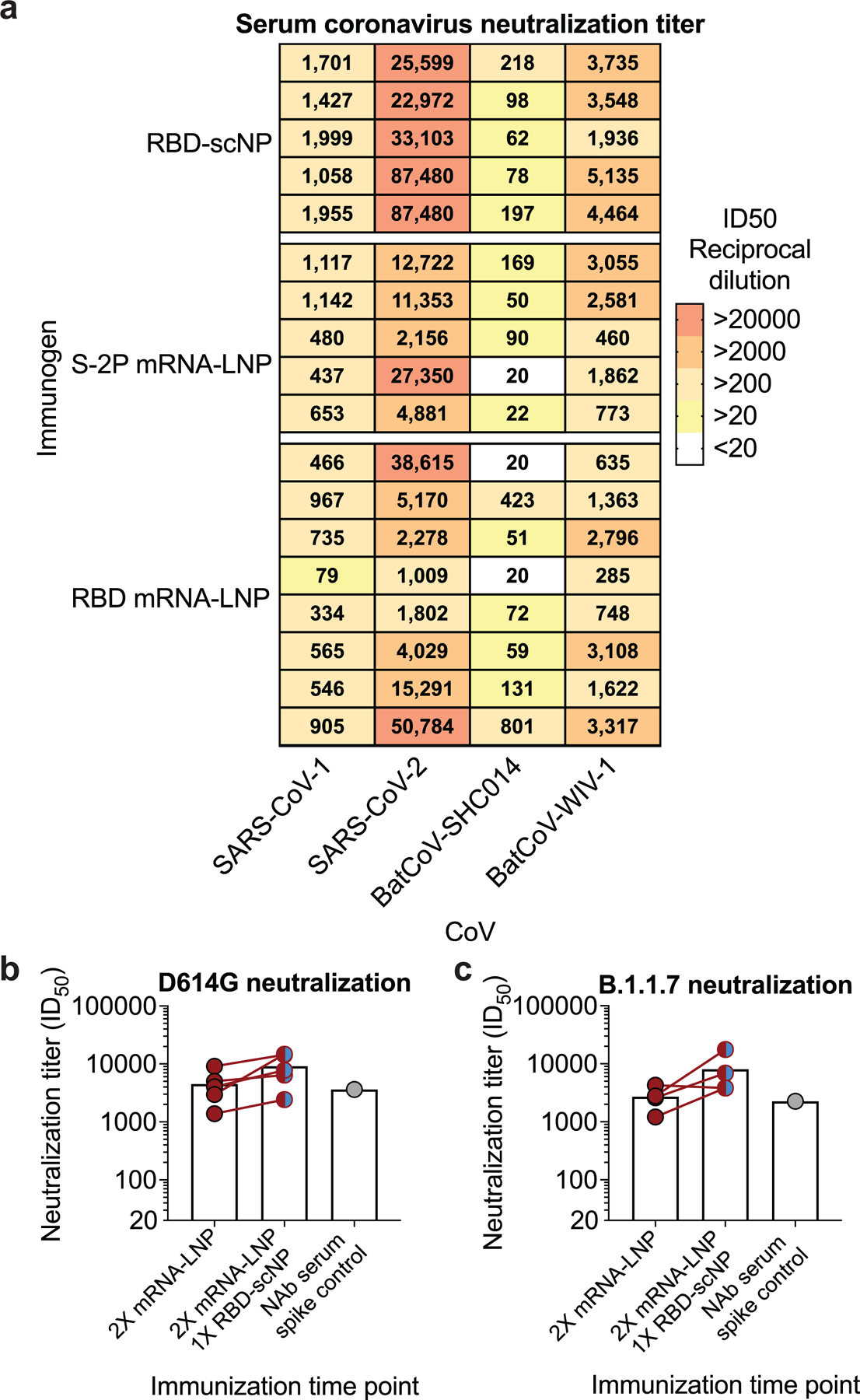
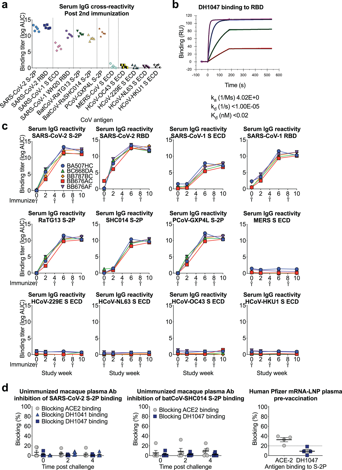
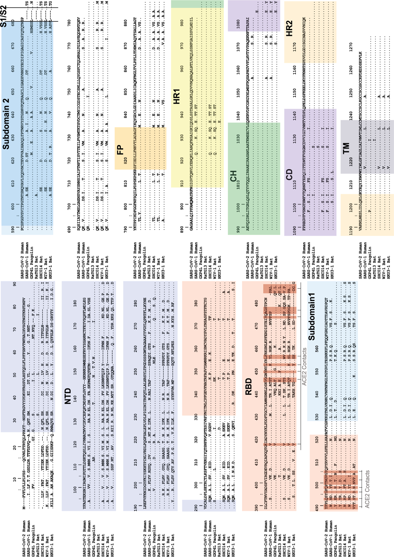
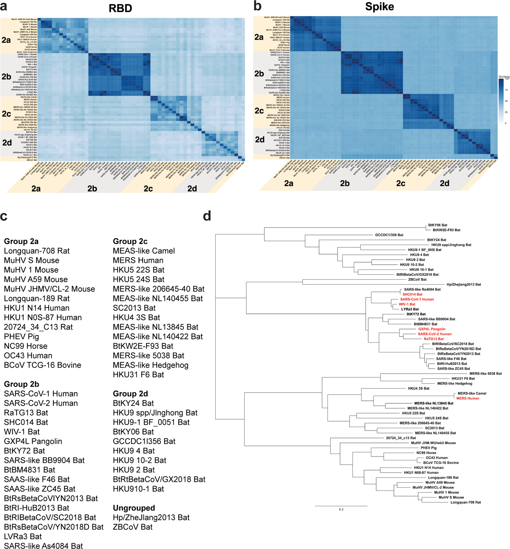
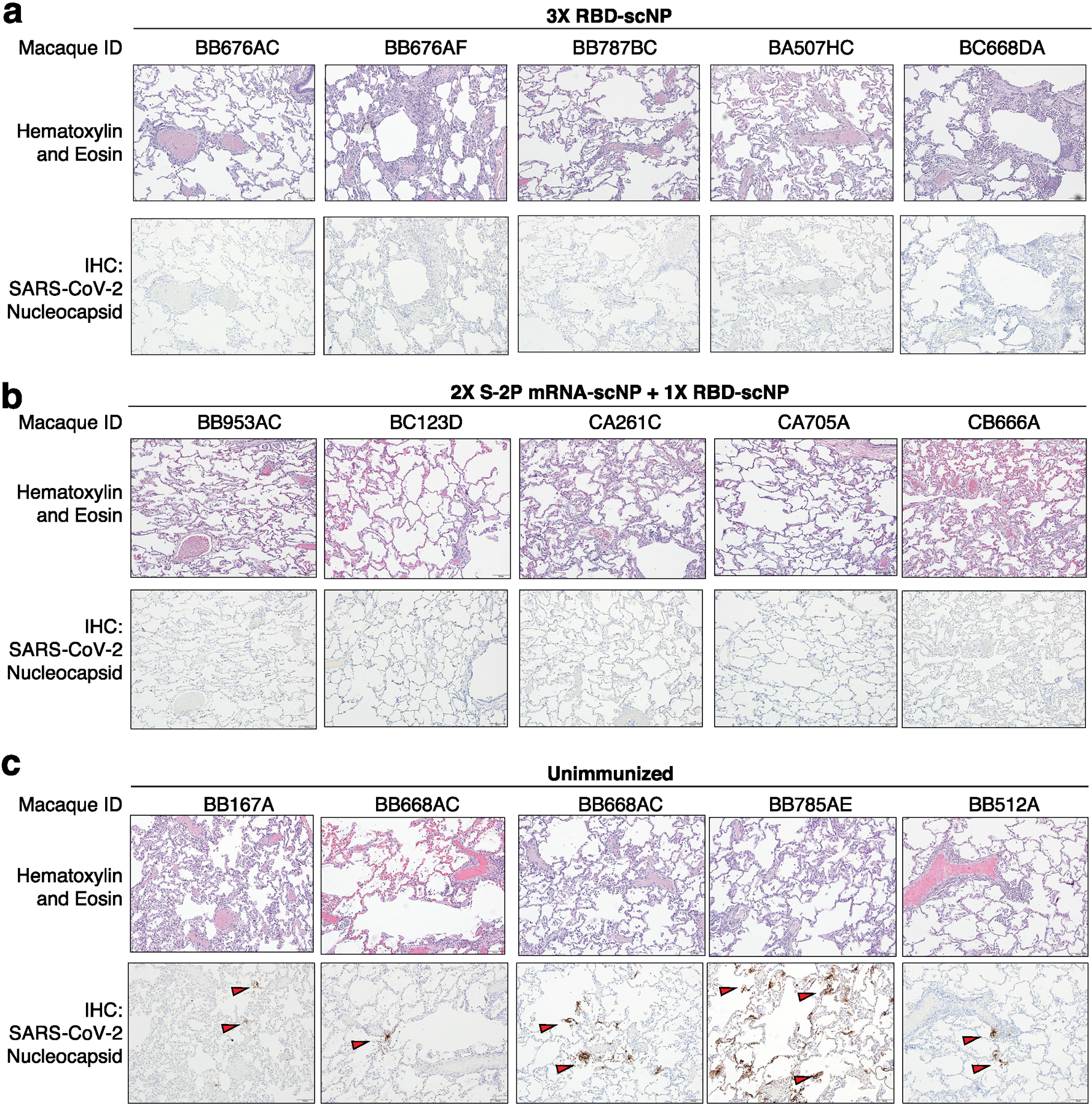
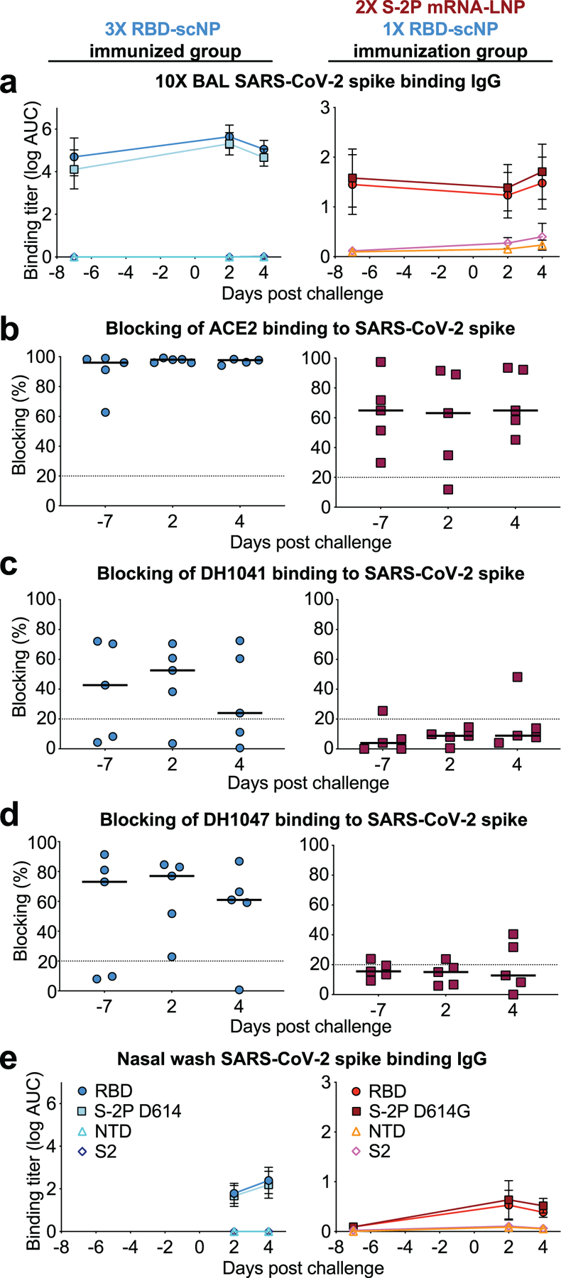
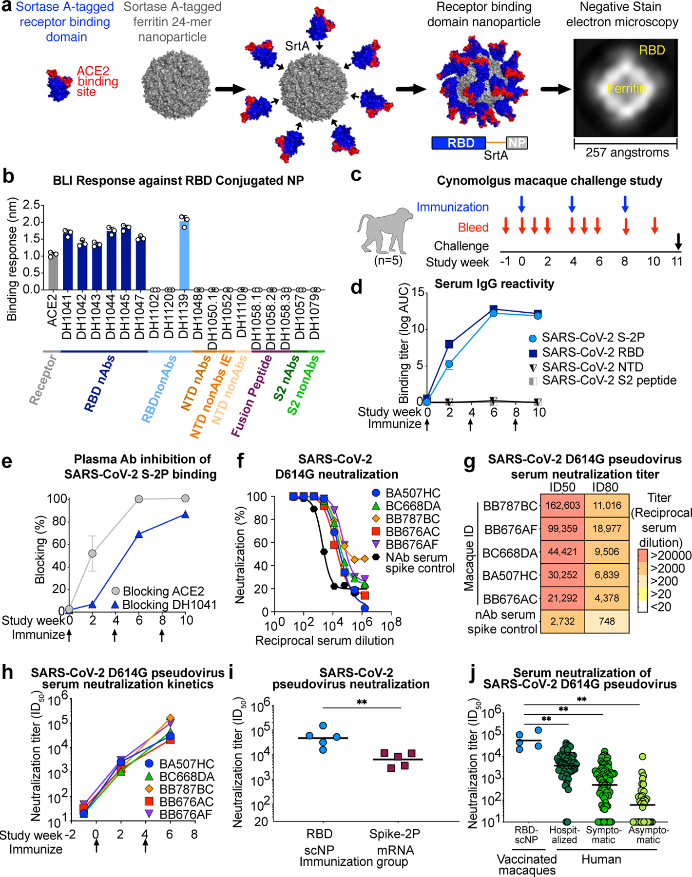
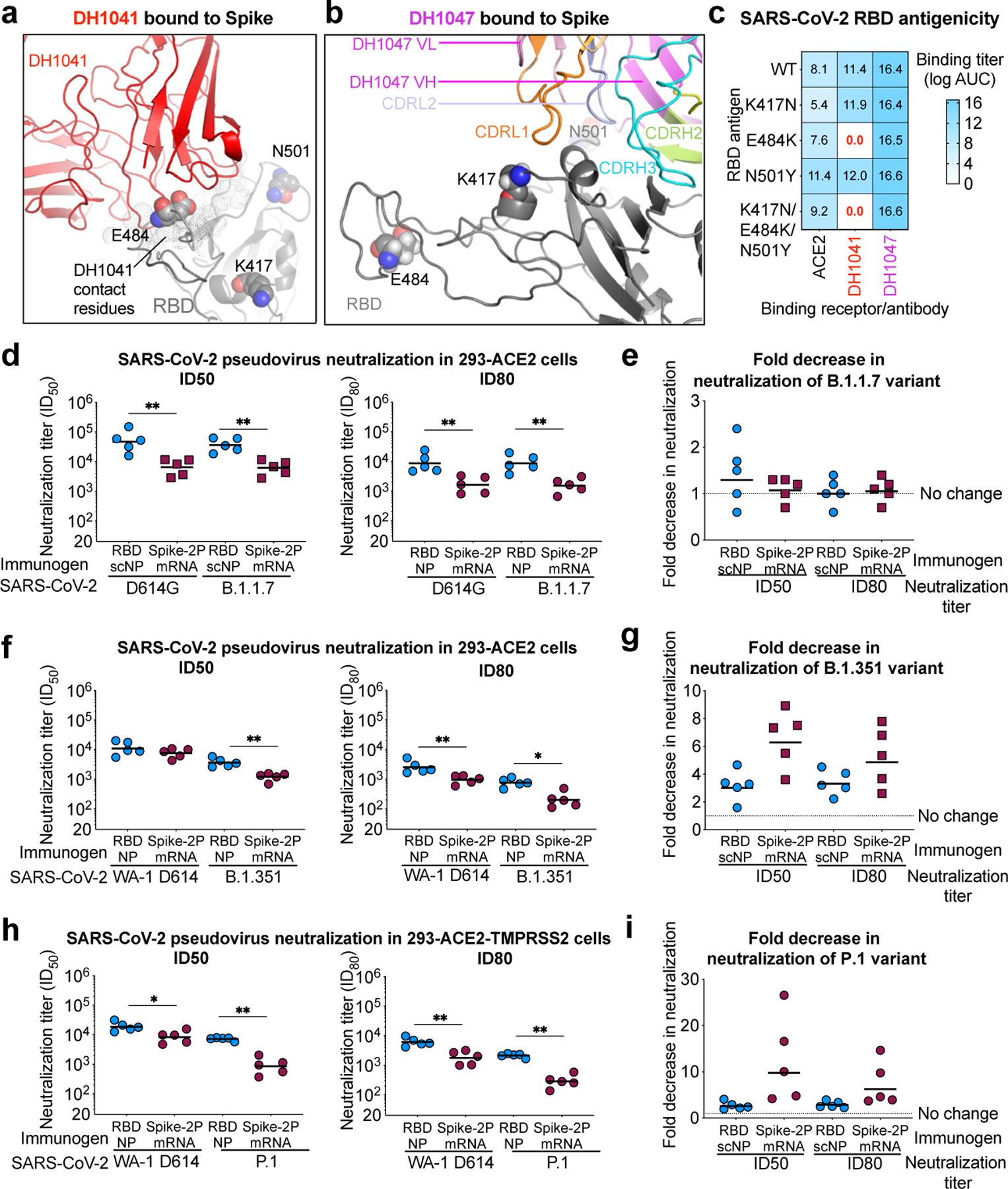
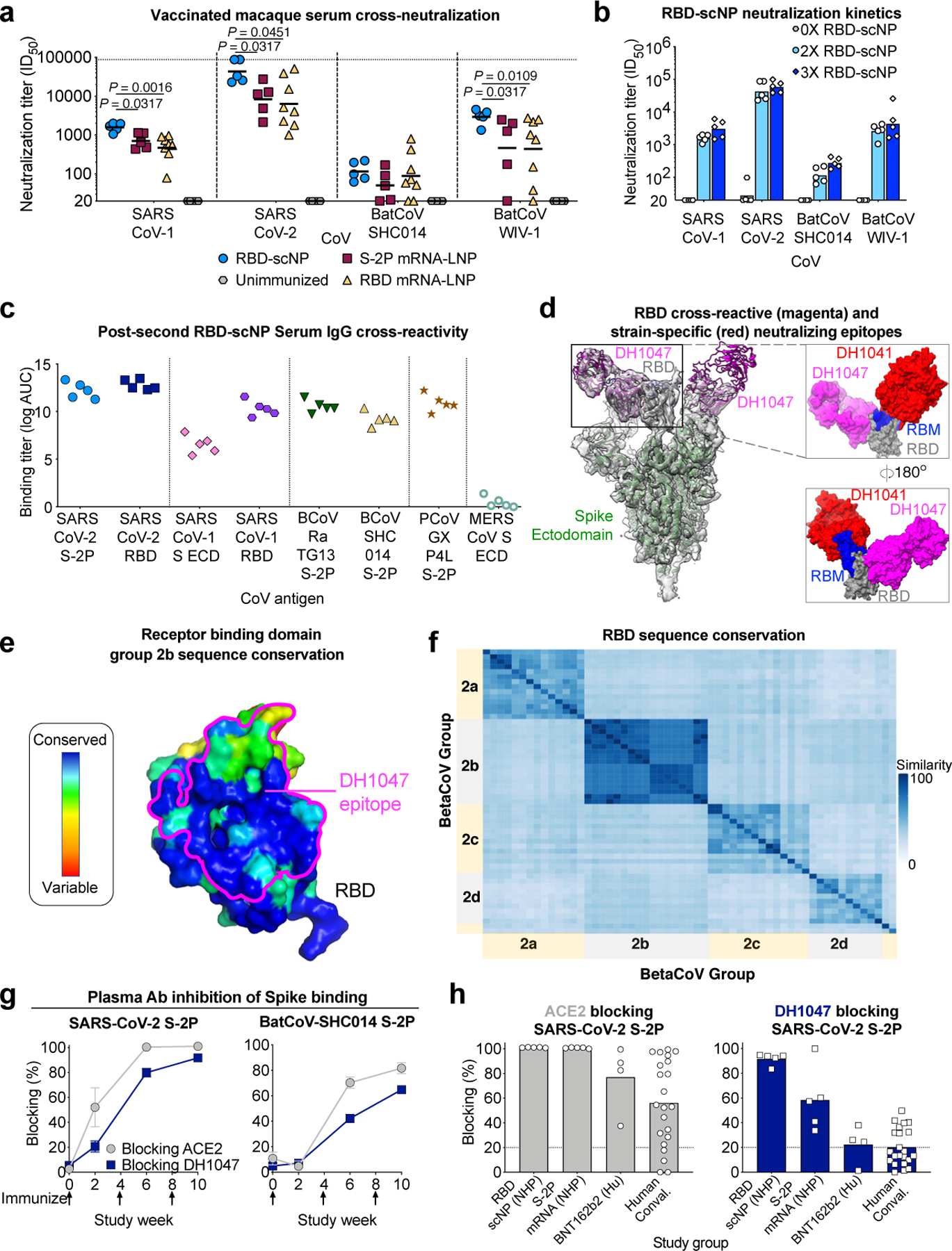
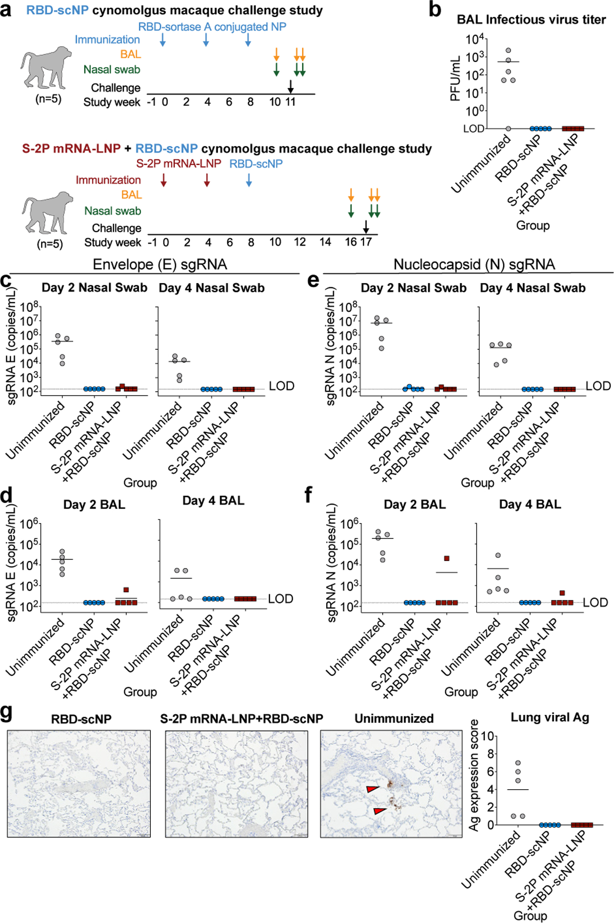
Betacoronaviruses caused the outbreaks of severe acute respiratory syndrome (SARS) and Middle East respiratory syndrome, as well as the current pandemic of SARS coronavirus 2 (SARS-CoV-2)1-4. Vaccines that elicit protective immunity against SARS-CoV-2 and betacoronaviruses that circulate in animals have the potential to prevent future pandemics. Here we show that the immunization of macaques with nanoparticles conjugated with the receptor-binding domain of SARS-CoV-2, and adjuvanted with 3M-052 and alum, elicits cross-neutralizing antibody responses against bat coronaviruses, SARS-CoV and SARS-CoV-2 (including the B.1.1.7, P.1 and B.1.351 variants). Vaccination of macaques with these nanoparticles resulted in a 50% inhibitory reciprocal serum dilution (ID50) neutralization titre of 47,216 (geometric mean) for SARS-CoV-2, as well as in protection against SARS-CoV-2 in the upper and lower respiratory tracts. Nucleoside-modified mRNAs that encode a stabilized transmembrane spike or monomeric receptor-binding domain also induced cross-neutralizing antibody responses against SARS-CoV and bat coronaviruses, albeit at lower titres than achieved with the nanoparticles. These results demonstrate that current mRNA-based vaccines may provide some protection from future outbreaks of zoonotic betacoronaviruses, and provide a multimeric protein platform for the further development of vaccines against multiple (or all) betacoronaviruses.


Conflict of interest statement

Competing financial interests BFH and KOS have filed patents regarding the nanoparticle vaccine, MAT and 3M company have patents filed on 3M052, and CF and IDRI have filed patents on the formulation of 3M052 and alum. 3M company had no role in the execution of the study, data collection, or data interpretation. DW is named on patents that describe the use of nucleoside-modified mRNA as a platform to deliver therapeutic proteins. DW and NP are also named on a patent describing the use of nucleoside-modified mRNA in lipid nanoparticles as a vaccine platform. All other authors declare no competing interests.

Figures

Extended Data Figure 1.
Extended Data Figure 1.. Molecular and structural characterization of the SARS-CoV-2 RBD sortase A conjugated nanoparticle.
a Size exclusion chromatography of RBD and ferritin sortase conjugation. The first peak shows conjugated protein. The second peak contains unconjugated RBD. b Analytical size exclusion trace shows a homogenous nanoparticle preparation. c Negative stain electron microscopy image of RBD-scNPs on a carbon grid. Inset shows a zoomed image of RBD-scNP. The zoomed image shows RBD molecules arrayed around the outside of the ferritin nanoparticle. A representative image from the 31 images taken of the micrograph to visualize 13,827 total particles is shown. d Chemical structure of toll-like receptor 7 and 8 agonist 3M-052. Alum formulation of 3M-052 was used to adjuvant RBD-scNP immunization. e RBD-scNP immunization regimen used for vaccination of cynomolgus macaques (N=5). Blue arrows indicate timepoints for intramuscular immunizations with RBD-scNP (100 µg) adjuvanted with 3M-052 (5 µg 3M-052 plus 500 µg Alum). Bronchoalveolar lavage (BAL, orange arrows) and nasal swab (green arrows) fluids were collected 7 days before, 2 days after, and 4 days after intratracheal/intranasal SARS-CoV-2 challenge (black arrow). f Transmembrane, diproline-stabilized spike (S-2P) mRNA-LNP prime, RBD-scNP boost vaccination of cynomolgus macaques (N=5). Maroon arrows indicate timepoints for S-2P mRNA-LNP immunization (50 µg mRNA dose). Blue arrows are the same as in a. Macaques were challenged 9 weeks after RBD-scNP boost (week 17 of the study). BAL and nasal swab fluids were collected as in a. Macaques were challenged at week 17 (black arrow). g Monomeric RBD mRNA-LNP immunization of rhesus macaques (N=8). Tan arrows indicate timepoints for RBD mRNA-LNP immunization (50 µg mRNA dose). Blood was collected throughout each study as shown by red arrows in all panels.
Extended Data Figure 2.
Extended Data Figure 2.. Blood chemical analysis and blood cell counts in RBD-scNP and S-2P mRNA-LNP-vaccinated macaques.
Each graph shows values for individual macaques before vaccination and 4 weeks after the RBD-scNP (week 8) or 6 weeks after the second S-2P mRNA-LNP immunization (week 10). RBD-scNP-immunized macaques are shown as blue symbols, and S-2P mRNA-LNP immunized macaques are shown as red symbols. The reference range for each value is shown as gray shaded area for female macaques and cyan shaded area for male macaques. Creatine kinase does not have a reference range indicated. Males are shown as circles and females are shown as triangles.
Extended Data Figure 3.
Extended Data Figure 3.. ACE-2, RBD neutralizing antibody, and post-vaccination macaque plasma IgG binding to SARS-CoV-2 Spike variants.
a,b Plasma IgG from macaques prior to immunization or after being immunized once with RBD-scNP adjuvanted with 3M-052-Alum (blue), RBD-scNP only (gray), or 3M-052-Alum only (white). Binding titers as log AUC were determined a before or b two weeks after a single immunization. Horizontal bars are the group mean. ECD, ectodomain; S-2P, diproline-stabilized spike. c ACE2 receptor and cross-nAbDH1047 ELISA binding to SARS-CoV-2 Spike ectodomain (ECD) based on a Danish mink (H69/V70deI/Y453F/D614G/I692V), B.1.351-like (K417N/E484K/N501Y/D614G), and B.1.1.7 (H69/V70del/Y144del/N501Y/A570D/D614G/P681H/T716I/S982A/D1118H) strains. Titers are shown as area under the log-transformed curve (log AUC). d RBD-scNP and S-2P mRNA-LNP-immunized macaque serum IgG ELISA binding to SARS-CoV-2 Spike variants shown in c. Serum was tested after two immunizations. Horizontal bars are the group mean. e ACE2 receptor (gray), cross-nAbDH1047 (navy), and ACE2 binding site-targeting neutralizing antibody DH1041 (green) ELISA binding to SARS-CoV-2 Spike RBD monomers. RBD variants contain a subset of mutations found in circulating B.1.351 and P.1 virus strains. Titers are shown as area under the log-transformed curve (log AUC). f RBD-scNP and S-2P mRNA-LNP-immunized macaque serum IgG ELISA binding to SARS-CoV-2 Spike RBD variants shown in e. Serum was tested after two immunizations. Horizontal bars are the group mean.
Extended Data Figure 4.
Extended Data Figure 4.. Cross-neutralizing antibodies are elicited by recombinant protein RBD-scNP and mRNA-LNP immunization.
a Each row shows neutralization titer for an individual macaque immunized with one of the three immunogens. A reciprocal serum dilution titer of 87,480 is the upper limit of detection and 20 is the lower limit of detection for this assay. Titers are derived from a nonlinear regression curve fit to the average of duplicate measurements. b,c Serum neutralization titers elicited by two S-2P mRNA-LNP immunizations were boosted by a subsequent RBD-scNP immunization. Serum neutralization of a SARS-CoV-2 D614G and b SARS-CoV-2 B.1.1.7 pseudovirus infection of ACE2-expressing 293 cells. Neutralization titers are ID50 as reciprocal serum dilution for serum collected two weeks after the second (week 6) and third immunization (week 10). Each symbol connected by a line represents the titer for an individual macaque before and after RBD-scNP immunization. Normal human serum spiked with DH1043 was used as a positive control.
Extended Data Figure 5.
Extended Data Figure 5.. Cross-reactive plasma antibody responses elicited by RBD-NP immunization in macaques.
a Plasma IgG from macaques immunized twice with RBD-scNP binds to Spike from human, bat, and pangolin SARS-related coronavirus Spike (S) in ELISA, but not endemic human coronaviruses or MERS-CoV. ECD, ectodomain. b Determination of DH1047 antigen binding fragment (Fab) binding kinetics to RBD monomer by surface plasmon resonance. Each curve shows a different concentration of DH1047 Fab. Binding kinetics are shown to the right from a 1:1 model fit. c Time course of vaccinated macaque plasma IgG binding to human, bat, and pangolin coronavirus S protein by ELISA. Each curve indicates the binding titer for an individual macaque. Arrows indicate immunization time points. d Unimmunized macaque plasma antibody blocking of SARS-CoV-2 S-2P (left) and batCoV-SHC014 (middle) binding to ACE2-Fc, RBD neutralizing antibody DH1041, and RBD cross-nAbDH1047. (Right) Blocking activity in the serum of humans immunized with Pfizer S-2P mRNA-LNP vaccine (N=4). Each symbol represents an individual human or macaque. Bars indicate group mean±s.e.m.
Extended Data Figure 6.
Extended Data Figure 6.. Multiple Sequence Alignment of Spike Protein from a Representative Set of Group 2b Betacoronaviruses.
SARS-CoV-2 Wuhan-1 spike protein numbering is shown. NTD=N-terminal domain; RBD=receptor binding domain; S1/S2=SARS2 furin cleavage site; FP=fusion peptide; HR1=heptad repeat 1; HR2=heptad repeat 2; CH=central helix; CD=connecting domain; TM=transmembrane domain. ACE2 contact positions in SARS2 (calculated from PDB coordinates 6MOJ and 6LZG) are highlighted in dark red.
Extended Data Figure 7.
Extended Data Figure 7.. Sequence conservation among SARS-related betaCoV, MERS-CoV, and endemic human CoVs.
a,b Sequence similarity of a RBD and b spike protein for representative betacoronaviruses. Heatmaps displaying pairwise amino acid sequence similarity for 57 representative betacoronaviruses. Dark blue shading indicates high sequence similarity. c List of viruses used for alignments in a and Fig. 3f. d Phylogenetic tree of representative betacoronavirus RBD sequences. Group 2b betaCoVs of interest are shown highlighted in red. Branch length units are substitutions per site.
Extended Data Figure 8.
Extended Data Figure 8.. Histology and immunohistochemistry of lung tissue collected seven days after SARS-CoV-2 WA-1 intratracheal and intranasal challenge.
a-c Macaques were immunized a thrice with RBD-scNP, b twice with S-2P mRNA-LNP and once with RBD-scNP, or c unimmunized. Each column shows results from an individual macaque. The macaque identification number is shown above each column. Hematoxylin and eosin stain of lung sections are shown on the top row, with nucleocapsid immunohistochemistry shown on the bottom row for each macaque. Red arrows indicate site of antigen positivity. All images are shown at 10X magnification with 100 micron scale bars shown in the bottom right corner.
Extended Data Figure 9.
Extended Data Figure 9.. Mucosal SARS-CoV-2 IgG responses in bronchoalveolar lavage (BAL) and nasal wash fluids before and after SARS-CoV-2 challenge.
a ELISA binding titers for SARS-CoV-2-specific IgG in 10X BAL fluid from macaques immunized with (blue symbols, left column) RBD-scNP three times or S-2P mRNA-LNP twice and RBD-scNP once (red symbols, left column). Day -7 BAL fluid was collected at week 10 or 16 for the RBD-scNP alone group or the S-2P mRNA-LNP/RBD-scNP group respectively. Group mean±s.e.m. are shown (N=5). b-d 10X BAL fluid blocking of ACE2, RBD neutralizing antibody DH1041, and cross-nAbDH1047 binding to SARS-CoV-2 D614G stabilized spike ectodomain. A black horizontal bar indicates the group mean blocking percentage. Blocking above 20% (above the dashed line) is considered positive. e Neat nasal wash fluid from RBD-scNP-immunized or S-2P mRNA-LNP/RBD-scNP-immunized macaques. Day -7 nasal wash fluid was collected at week 16 and 2 and 4 days post challenge for the S-2P mRNA-LNP/RBD-scNP group. Nasal wash fluid was unavailable for the RBD-scNP before challenge, but was collected 2 and 4 days after the week 11 challenge. Group mean±s.e.m. are shown (N=5).
Figure 1.
Figure 1.. SARS-CoV-2 receptor binding domain (RBD) sortase-conjugated nanoparticles (scNPs) elicits extremely high titers of SARS-CoV-2 pseudovirus neutralizing antibodies (nAbs).
a SARS-CoV-2 RBD (blue and red) Helicobacter pylori ferritin (gray) nanoparticle sortase conjugation. A model and two-dimensional class average of negative stain electron microscopy of the resultant RBD nanoparticle are shown. b Biolayer interferometry SARS-CoV-2 antibody and ACE2 receptor binding to RBD nanoparticles. N-terminal domain (NTD), infection enhancing non-neutralizing antibody (nonAbs IE), non-neutralizing antibody (nonAb). Symbols represent values from 3 independent experiments and bars represent the mean and standard error of the mean (s.e.m.). c Cynomolgus macaque immunogenicity and challenge study design. d Macaque serum IgG binding titer as area-under-the curve of the log10-transformed curve (log AUC) to recombinant SARS-CoV-2 stabilized Spike ectodomain (S-2P), RBD, NTD, and Fusion peptide (FP). Group mean±s.e.m. are shown in d and e (n = 5 macaques). e Plasma antibody blocking of SARS-CoV-2 S-2P binding to ACE2-Fc and RBD neutralizing antibody DH1041. f,g f Dose-dependent serum neutralization of SARS-CoV-2 D614G pseudovirus infection of ACE2-expressing 293T cells and g neutralization ID50 and ID80 titers. Serum was examined after two immunizations. The mean value of duplicates is shown in f. h SARS-CoV-2 D614G pseudovirus serum neutralization titer over time for individual macaques. i Serum neutralization ID50 titers from macaques immunized twice with protein RBD nanoparticles (blue) or nucleoside-modified mRNA-LNP expressing S-2P (burgundy) (**P = 0.0079, Two-tailed Exact Wilcoxon test, n = 5 macaques). j Serum neutralization titers for macaques immunized twice with RBD-scNP (blue, n =5 macaques) or humans with asymptomatic infection (n=34 individuals), symptomatic infection (n=71 individuals), or hospitalized (n=60 individuals) (**P<0.01, Two-tailed Wilcoxon test). Horizontal bars are the group geometric mean in i and j. Pre-vaccination serum or nAb spiked serum were used as controls in f, g, and h.
Figure 2.
Figure 2.. RBD-scNP immunization elicits higher titers of nAbs against more transmissible or neutralization-resistant SARS-CoV-2 variants than stabilized spike mRNA-LNP vaccination.
a,b The location of K417, E484, and N501 (spheres) present in the B.1.351 variant are shown in the cryo-EM structures of RBD nAbs a DH1041 (red) and b DH1047 (magenta) bound to the RBD (gray) of S trimers (PDB: 7LAA and 7LD1). c ACE2 receptor, DH1041, and DH1047 ELISA binding titer as log AUC for wildtype and mutant SARS-CoV-2 Spike RBD monomers. d Serum neutralization ID50 (left) and ID80 titers for SARS-CoV-2 D614G and SARS-CoV-2 B.1.1.7 pseudoviruses from immunized macaques. Symbols represent individual macaques and horizontal bars are group means (**P = 0.0079, Two-tailed Exact Wilcoxon test, n = 5 macaques). e Fold decrease in neutralization potency between neutralization of SARS-CoV-2 D614G and SARS-CoV-2 B.1.1.7 pseudoviruses. Fold change is shown for RBD-scNP-immunized and mRNA-LNP-immunized macaques based ID50 (left) and ID80 (right) titers. Horizontal bars are the group mean. f Vaccinated macaque serum neutralization ID50 (left) and ID80 titers (right) against WA-1 and B.1.351 pseudovirus. Symbols and horizontal bars are shown the same as in d (*P = 0.0159 and **P = 0.0079, Two-tailed Exact Wilcoxon test, n = 5 macaques). g Fold decrease in neutralization potency between neutralization of SARS-CoV-2 WA-1 and B.1.351 pseudoviruses. Fold change is shown the same as in e . h Vaccine-induced neutralization of SARS-CoV-2 WA-1 and P.1 pseudovirus infection of ACE2 and TMPRSS2-expressing 293 cells. Symbols and horizontal bars are shown the same as in d (*P = 0.0159 and **P = 0.0079, Two-tailed Exact Wilcoxon test, n = 5 macaques). i Fold decrease in neutralization potency as shown in e between neutralization of SARS-CoV-2 WA-1 and P.1 pseudoviruses.
Figure 3.
Figure 3.. RBD-scNP vaccine induction of serum cross-neutralization of SARS-related betacoronavirus infection.
a Serum neutralization ID50 titers from macaques immunized twice with RBD-scNP, S-2P mRNA-LNP, or RBD mRNA-LNP for SARS-CoV-1 and SARS-CoV-2 and SARS-related batCoVs (WIV-1 and SHC014). Symbols indicate individual macaques and black bars show the group geometric mean (Two-tailed Exact Wilcoxon test, n = 5 or 8 macaques). b Serum cross-neutralization ID50 titers before (gray), after two (light blue) or after three (blue) RBD-scNP immunizations. Bars represent the group geometric mean. c Human, bat, and pangolin SARS-related betaCoV S protein ELISA log AUC titer for plasma IgG from macaques immunized twice with RBD-scNP. ECD, ectodomain. d Structural comparison of the epitopes of SARS-CoV-2-specific neutralizing RBD antibody (DH1041, red, PDB ID: 7LAA) and cross-neutralizing RBD antibody (DH1047, magenta, PDB ID: 7LD1). (Left) Cartoon view of Spike (green), RBD (gray), Receptor Binding Motif (RBM, blue). (Right) Overlay of the RBDs of the two complexes from their respective cryo-EM structures. e RBD colored by conservation within group 2b betacoronaviruses. DH1047 epitope is shown in magenta outline. f Heatmaps displaying pairwise amino acid sequence similarity for 57 representative betaCoVs. g, h Plasma/serum antibody blocking of S-2P binding to ACE2 (gray) and DH1047 (navy blue). g Kinetics of SARS-CoV-2 or batCoV-SHC014 blocking by serum from macaques immunized twice with RBD-scNP. Group mean±s.e.m. are shown (n = 5 macaques). h Blocking activity by serum from macaques immunized twice with RBD-scNP or S-2P mRNA-LNP and humans immunized twice with Pfizer BNT162b2 or naturally infected with SARS-CoV-2. Each symbol represents an individual subject and filled bars indicate the group mean in h. Positivity threshold (dashed line) is greater than 20% in g and h.
Figure 4.
Figure 4.. RBD-scNP vaccination alone or as a boost completely prevents virus replication in the upper and lower respiratory tract after intranasal and intratracheal SARS-CoV-2 challenge in nearly all macaques.
a. Macaque intranasal/intratracheal SARS-CoV-2 challenge study design. Blue and maroon arrows indicate the time points for RBD-scNP and mRNA-LNP immunizations respectively. b Infectious virus in macaque BAL fluid two days after challenge. c-f. Quantification of viral envelope (E) gene or nucleocapsid (N) gene subgenomic RNA (sgRNA) in unimmunized (gray) and RBD-scNP-immunized (blue), and S-2P mRNA-LNP prime/RBD-scNP boosted (burgundy) macaques. sgRNA in nasal swabs and bronchoalveolar lavage (BAL) was quantified two (left) and four (right) days after challenge. Limit of detection (LOD) for the assay is 150 copies/mL. Symbols and bars are shown as in b. g Nucleocapsid immunohistochemistry of lung tissue sections seven days post challenge. (Left) A representative image from 1 macaque from each group of 5 macaques is shown. Red arrows indicate site of antigen positivity. All images are shown at 10X magnification with 100 micron scale bars. (Right) Quantification of antigen positivity. In each panel symbols represent individual macaques with the group mean shown as a black horizontal bar.

Comment in

References

    1. Wang C, Horby PW, Hayden FG & Gao GF A novel coronavirus outbreak of global health concern. Lancet 395, 470–473, doi:10.1016/s0140-6736(20)30185-9 (2020). - DOI - PMC - PubMed
    1. Zaki AM, van Boheemen S, Bestebroer TM, Osterhaus AD & Fouchier RA Isolation of a novel coronavirus from a man with pneumonia in Saudi Arabia. N Engl J Med 367, 1814–1820, doi:10.1056/NEJMoa1211721 (2012). - DOI - PubMed
    1. Zhong NS et al. Epidemiology and cause of severe acute respiratory syndrome (SARS) in Guangdong, People’s Republic of China, in February, 2003. Lancet 362, 1353–1358, doi:10.1016/s0140-6736(03)14630-2 (2003). - DOI - PMC - PubMed
    1. Zhu N et al. A Novel Coronavirus from Patients with Pneumonia in China, 2019. N Engl J Med 382, 727–733, doi:10.1056/NEJMoa2001017 (2020). - DOI - PMC - PubMed
    1. Zhou P et al. A pneumonia outbreak associated with a new coronavirus of probable bat origin. Nature 579, 270–273, doi:10.1038/s41586-020-2012-7 (2020). - DOI - PMC - PubMed

METHODS REFERENCES

    1. Laczko D et al. A Single Immunization with Nucleoside-Modified mRNA Vaccines Elicits Strong Cellular and Humoral Immune Responses against SARS-CoV-2 in Mice. Immunity 53, 724–732 e727, doi:10.1016/j.immuni.2020.07.019 (2020). - DOI - PMC - PubMed
    1. Pardi N et al. Zika virus protection by a single low-dose nucleoside-modified mRNA vaccination. Nature 543, 248–251, doi:10.1038/nature21428 (2017). - DOI - PMC - PubMed
    1. Wrapp D et al. Cryo-EM structure of the 2019-nCoV spike in the prefusion conformation. Science 367, 1260–1263, doi:10.1126/science.abb2507 (2020). - DOI - PMC - PubMed
    1. Hsieh CL et al. Structure-based Design of Prefusion-stabilized SARS-CoV-2 Spikes. bioRxiv, doi:10.1101/2020.05.30.125484 (2020). - DOI - PMC - PubMed
    1. Zhou T et al. Structure-Based Design with Tag-Based Purification and In-Process Biotinylation Enable Streamlined Development of SARS-CoV-2 Spike Molecular Probes. Cell Rep 33, 108322, doi:10.1016/j.celrep.2020.108322 (2020). - DOI - PMC - PubMed

Publication types

MeSH terms