. 2021 May 11;18(1):108.
doi: 10.1186/s12974-021-02156-5.

IL-9-triggered lncRNA Gm13568 regulates Notch1 in astrocytes through interaction with CBP/P300: contribute to the pathogenesis of experimental autoimmune encephalomyelitis

Affiliations

IL-9-triggered lncRNA Gm13568 regulates Notch1 in astrocytes through interaction with CBP/P300: contribute to the pathogenesis of experimental autoimmune encephalomyelitis

Xiaomei Liu et al. J Neuroinflammation. .

Abstract

Background: Interleukin 9 (IL-9), produced mainly by T helper 9 (Th9) cells, has been recognized as an important regulator in multiple sclerosis (MS) and its animal model, experimental autoimmune encephalomyelitis (EAE). Astrocytes respond to IL-9 and reactive astrocytes always associate with blood-brain barrier damage, immune cell infiltration, and spinal injury in MS and EAE. Several long non-coding RNAs (lncRNAs) with aberrant expression have been identified in the pathogenesis of MS. Here, we examined the effects of lncRNA Gm13568 (a co-upregulated lncRNA both in EAE mice and in mouse primary astrocytes activated by IL-9) on the activation of astrocytes and the process of EAE.

Methods: In vitro, shRNA-recombinant lentivirus with glial fibrillary acidic protein (GFAP) promoter were performed to determine the relative gene expression and proinflammatory cytokines production in IL-9 treated-astrocytes using Western blot, real-time PCR, and Cytometric Bead Array, respectively. RIP and ChIP assays were analyzed for the mechanism of lncRNA Gm13568 regulating gene expression. Immunofluorescence assays was performed to measure the protein expression in astrocytes. In vivo, H&E staining and LFB staining were applied to detect the inflammatory cells infiltrations and the medullary sheath damage in spinal cords of EAE mice infected by the recombinant lentivirus. Results were analyzed by one-way ANOVA or Student's t test, as appropriate.

Results: Knockdown of the endogenous lncRNA Gm13568 remarkably inhibits the Notch1 expression, astrocytosis, and the phosphorylation of signal transducer and activator of transcription 3 (p-STAT3) as well as the production of inflammatory cytokines and chemokines (IL-6, TNF-α, IP-10) in IL-9-activated astrocytes, in which Gm13568 associates with the transcriptional co-activators CBP/P300 which are enriched in the promoter of Notch1 genes. More importantly, inhibiting Gm13568 with lentiviral vector in astrocytes ameliorates significantly inflammation and demyelination in EAE mice, therefore delaying the EAE process.

Conclusions: These findings uncover that Gm13568 regulates the production of inflammatory cytokines in active astrocytes and affects the pathogenesis of EAE through the Notch1/STAT3 pathway. LncRNA Gm13568 may be a promising target for treating MS and demyelinating diseases.

Keywords: Astrocytes; Experimental autoimmune encephalomyelitis (EAE); IL-9; Inflammatory cytokines; LncRNA Gm13568; Notch1.


Conflict of interest statement

None of the authors have any conflicts of interest in this study.

Figures

Fig. 1
Fig. 1
Upregulation of IL-9 and inflammatory cytokines as well as the activation of Notch1 signaling during EAE process. a The level of IL-9 in spinal cords of EAE mice with different clinical scores was measured by real-time PCR and Western blot assay, respectively. b The expressions of GFAP, NICD and p-STAT3 in spinal cords along with EAE process were detected by Western blot assay. c The mRNA levels of IL-6, TNF-α, IP-10, and MCP-1 in spinal cords were determined using real-time PCR. d The secretion levels of IL-6, TNF-α, IP-10 and MCP-1 in the sera were detected by Cytometric Bead Array (CBA) during the EAE process. *p < 0.05, **p < 0.01 and ***p < 0.001 versus PBS group (n = 6/group, one-way ANOVA). These results were repeated four times. Data are represented as the mean ± SEM
Fig. 2
Fig. 2
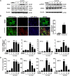
IL-9 activates the Notch1 pathway and promotes inflammatory cytokines production in astrocytes. Primary mouse astrocytes were incubated in a serum-free medium overnight followed by treating with IL-9 at the indicated time point. a The expression changes of GFAP, NICD, and p-STAT3 were analyzed by Western blot assay. b Immunofluorescent staining for GFAP (green), Notch1 (red), and nuclear staining of DAPI (blue) in cultured astrocytes with IL-9 treatment for 6 h. Scale bars, 50 μm. c The mRNA levels of IL-6, TNF-α, IP-10, and MCP-1 in astrocytes were detected using real-time PCR assay. d The secretion levels of IL-6, TNF-α, IP-10, and MCP-1 in the supernatant of astrocytes were measured by CBA assay. *p < 0.05, **p < 0.01 and ***p < 0.001 versus DMEM group (one-way ANOVA). The data are from three independent experiments and represented as the mean ± SEM
Fig. 3
Fig. 3
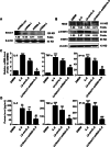
Knockdown of Notch1 reduces inflammatory cytokine production in activated-astrocytes by IL-9. a The 3 recombinant lentivirus plasmids of Notch1-shRNA targeting astrocytes were transduced into mouse primary astrocytes using the Neon™ electron transfection system (MPK5000) for 48 h. The expression of Notch1 protein in astrocytes was evaluated by Western blot assay. b Primary mouse astrocytes were infected with recombinant lentivirus (LV-Notch1-shRNA, LV-ctrl) for 72 h, and then incubated in a serum-free medium overnight followed by treating with IL-9 for 6 h. Western blot analysis for the proteins of Notch1 pathway. c The mRNA levels of IL-6, TNF-α, and IP-10 in astrocytes were measured by real-time PCR. d The secretion levels of IL-6, TNF-α, and IP-10 in the supernatant of astrocytes were detected by CBA assay. **p < 0.01 and ***p < 0.001 versus DMEM group; #p < 0.05 and ##p < 0.01 and ###p < 0.001 versus LV-ctrl+IL-9 group (one-way ANOVA, the two-tailed Student’s t test). The data are from three independent experiments and represented as mean ± SEM
Fig. 4
Fig. 4
Identification of lncRNA targeting the Notch1 gene. a The sequence of lncRNA Gm13568 with 446 base length. b The sequence of Gm13568 is complementary to the sense chain of Notch1 gene at 9075-9497 locus. c Primary mouse astrocytes were treated in a serum-free medium overnight followed by stimulating with IL-9 at different time period. Real-time PCR was performed to detect the change levels of Gm13568 and Notch1 in astrocytes. *p < 0.05 and **p < 0.01 and ***p < 0.001 versus DMEM (one-way ANOVA). Results are represented as mean ± SEM
Fig. 5
Fig. 5
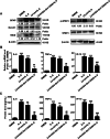
Inhibition of Gm13568 downregulates Notch1 signaling activation as well as inflammatory cytokine production in astrocytes by IL-9. a Primary mouse astrocytes were infected with recombinant lentivirus, LV-Inhibit-Gm13568 and LV-ctrl, for 72 h, respectively. Then, the astrocytes were incubated in a serum-free medium overnight followed by IL-9 stimulation for 6 h. Western blot assay was used for measuring the protein expressions of GFAP, Notch1/NICD, and p-STAT3. b The mRNA levels of IL-6, TNF-α and IP-10 in astrocytes were measured by real-time PCR assay. c The secretion of IL-6, TNF-α, and IP-10 in the supernatant of astrocytes were analyzed by CBA assay. The data are represented as the mean ± SEM. *p < 0.05, **p < 0.01, and ***p < 0.001 versus DMEM group; ##p < 0.01 and ###p < 0.001 versus LV-ctrl+IL-9 group (one-way ANOVA, the two-tailed Student’s t test)
Fig. 6
Fig. 6
LncRNA Gm13568 interacts with CBP/P300 to regulate the expression of Notch1 in astrocytes. a RIP assay was performed for the interaction of Gm13568 with NF-κB p65 and CBP/P300 in astrocytes treatment with IL-9 for 6 h. The data were from three independent experiments. b ChIP assay analysis of NF-κB p65, CBP/P300, RNA Pol II, and H3K27ac enrichment on the promoter of Notch1 gene in astrocytes stimulation with IL-9 for 6 h. *p < 0.05, **p < 0.01, ***p < 0.001 vs DMEM (one-way ANOVA). The data are from three independent experiments. Error bars represent mean ± SEM. c ChIP assay analysis of NF-κB p65, CBP/P300, and H3K27ac enrichment on the promoter of Notch1 gene in astrocytes infected by LV-Inhibit-Gm13568 for 72 h, followed by stimulation with IL-9 for 6 h. #p < 0.05 versus LV-ctrl. The data were from three independent experiments
Fig. 7
Fig. 7
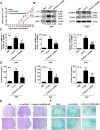
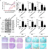
Knockdown of Notch1 in astrocytes suppresses inflammation and alleviates EAE in mice. Mice were subjected to recombinant lentiviruses, LV-ctrl or LV-Notch1-shRNA, for 7 days, followed by MOG35-55 immunization for 23 days (n = 10 mice per group). a The clinical scores of EAE mice with LV-ctrl and LV-Notch1-shRNA. b The expressions of IL-9, GFAP, NICD, and p-STAT3 in the spinal cords were detected using western blot assay. c, d The changes of IL-6, TNF-α, and IP-10 in the spinal cords and peripheral blood of the LV-ctrl and LV-Notch1-shRNA mice were evaluated by real-time PCR and CBA assay, respectively. *p < 0.05, **p < 0.01, ***p < 0.001 vs. PBS group; #p < 0.05, ##p < 0.01, ###p < 0.001 versus LV-ctrl group (one-way ANOVA, the two-tailed Student’s t test). Results are represented as mean ± SEM. e The infiltration of inflammatory cells in spinal cords was investigated using hematoxylin and eosin (H&E) staining (scale bars, 50 μm). f The medullary sheath damages from spinal cords were observed via luxol fast blue (LFB) staining (scale bars, 50 μm). Boxed areas in the upper rows are presented enlarged underneath
Fig. 8
Fig. 8
The inhibition of Gm13568 in astrocytes decreases Notch1 pathway activation and ameliorates the pathological process in EAE mice. On day 7 after administration with LV-ctrl or LV-Inhibit Gm13568, the mice were immunized with MOG35-55 for 23 days. a The clinical scores for EAE mice infected by LV-ctrl or LV-Inhibit Gm13568 (n = 10 mice per group). b The expression of IL-9 and GFAP and the activation of Notch1 pathway in the spinal cords were evaluated by Western blot assay. The results of a typical experiment are presented. c, d The levels of IL-6, TNF-α, and IP-10 from the spinal cords and peripheral blood in lentivirus-infected mice were analyzed by real-time PCR and CBA assay, respectively. *p < 0.05, **p < 0.01, ***p < 0.001 vs. PBS group; #p < 0.05, ##p < 0.01, ###p < 0.001versus LV-ctrl group (one-way ANOVA, the two-tailed Student’s t test). e H&E staining for inflammatory cells infiltrations in spinal cords of mice (Scale bars, 50 μm). f LFB staining for the medullary sheath damage in spinal cords of mice (scale bars, 50 μm). Boxed areas in the upper rows are presented enlarged underneath
Fig. 9
Fig. 9
Schematic representation of lncRNA Gm13568 contributing to the pathogenesis of EAE mice via regulation of the Notch1 pathway. Under IL-9 stimulation, Gm13568 is upregulated in astrocytes, which promotes the expression and activation of Notch1 via interacting with CBP/P300, in turn contributing to the release of inflammatory cytokines, thus aggravating the EAE development

Similar articles

Cited by

References

    1. Faissner S, Plemel JR, Gold R, Yong VW. Progressive multiple sclerosis: from pathophysiology to therapeutic strategies. Nat Rev Drug Discov. 2019;1812:905–922. doi: 10.1038/s41573-019-0035-2. - DOI - PubMed
    1. Dong Y, Yong VW. When encephalitogenic T cells collaborate with microglia in multiple sclerosis. Nat Rev Neurol. 2019;1512:704–717. doi: 10.1038/s41582-019-0253-6. - DOI - PubMed
    1. Yi W, Schlüter D, Wang X. Astrocytes in multiple sclerosis and experimental autoimmune encephalomyelitis: star-shaped cells illuminating the darkness of CNS autoimmunity. Brain Behav Immun. 2019;80:10–24. doi: 10.1016/j.bbi.2019.05.029. - DOI - PubMed
    1. Baecher-Allan C, Kaskow BJ, Weiner HL. Multiple sclerosis: mechanisms and immunotherapy. Neuron. 2018;974:742–768. doi: 10.1016/j.neuron.2018.01.021. - DOI - PubMed
    1. Elyaman W, Khoury SJ. Th9 cells in the pathogenesis of EAE and multiple sclerosis. Semin Immunopathol. 2017;391:79–87. doi: 10.1007/s00281-016-0604-y. - DOI - PubMed

MeSH terms

LinkOut - more resources