Review
. 2021 May 10;19(1):132.
doi: 10.1186/s12951-021-00870-z.

Recent advances in near-infrared II imaging technology for biological detection

Affiliations
Review

Recent advances in near-infrared II imaging technology for biological detection

Nan-Nan Zhang et al. J Nanobiotechnology. .

Abstract

Molecular imaging technology enables us to observe the physiological or pathological processes in living tissue at the molecular level to accurately diagnose diseases at an early stage. Optical imaging can be employed to achieve the dynamic monitoring of tissue and pathological processes and has promising applications in biomedicine. The traditional first near-infrared (NIR-I) window (NIR-I, range from 700 to 900 nm) imaging technique has been available for more than two decades and has been extensively utilized in clinical diagnosis, treatment and scientific research. Compared with NIR-I, the second NIR window optical imaging (NIR-II, range from 1000 to 1700 nm) technology has low autofluorescence, a high signal-to-noise ratio, a high tissue penetration depth and a large Stokes shift. Recently, this technology has attracted significant attention and has also become a heavily researched topic in biomedicine. In this study, the optical characteristics of different fluorescence nanoprobes and the latest reports regarding the application of NIR-II nanoprobes in different biological tissues will be described. Furthermore, the existing problems and future application perspectives of NIR-II optical imaging probes will also be discussed.

Keywords: Biomedical applications; Fluorescence imaging; Second near-infrared (NIR-II) window.


Conflict of interest statement

The authors have declared that no competing interests exist.

Figures
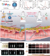
Fig. 1
Fig. 1
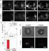
Dual-NIR-IIa and NIR-IIb fluorescence imaging-guided sentinel LN resection. ac The optical properties of DAD dye and PbS QDs. d Schematic illustration of imaging-guided sentinel LN resection. The DAD probe was injected intravenously, and then the QDs were injected into the tumor. e The photostability of the DAD dye and PbS QDs. f Color testing of the dual-NIR-IIa and NIR-IIb probes. g Fluorescence images of QDs and ICG. [73]
Fig. 2
Fig. 2
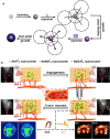
Scheme of the NIR-II fluorescence/dual bioluminescence multifunctional optical imaging system. a Schematic illustration of the multifunctional optical imaging strategy and b fluorescence imaging equipment with a 400–1700 nm wide spectral detection range in vivo [76]
Fig. 3
Fig. 3
Scheme of NPs@BOD/CPT for tumor NIR-II fluorescence imaging and drug release [80]
Fig. 4
Fig. 4
Scheme of the preparation of AIE@NE and its application for brain inflammation imaging [83]
Fig. 5
Fig. 5
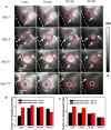
AIE@NE for brain inflammation NIR-II imaging. a Time-dependent NIR-II fluorescence imaging of brain inflammation. b The average fluorescence signal of the inflammatory area at different time points. c The SBR of the inflammatory area at 12 h. d Bright field and fluorescent field photos of the brain tissue [83]
Fig. 6
Fig. 6
a Schematic illustration of the preparation of the NaYF4:5%Nd@NaGdF4 nanoprobe and b its application in NIR II, MRI and CT imaging of tumors during tumorigenesis, growth and necrosis [87]
Fig. 7
Fig. 7
NIR II imaging monitors the changes in the tumor vasculature. a NIR II fluorescence imaging of tumors in a 4T1 breast tumor model. b, c The tumor-to-background ratios at different times [87]

Similar articles

Cited by

References

    1. Luo S, Zhang E, Su Y, et al. A review of NIR dyes in cancer targeting and imaging. Biomaterials. 2011;32:7127–7138. doi: 10.1016/j.biomaterials.2011.06.024. - DOI - PubMed
    1. Peng Y, Xiong B, Peng L, et al. Recent advances in optical imaging with anisotropic plasmonic nanoparticles. Anal Chem. 2015;87:200–215. doi: 10.1021/ac504061p. - DOI - PubMed
    1. Hu X, Wang Q, Liu Y, et al. Optical imaging of articular cartilage degeneration using near-infrared dipicolylamine probes. Biomaterials. 2014;35:7511–7521. doi: 10.1016/j.biomaterials.2014.05.042. - DOI - PubMed
    1. Chen G, Qiu H, Prasad PN, et al. Upconversion nanoparticles: design, nanochemistry, and applications in theranostics. Chem Rev. 2014;114:5161–5214. doi: 10.1021/cr400425h. - DOI - PMC - PubMed
    1. Zhang F, Zhao M, Wang R, et al. Precise in vivo inflammation imaging using in situ responsive cross-linking of glutathione-modified ultra-small NIR-II lanthanide nanoparticles. Angew Chem Int Ed Engl. 2019;58:2050–2054. doi: 10.1002/anie.201812878. - DOI - PubMed

LinkOut - more resources