. 2021 May 10;12(1):5.
doi: 10.1186/s13227-021-00176-z.

Early embryogenesis and organogenesis in the annelid Owenia fusiformis

Affiliations

Early embryogenesis and organogenesis in the annelid Owenia fusiformis

Allan Martín Carrillo-Baltodano et al. Evodevo. .

Abstract

Background: Annelids are a diverse group of segmented worms within Spiralia, whose embryos exhibit spiral cleavage and a variety of larval forms. While most modern embryological studies focus on species with unequal spiral cleavage nested in Pleistoannelida (Sedentaria + Errantia), a few recent studies looked into Owenia fusiformis, a member of the sister group to all remaining annelids and thus a key lineage to understand annelid and spiralian evolution and development. However, the timing of early cleavage and detailed morphogenetic events leading to the formation of the idiosyncratic mitraria larva of O. fusiformis remain largely unexplored.

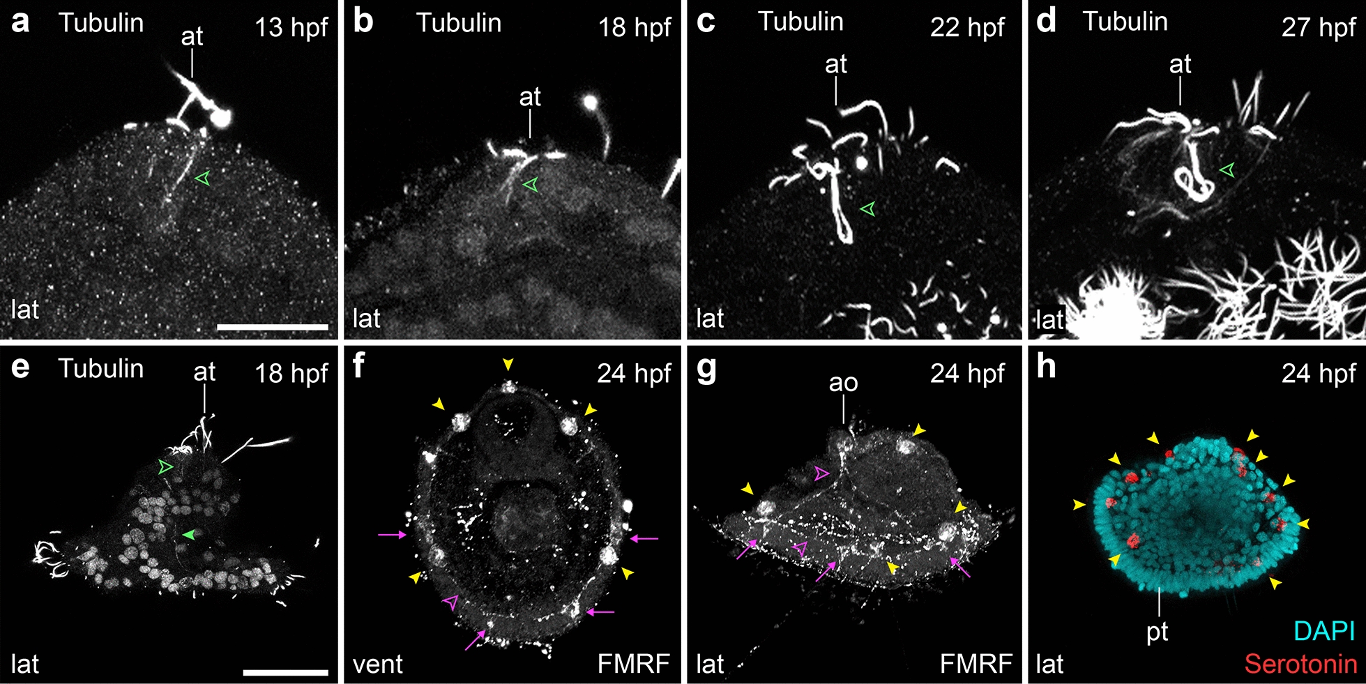
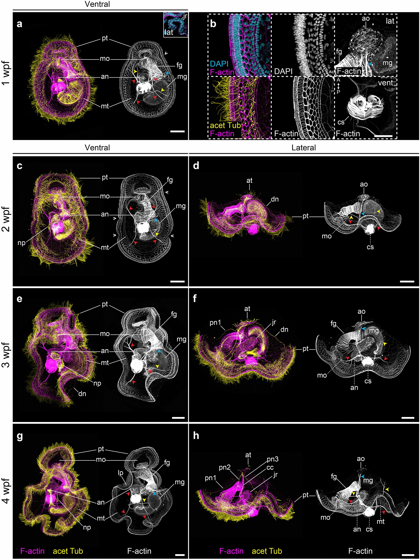
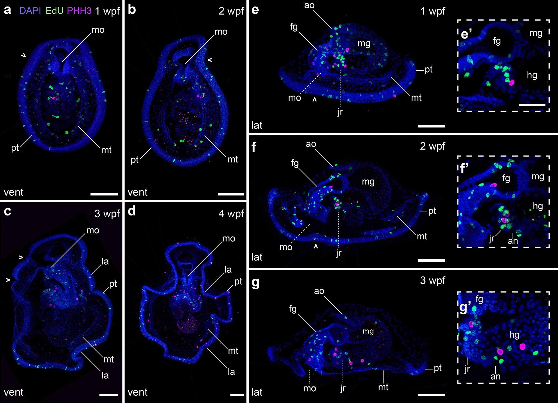
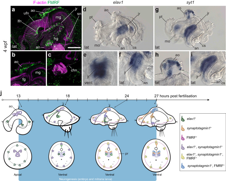
Results: Owenia fusiformis undergoes equal spiral cleavage where the first quartet of animal micromeres are slightly larger than the vegetal macromeres. Cleavage results in a coeloblastula approximately 5 h post-fertilization (hpf) at 19 °C. Gastrulation occurs via invagination and completes 4 h later, with putative mesodermal precursors and the chaetoblasts appearing 10 hpf at the dorso-posterior side. Soon after, at 11 hpf, the apical tuft emerges, followed by the first neurons (as revealed by the expression of elav1 and synaptotagmin-1) in the apical organ and the prototroch by 13 hpf. Muscles connecting the chaetal sac to various larval tissues develop around 18 hpf and by the time the mitraria is fully formed at 22 hpf, there are FMRFamide+ neurons in the apical organ and prototroch, the latter forming a prototrochal ring. As the mitraria feeds, it grows in size and the prototroch expands through active proliferation. The larva becomes competent after ~ 3 weeks post-fertilization at 15 °C, when a conspicuous juvenile rudiment has formed ventrally.

Conclusions: Owenia fusiformis embryogenesis is similar to that of other equal spiral cleaving annelids, supporting that equal cleavage is associated with the formation of a coeloblastula, gastrulation via invagination, and a feeding trochophore-like larva in Annelida. The nervous system of the mitraria larva forms earlier and is more elaborated than previously recognized and develops from anterior to posterior, which is likely an ancestral condition to Annelida. Altogether, our study identifies the major developmental events during O. fusiformis ontogeny, defining a conceptual framework for future investigations.

Keywords: Annelida; Equal cleavage; Larva; Mitraria; Nervous system; Neural development; Owenia fusiformis; Spiral cleavage; Spiralia; Trochophore.


Conflict of interest statement

The authors declare that they have no competing interests.

Figures

Fig. 1
Fig. 1
Owenia fusiformis, phylogenetic position, sampling location, and embryonic development. a O. fusiformis (blue box) is a member of Palaeoannelida, the sister clade to all remaining annelids. Annelid phylogeny according to [11]; b, c as adult, O. fusiformis dwells inside a self-built sand tube. The adult body is divided into a head with tentacles, three thoracic segments, abdominal segments and a pygidium (c); d embryological studies on O. fusiformis have focused on specimens collected from the English Channel, with the most recent ones studying a population near the Marine Biological Station of Roscoff, France. e Time course of developmental from oocyte to late mitraria larvae. Top row: Owenia fusiformis undergoes stereotypical equal spiral cleavage, formation of a coeloblastula and gastrulation by invagination. Bottom row: organogenesis starts with the formation of the mesodermal precursors and chaetoblasts, followed by the apical tuft and prototroch. By 13 hpf the blastopore elongates and a neurotroch forms posterior to the blastopore. Myogenesis starts when different larval muscles extend from the chaetal sac to the apical organ and gut. As the larva continues to grow until competence, several other muscles form. Red in e marks the first appearance of the most relevant cells or tissues at that stage. an: anus; ao: apical organ; at: apical tuft; as: abdominal segments; at: apical tuft; bp: blastopore; cb: chaetoblasts; cht: chaetae; dl: dorsal levator muscles; dlh: dorsolateral hyposphere muscles; em: esophageal muscle; en: endomesoderm; fgm: foregut muscle; ho: head opening; jr: juvenile rudiment; mo: mouth; ms: mesentoblasts; mt: metatroch; np: nephridia; nt: neurotroch; p: pygidium; pb: polar bodies; pt: protrotroch; rm: retractor muscles; st: sand tube; tc: tentacles; to: tail opening; ts: thoracic segments; vlh: ventrolateral hyposphere muscles. Scale bar is 2 cm
Fig. 2
Fig. 2
Owenia fusiformis undergoes equal spiral cleavage. Confocal laser scanning microscopy (CLSM) images of early embryonic stages from a oocyte to p blastula stage (approx. 64-cell). a The oocyte activates naturally in sea water and after the germinal vesicle breaks down. b After fertilization, the embryo undergoes equal cleavage resulting in two and later four equal blastomeres (cf). Open arrowheads in (d) and (e) points to the spiral deformation of the actin cytoskeleton in preparation for the dextral cleavage. fk The spiral cleavage initiates with the formation of four oblique micromeres (1q) that are larger than the macromeres (1Q). lo Cleavage continues with the stereotypical alternation of the spindles back and forward from counterclockwise to clockwise to give rise to the second (2q) (l), third (3q) (m, n) and fourth (4q) quartet of micromeres (o, p), until the formatin of the coeloblastula (o). bl: blastocoel; gv: germinal vesicle; pb: polar bodies; vf: vegetal cross furrow. Scale bar is 50 µm
Fig. 3
Fig. 3
Gastrulation via invagination in O. fusiformis. CLSM images of early embryonic stages from a 6 hpf (beginning of gastrulation) to h 10 hpf (post gastrulation). a At 6 hpf the gastral plate starts invaginating with the 5Q macromeres at the front of the archenteron roof (b). c By 7 hpf the archenteron roof connects with the animal ectoderm (closed arrowhead) (inset), and the polar bodies have internalized into the blastocoel (c, d). e Gastrulation ends by 9 hpf, in which the two germinal layers: endoderm and ectoderm are formed; while the mesoderm precursors and chaetoblasts form at the dorso-posterior end of the embryo (g, h). Arrows in (h) show the row of presumptive prototrochal cells dividing. bl: blastocoel; bp: blastopore; cb: chaetoblast; gv: germinal vesicle; ms: mesodermal precursor cells; pb: polar bodies; vf: vegetal cross furrow. Scale bar is 50 µm
Fig. 4
Fig. 4
Early mitraria larvae and the beginning of organogenesis. CLSM images of early larval stages from a 11 hpf to k 27 hpf. Images on the left column are ventral views with anterior facing up, while those on the right are lateral views with anterior facing left, except for (b) which is an apical view. ac Ciliogenesis starts at 11 hpf with the formation of the apical tuft and the prototroch dividing the embryo into an apical episphere and a vegetal hyposphere. d, e A short neurotroch then forms on the posterior side of the blastopore by 13 hpf. By this stage the blastopore elongates to form the presumptive mouth, but still remains open. f, g By 18 hpf, the early mitraria now has a secondary ciliary band, the metatroch, in addition to a complete gut, larval muscles and chaetae. hk Soon after, the larva grows and expands the metatroch throughout the hyposphere, while more muscle develops, including circular muscles around the foregut. an: anus; at: apical tuft; bl: blastocoel; bp: blastopore; cb: chaetoblast; cht: chaetae; cs: chaetal sac; dl: dorsal levator; em: esophageal muscle; fg: foregut; fgm: foregut circular muscle; hg: hindgut; mg: midgut; mo: mouth; mt: metatroch; np: nephridia; nt: neurotroch; pt: prototroch; rg: refringent globules; rm: retractor muscle; tm: membrane between chaetal sac and blastocoel. Scale bar is 50 µm
Fig. 5
Fig. 5
Cell proliferation during early mitraria. CLSM images of EdU and phospho-Histone 3 (PHH3) labeling on early mitraria larvae from a 13 hpf to h 27 hpf. a, c, e, g Are ventral views with anterior facing up, while b, d, f, h are lateral views with anterior facing left. Cell proliferation is widespread throughout the body, overlapping with cells that are mitotically active in the ciliary bands and the developing gut. an: anus; bp: blastopore; cht: chaetae; cs: chaetal sac; mg: midgut; mo: mouth; mt: metatroch; pt: prototroch. Scale bar is 50 µm
Fig. 6
Fig. 6
Early neural development during elongation and early mitraria. CLSM images of F-actin and FMRFamide+ elements and Differential Interference Contrast (DIC) images showing expression of elav1 and syt1 from a 13 hpf to x 27 hpf. Insets in (c, g, i, k, or, u) are close ups of the apical organ in the same view as the larger image. a, b, g, h There are no FMRFamide+ cells except for ten refringent globules (yellow arrowheads) in the apical organ and the prototroch either at 13 or 18 hpf. c, d A V-shaped apical organ composed of three elav1+ and syt1+ cells and seven prototrochal cells (black arrowheads) expressing elav1 are the first neurons to appear. i, j By early mitraria larva (18 hpf) elav1+ cells are positioned anterior and lateral to the gut reaching the ventral side of the larva, and k, l syt+ cells are also now fully expressed in seven prototrochal cells (black arrowheads). m, n The first FMRFamide+ cells are present by 22 hpf in the apical organ and by 24 hpf axons extend from the apical organ to an FMRFamide+ prototrochal ring connecting seven FMRFamide+ cells in the prototroch (s, t) (see Fig. 7f–g). or elav1+ and syt1+ expression remains very similar from 18 hpf up to 24 hpf. ux By 27 hpf, more cells express syt1 in the apical organ, but elav1 is now mostly restricted to neurons on the ventral side where neurons of the juvenile will start forming. an: anus; ao: apical organ; cs: chaetal sac; mo: mouth; mt: metatroch; nt: neurotroch; pt: prototroch. Scale bar is 50 µm
Fig. 7
Fig. 7
Tubulin+ and FMRFamide+ elements during neurogenesis. CLSM images of early larval stages from a 13 hpf to h 24 hpf. ad Close ups of the apical tuft innervation into the apical organ (green open arrowhead). e By 18 hpf an axon connects the apical organ with the prototroch (green closed arrowhead). f, g The apical organ connects with the neurons along the prototroch (magenta arrows), which start to be FMRFamide immunoreactive by 22–24 hpf (see Fig. 6m, n). These neurons eventually form an FMRFamide+ prototrochal ring (magenta arrowheads) which connects with the apical organ via an FMRFamide+ axon (magenta arrowheads). g, h The refringent globules that develop from 11 hpf, by 24 hpf are both FMRFamide+ and serotonin+. ao: apical organ; at: apical tuft; pt: prototroch. Scale is 25 µm in (ad). Scale bar in (eh) is 50 µm
Fig. 8
Fig. 8
Late mitraria larvae and juvenile rudiment development. CLSM images of late larval stages from a 1 wpf to h 4 wpf. a, c, e and g Are ventral views with anterior facing up, while d, f and h are lateral views with anterior facing left. b Close ups of specimens at 1 wpf. a The mature larva continues to grow and develop more musculature to connect the chaetal sac to different parts of the episphere and the hyposphere. The prototroch starts making bends (open arrowheads), and the metatroch is cleared from the chaetal sac and the ventral area where the juvenile rudiment is developing. b Both the hyposphere and episphere epithelial cells enlarge, the apical organ becomes more prominent and is full of microvilli, and the chaetal sac muscles become more robust. ch The apical organ now is connected to the prototrochal ring via peripheral and dorsal nerves. Cyan arrowheads point to the retractor muscles. Yellow arrowheads point to esophageal and dorsal levator muscles. Red arrowheads point to branching muscle connecting the peripheral regions of the ventrolateral hyposhere and the dorsolateral hyposphere muscles. an: anus; ao: apical organ; at: apical tuft; cc: circumesophageal connectives; cs: chaetal sac; dl: dorsal levator; dn: dorsal nerve; fg: foregut; jr: juvenile rudiment; lp: lappet; mg: midgut; mo: mouth; mt: metatroch; np: nephridia; nt: neurotroch; pn1–pn3: peripheral nerves 1 to 3; pt: prototroch. Scale bar is 50 µm. In b the scale bar is 25 µm
Fig. 9
Fig. 9
Growth of the late mitraria larvae. CLSM images of EdU and phosphoHistone 3 (PHH3) labeling on mitraria larvae from a, e 1 wpf to d 4 wpf. Images in ad are ventral views with anterior facing up, while those in eg are lateral views with anterior facing left. Are close-up sections of (eg). ag Proliferation in the late mitraria continues mostly in the ciliary bands, the gut and the developing juvenile rudiment. Open arrowheads point to the flexions of the ciliary band. eʹ–gʹ The juvenile rudiment has proliferative cells and a terminal mitotic cell, in what it could represent a posterior growth zone. an: anus; ao: apical organ; bp: blastopore; cs: chaetal sac; fg: foregut; hg: hindgut; jr: juvenile rudiment; la: lappet; mg: midgut; mo: mouth; mt: metatroch; nt: neurotroch; pt: prototroch. Scale bar is 50 µm. In the scale bar is 25 µm
Fig. 10
Fig. 10
Neural development in the late mitraria larvae. CLSM images of F-actin and FMRFamide+ elements and Differential Interference Contrast (DIC) images showing expression of elav1 and syt1 at 4 wpf. Images are in lateral view, except for (e) which is in ventral view. Bottom row are close-up sections of the animals from the top row. ac More FMRFamide+ neurons and neurites continue to form as the mitraria matures, including those innervating the foregut and the peripheral nerves of the juvenile rudiment (a), and the chaetal sac (c). See Helm et al. [14] for an extended description of the development of the FMRFamidergic and serotonergic nervous system of the late mitraria. elav1 expression has declined from the apical organ and is now restricted to the juvenile rudiment (ac), instead, syt1 is expressed in the mature neurons of the apical organ and the juvenile rudiment. j Diagram of neurogenesis in the mitraria larvae. At 13 hpf, neurogenesis starts from anterior to posterior, with elav1+ and syt1+ cells at the apical organ and as the embryo develops, the nervous system differentiates in a ventral/posterior progression where the nerve cord of the juvenile rudiment will form. FMRFamide+ cells appear by 22 to 24 hpf in the apical organ and in the prototroch. an: anus; ao: apical organ; chn: chaetal sac nerve; cs: chaetal sac; dlh: dorsolateral hyposphere muscles; em: esophageal muscle; en: endoderm; fg: foregut; hg: hindgut; jr: juvenile rudiment; lfn: lower foregut nerve; mg: midgut; mo: mouth; ms: mesentoblasts; mt: metatroch; np: nephridia; pr: prototrochal ring; pt: prototroch; sn: sphincter nerve; ufn: upper foregut nerve. Scale bar is 50 µm

Similar articles

Cited by

References

    1. Laumer CE, Bekkouche N, Kerbl A, Goetz F, Neves RC, Sørensen MV, et al. Spiralian phylogeny informs the evolution of microscopic lineages. Curr Biol. 2015;25:2000–2006. doi: 10.1016/j.cub.2015.06.068. - DOI - PubMed
    1. Marlétaz F, Peijnenburg KTCA, Goto T, Satoh N, Rokhsar DS. A new spiralian Phylogeny places the enigmatic arrow worms among gnathiferans. Curr Biol. 2019;29(312–318):e3. - PubMed
    1. Henry JQ. Spiralian model systems. Int J Dev Biol. 2014;58:389–401. doi: 10.1387/ijdb.140127jh. - DOI - PubMed
    1. Seaver EC. Variation in spiralian development: insights from polychaetes. Int J Dev Biol. 2014;58:457–467. doi: 10.1387/ijdb.140154es. - DOI - PubMed
    1. Martin-Duran JM, Marletaz F. Unravelling spiral cleavage. Development. 2020;147(1):dev181081. doi: 10.1242/dev.181081. - DOI - PubMed

LinkOut - more resources