. 2021 May 10;12(1):281.
doi: 10.1186/s13287-021-02349-y.

DPSCs treated by TGF-β1 regulate angiogenic sprouting of three-dimensionally co-cultured HUVECs and DPSCs through VEGF-Ang-Tie2 signaling

Affiliations

DPSCs treated by TGF-β1 regulate angiogenic sprouting of three-dimensionally co-cultured HUVECs and DPSCs through VEGF-Ang-Tie2 signaling

Yuchen Zhang et al. Stem Cell Res Ther. .

Abstract

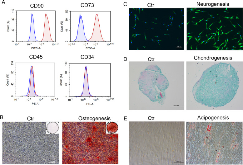
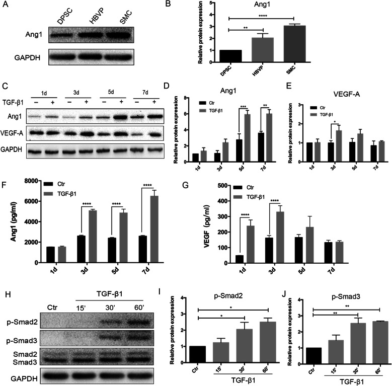
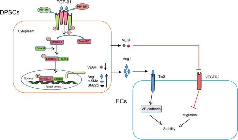
Background: Maintaining the stability and maturation of blood vessels is of paramount importance for the vessels to carry out their physiological function. Smooth muscle cells (SMCs), pericytes, and mesenchymal stem cells (MSCs) are involved in the maturation process of the newly formed vessels. The aim of this study was to investigate whether transforming growth factor beta 1 (TGF-β1) treatment could enhance pericyte-like properties of dental pulp stem cells (DPSCs) and how TGF-β1-treated DPSCs for 7 days (T-DPSCs) stabilize the newly formed blood vessels.

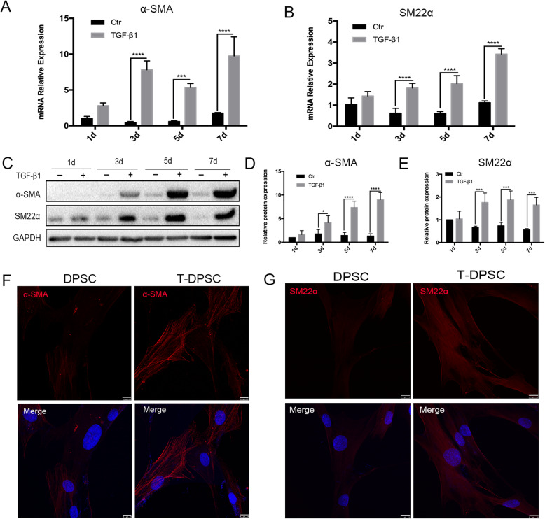
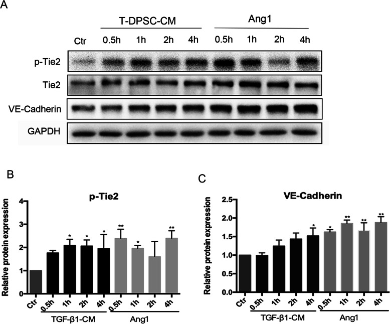
Methods: We utilized TGF-β1 to treat DPSCs for 1, 3, 5, and 7 days. Western blotting and immunofluorescence were used to analyze the expression of SMC markers. Functional contraction assay was conducted to assess the contractility of T-DPSCs. The effects of T-DPSC-conditioned media (T-DPSC-CM) on human umbilical vein endothelial cell (HUVEC) proliferation and migration were examined by MTT, wound healing, and trans-well migration assay. Most importantly, in vitro 3D co-culture spheroidal sprouting assay was used to investigate the regulating role of vascular endothelial growth factor (VEGF)-angiopoietin (Ang)-Tie2 signaling on angiogenic sprouting in 3D co-cultured spheroids of HUVECs and T-DPSCs. Angiopoietin 2 (Ang2) and VEGF were used to treat the co-cultured spheroids to explore their roles in angiogenic sprouting. Inhibitors for Tie2 and VEGFR2 were used to block Ang1/Tie2 and VFGF/VEGFR2 signaling.

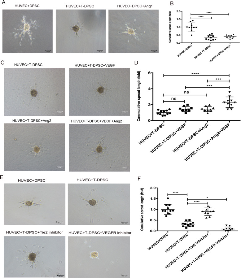
Results: Western blotting and immunofluorescence showed that the expression of SMC-specific markers (α-SMA and SM22α) were significantly increased after treatment with TGF-β1. Contractility of T-DPSCs was greater compared with that of DPSCs. T-DPSC-CM inhibited HUVEC migration. In vitro sprouting assay demonstrated that T-DPSCs enclosed HUVECs, resembling pericyte-like cells. Compared to co-culture with DPSCs, a smaller number of HUVEC sprouting was observed when co-cultured with T-DPSCs. VEGF and Ang2 co-stimulation significantly enhanced sprouting in HUVEC and T-DPSC co-culture spheroids, whereas VEGF or Ang2 alone exerted insignificant effects on HUVEC sprouting. Blocking Tie2 signaling reversed the sprouting inhibition by T-DPSCs, while blocking VEGF receptor (VEGFR) signaling boosted the sprouting inhibition by T-DPSCs.

Conclusions: This study revealed that TGF-β1 can induce DPSC differentiation into functional pericyte-like cells. T-DPSCs maintain vessel stability through Ang1/Tie2 and VEGF/VEGFR2 signaling.

Keywords: Ang1/Tie2 signaling; Angiogenesis; Dental pulp stem cells; Smooth muscle cells; Vessel stability.


Conflict of interest statement

The authors declare that they have no competing interests.

Figures

Fig. 1
Fig. 1
Characterization of DPSCs. a Specific marker analysis of DPSCs by flow cytometry. b Alizarin red staining after osteogenic induction for 21 days. c Immunofluorescence staining after neurogenic induction for 7 days (green: Tuj1, blue: DAPI). d Alcian blue staining after chondrogenic induction for 28 days. e Oil red O staining after adipogenic induction for 28 days
Fig. 2
Fig. 2
TGF-β1 induced the expression of α-SMA and SM22α in DPSCs. a, b RT-qPCR analysis of the relative mRNA expression of α-SMA and SM22α in DPSCs after treatment with TGF-β1 (10 ng/mL) for 1, 3, 5, and 7 days. ce Western blot analysis of the expression of α-SMA and SM22α in DPSCs after treatment with TGF-β1 (10 ng/mL) for 1, 3, 5, and 7 days. f, g Representative immunofluorescence images of α-SMA (red) and SM22α (red) in DPSCs treated with TGF-β1 for 7 days. Scale bar represents 10 μm. Data are mean ± standard error for n = 3 replicates, *p < 0.05, ***p <0.001, ****p <0.0001
Fig. 3
Fig. 3
T-DPSCs exhibited similar functional properties as SMCs. a Contraction assay of the contractility of T-DPSCs in collagen gel. 1, 2, and 3 are three replicates. b The gel diameter was measured and analyzed using ImageJ software after T-DPSC seeding in the gels for 48 h. c Confocal laser microscopy images showed the location of HUVECs and T-DPSCs in sprouting structures. HUVECs and T-DPSCs were labeled with green and red cell tracker dyes, respectively. Scale bar represents 100 μm. d, e T-DPSCs significantly inhibited the sprouting of HUVECs in a 3D spheroid model. Data are mean ± standard error for n = 3 replicates, **p <0.01, ****p <0.0001
Fig. 4
Fig. 4
TGF-β1 regulated Ang1 and VEGF expression and secretion in DPSCs. a, b Western blot analysis of the expression of Ang1 in DPSCs, HBVPs, and SMCs. ce Western blot analysis of the expression of Ang1 and VEGF in DPSCs treated with TGF-β1 (10 ng/mL) for 1, 3, 5, and 7 days. f, g ELISA analysis of the level of Ang1 and VEGF in conditioned media from DPSCs and T-DPSCs at 1, 3, 5, and 7 days. hj Western blot analysis of the expression of p-Smad2 and p-Smad3 in DPSCs treated with TGF-β1 (10 ng/mL) at 15, 30, and 60 min. Data are mean ± standard error for n = 3 replicates, *p <0.05, **p <0.01, ***p <0.001, ****p <0.0001
Fig. 5
Fig. 5
Conditioned media from T-DPSCs inhibited HUVEC migration. a HUVEC proliferation in DPSC-CM and T-DPSC-CM was determined by MTT assay. b, c Trans-well migration assay was performed to evaluate the effect of DPSC-CM and T-DPSC-CM on HUVEC migration ability. d, e HUVEC migration was determined by wound healing assay under DPSC-CM and T-DPSC-CM for 24 h. Data are mean ± standard error for n = 3 replicates, ****p <0.0001
Fig. 6
Fig. 6
Conditioned media from T-DPSCs activated Tie2 signaling. ac Western blot analysis of the expression of p-Tie2 and VE-Cadherin in HUVECs after treatment with T-DPSC-CM or Ang1 at different time points. Data are mean ± standard error for n = 3 replicates, *p <0.05, **p <0.01
Fig. 7
Fig. 7
T-DPSCs inhibited HUVEC sprouting through Ang1/Tie2 and VEGF/VEGFR2 signaling. a T-DPSCs and exogenous Ang1 inhibited endothelial sprouting. Representative images of in vitro sprouting assay using HUVECs + DPSCs, HUVECs + T-DPSCs, and HUVECs + DPSCs + Ang1 co-culture spheroids, respectively. b Quantification of cumulative length of sprouting assay using ImageJ software. c, d Sprouting assay of HUVEC and T-DPSC co-culture spheroids stimulated with VEGF (20 ng/mL) and Ang2 (1000 ng/mL). Representative images (c) and quantification of cumulative length of sprouting (d). e, f HUVECs were pretreated with the Tie2 inhibitor (5 μM) or VEGFR2 inhibitor (5μM) for 12 h before co-cultured with T-DPSCs to form spheroids. Sprouting images (e) and quantification of cumulative length (f). Data are mean ± standard error for n = 3 replicates, *p <0.05, ***p <0.001, ****p <0.0001
Fig. 8
Fig. 8
The expression of p-Tie2 and p-VEGFR2 in HUVECs. ac Western blot analysis of the expression of p-Tie2 and Tie2 in HUVEC/T-DPSC co-culture with or without high concentration Ang2 (1000 ng/mL) for 24 h. d, e HUVECs were pretreated with the Tie2 inhibitor (5 μM) for 12h, then treated with T-DPSC-CM or Ang1 for 30 min. Western blot analysis of the expression of p-Tie2 in HUVECs. f, g HUVECs were pretreated with the VEGFR2 inhibitor (5 μM) for 12h, then treated with VEGF for 2, 5, and 10 min. Western blot analysis of the expression of p-VEGFR2 in HUVECs. Data are mean ± standard error for n = 3 replicates, *p <0.05, **p <0.01, ***p <0.001, ****p <0.0001
Fig. 9
Fig. 9
The proposed molecular mechanism on how TGF-β1 regulate the role of DPSCs in vascular stabilization. TGF-β1 treatment activated Smad2 and Smad3 via phosphorylation, and the activated Smad2 and Smad3 associated with Smad4 regulated Ang1 and VEGF expression besides α-SMA, SM22α. After secretion, Ang1 derived from DPSCs activated its receptor, Tie2, on the HUVEC membrane surface. The activated Ang1/Tie2 promoted VE-Cadherin expression, enhancing cell-cell adhesion and blood vessel stability. The secreted VEGF from DPSCs decreased gradually as DPSCs differentiated into pericyte-like cells, which in turn attenuated VEGF/VEGFR2 signaling, inhibiting HUVEC migration

Similar articles

Cited by

References

    1. Sloan AJ, Waddington RJ. Dental pulp stem cells: what, where, how? Int. J. Paediatr. Dent. 2009;19(1):61–70. doi: 10.1111/j.1365-263X.2008.00964.x. - DOI - PubMed
    1. Ledesma-Martínez E, Mendoza-Núñez VM, Santiago-Osorio E. Mesenchymal stem cells derived from dental pulp: a review. Stem Cells Int. 2016;2016:4709572. doi: 10.1155/2016/4709572. - DOI - PMC - PubMed
    1. Janebodin K, Zeng Y, Buranaphatthana W, Ieronimakis N, Reyes M. VEGFR2-dependent angiogenic capacity of pericyte-like dental pulp stem cells. J Dent Res. 2013;92(6):524–531. doi: 10.1177/0022034513485599. - DOI - PubMed
    1. Dissanayaka WL, Zhan X, Zhang C, Hargreaves KM, Jin L, Tong EH. Coculture of dental pulp stem cells with endothelial cells enhances osteo-/odontogenic and angiogenic potential in vitro. J Endod. 2012;38(4):454–463. doi: 10.1016/j.joen.2011.12.024. - DOI - PubMed
    1. Xu JG, Zhu SY, Heng BC, Dissanayaka WL, Zhang CF. TGF-β1-induced differentiation of SHED into functional smooth muscle cells. Stem Cell Res Ther. 2017;8(1):10. doi: 10.1186/s13287-016-0459-0. - DOI - PMC - PubMed

Publication types

MeSH terms

Substances

LinkOut - more resources