

COVID-19: The experience from Italy
- PMID: 33972039
- PMCID: PMC7746129
- DOI: 10.1016/j.clindermatol.2020.12.008
COVID-19: The experience from Italy
Abstract
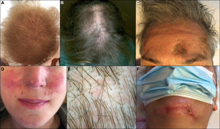
A wide range of cutaneous signs are attributed to COVID-19 infection. This retrospective study assesses the presence and impact of dermatologic manifestations related to the spread of COVID-19 in Lombardy, the geographic district with the first outbreak in Italy. A cohort of 345 patients with laboratory confirmed COVID-19 was collected from February 1, 2020 to May 31, 2020. Cutaneous signs and dermatologic diagnoses were recorded on admission, and during the course of the disease. Of the 345 patients included in the study, 52 (15%) had new-onset dermatologic conditions related to COVID-19. We observed seven major cutaneous clinical patterns, merged under 3 main groups: Exanthems, vascular lesions, and other cutaneous manifestations. Each subset was detailed with prevalence, age, duration, prognosis, and histology. Cutaneous findings can lead to suspect COVID-19 infection and identify potentially contagious cases with indolent course.
Copyright © 2021 Elsevier Ltd. All rights reserved.
Conflict of interest statement
Conflict of interest The authors have no conflicts of interest to declare.
Figures


Similar articles
-
Clinicopathologic correlations of COVID-19-related cutaneous manifestations with special emphasis on histopathologic patterns.Clin Dermatol. 2021 Jan-Feb;39(1):149-162. doi: 10.1016/j.clindermatol.2020.12.004. Epub 2020 Dec 14. Clin Dermatol. 2021. PMID: 33972045 Free PMC article. Review.
-
Skin manifestations of COVID-19 in children: Part 2.Clin Exp Dermatol. 2021 Apr;46(3):451-461. doi: 10.1111/ced.14482. Epub 2020 Nov 9. Clin Exp Dermatol. 2021. PMID: 33166429 Free PMC article. Review.
-
Cutaneous manifestations of COVID-19.Dermatol Online J. 2021 Jan 15;27(1):13030/qt2m54r7nv. Dermatol Online J. 2021. PMID: 33560783 Review.
-
COVID 19-associated chilblain-like acral lesions among children and adolescents: an Italian retrospective, multicenter study.Ital J Dermatol Venerol. 2023 Apr;158(2):117-123. doi: 10.23736/S2784-8671.23.07539-4. Ital J Dermatol Venerol. 2023. PMID: 37153946
-
Pernio-like skin lesions associated with COVID-19: A case series of 318 patients from 8 countries.J Am Acad Dermatol. 2020 Aug;83(2):486-492. doi: 10.1016/j.jaad.2020.05.109. Epub 2020 May 30. J Am Acad Dermatol. 2020. PMID: 32479979 Free PMC article.
Cited by
-
Dermatology and COVID-19: The Hidden Pandemic.J Clin Med. 2022 Jul 28;11(15):4397. doi: 10.3390/jcm11154397. J Clin Med. 2022. PMID: 35956013 Free PMC article.
-
How Coronavirus Disease 2019 Changed Dermatology Practice in 1 Year Around the World: Perspectives from 11 Countries.Dermatol Clin. 2021 Oct;39(4):639-651. doi: 10.1016/j.det.2021.05.014. Epub 2021 May 31. Dermatol Clin. 2021. PMID: 34556253 Free PMC article. Review.
-
Urticarial rash as the initial presentation of COVID-19 infection: A case report.Clin Case Rep. 2022 Jul 14;10(7):e6076. doi: 10.1002/ccr3.6076. eCollection 2022 Jul. Clin Case Rep. 2022. PMID: 35846919 Free PMC article.
-
Value of the Lymphocyte Transformation Test for the Diagnosis of Drug-Induced Hypersensitivity Reactions in Hospitalized Patients with Severe COVID-19.Int J Mol Sci. 2023 Jul 17;24(14):11543. doi: 10.3390/ijms241411543. Int J Mol Sci. 2023. PMID: 37511302 Free PMC article.
-
Chilblains-Like Lesions in Pediatric Patients: A Review of Their Epidemiology, Etiology, Outcomes, and Treatment.Front Pediatr. 2022 Jun 23;10:904616. doi: 10.3389/fped.2022.904616. eCollection 2022. Front Pediatr. 2022. PMID: 35813389 Free PMC article. Review.
References
-
- Recalcati S. Cutaneous manifestations in COVID-19: a first perspective. J Eur Acad Dermatol Venereol. 2020;34 e212-e213. - PubMed
MeSH terms
LinkOut - more resources
Full Text Sources
Other Literature Sources
Medical
