. 2021 Jan-Feb;39(1):12-22.
doi: 10.1016/j.clindermatol.2020.12.008. Epub 2020 Dec 17.

COVID-19: The experience from Italy

Affiliations

COVID-19: The experience from Italy

Sebastiano Recalcati et al. Clin Dermatol. 2021 Jan-Feb.

Abstract

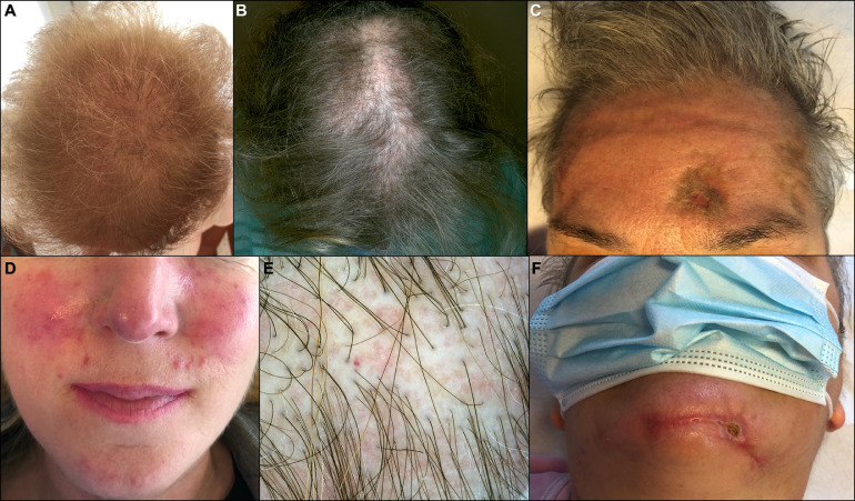
A wide range of cutaneous signs are attributed to COVID-19 infection. This retrospective study assesses the presence and impact of dermatologic manifestations related to the spread of COVID-19 in Lombardy, the geographic district with the first outbreak in Italy. A cohort of 345 patients with laboratory confirmed COVID-19 was collected from February 1, 2020 to May 31, 2020. Cutaneous signs and dermatologic diagnoses were recorded on admission, and during the course of the disease. Of the 345 patients included in the study, 52 (15%) had new-onset dermatologic conditions related to COVID-19. We observed seven major cutaneous clinical patterns, merged under 3 main groups: Exanthems, vascular lesions, and other cutaneous manifestations. Each subset was detailed with prevalence, age, duration, prognosis, and histology. Cutaneous findings can lead to suspect COVID-19 infection and identify potentially contagious cases with indolent course.


Conflict of interest statement

Conflict of interest The authors have no conflicts of interest to declare.

Figures

Fig 1
Fig. 1
Clinical characteristics of COVID-19 exanthems. A-C, Maculopapular dermatitis. D,E, Urticarial dermatitis in case 25. F, Vesicular dermatitis.
Fig 2
Fig. 2
Histopathological findings. A, Urticarial dermatitis: cuffs of lymphocytes surrounding blood vessels (red arrow) and dermal ducts (black arrow). B, Maculopapular dermatitis: superficial and deep perivascular dermatitis with lichenoid band-like pattern. C, Maculopapular dermatitis: exocytosis of lymphocytes and necrotic keratinocytes in the epidermis (black arrow); perivascular cuffs of lymphocytes in the dermis are found (red arrow). D, Maculopapular dermatitis: lymphocytic infiltrate surrounding dermal ducts and blood vessels (red arrow). E, Maculopapular dermatitis: dense patchy band-like infiltrate surrounding acrosyringeal ducts. F, Maculopapular dermatitis: lymphocytes surrounding an acrosyringeal duct (red arrow). G, Vesicular dermatitis: superficial spongiotic and perivascular dermatitis with intraepidermal vesicles (red arrow); heavy lymphocytic cuff around dermal vessels (black arrows). H, Vesicular dermatitis: superficial perivascular dermatitis with large intraepidermal nests of Langerhans cells (red arrow).
Fig 3
Fig. 3
Clinical characteristics of COVID-19 exanthems: erythema multiforme. A,B, Targetoid lesions on the hands in case 32. C,D, Lesions on the upper extremities in case 35.
Fig 4
Fig. 4
Histopathological findings. A, Erythema multiforme: superficial band-like perivascular dermatitis with necrotic keratinocytes (black arrow). In the mid-dermis, dilated blood vessels surrounded by lymphocytic cuff (red arrow). B, Livedo reticularis: large thrombus in the mid-dermis (red arrow). C, Livedo: numerous thrombosed vessels with marked endothelial hyperplasia in the superficial and deep dermis (red arrows). D, Chilblain-like lesions: dense coat-sleeve-like lymphoid infiltrate around medium- and small-caliber dermal vessels, as well as around dermal glands. E, Lichen planopilaris of the scalp: ulcerated epidermis with dense lymphocytic infiltration around hair follicles (red arrow); the dermis is diffusely fibrotic. F, Lichen planopilaris: diffuse thrombosis of the upper dermal vessels (red arrows).
Fig 5
Fig. 5
Clinical characteristics of COVID-19 vascular lesions. A, Vasculitis. B, Thrombosis. C, Acro-ischemia. D,E, Chilblain-like lesions in 2 cases. F, Livedo reticularis. b c d e should be rotated 180 degrees
Fig 6
Fig. 6
Indirect cutaneous manifestations related to COVID-19. A, Telogen effluvium. B, Lichen planopilaris. C,F, Pressure sores and telogen effluvium. D, Rosacea. E, Trichoscopy of lichen planopilaris.

References

    1. Li Q, Guan X, Wu P. Early transmission dynamics in Wuhan, China, of novel coronavirus-infected pneumonia. N Engl J Med. 2020;382:1199–1207. - PMC - PubMed
    1. Wang T, Du Z, Zhu F. Comorbidities and multi-organ injuries in the treatment of COVID-19. Lancet. 2020;395:e52. - PMC - PubMed
    1. Recalcati S. Cutaneous manifestations in COVID-19: a first perspective. J Eur Acad Dermatol Venereol. 2020;34 e212-e213. - PubMed
    1. De Giorgi V, Recalcati S, Jia Z. Cutaneous manifestations related to coronavirus disease 2019 (COVID-19): a prospective study from China and Italy. J Am Acad Dermatol. 2020;83:674–675. - PMC - PubMed
    1. Recalcati S, Barbagallo T, Frasin LA. Acral cutaneous lesions in the time of COVID-19. J Eur Acad Dermatol Venereol. 2020;34 e346-e347. - PMC - PubMed

MeSH terms