

Clinicopathologic correlations of COVID-19-related cutaneous manifestations with special emphasis on histopathologic patterns
- PMID: 33972045
- PMCID: PMC7832768
- DOI: 10.1016/j.clindermatol.2020.12.004
Clinicopathologic correlations of COVID-19-related cutaneous manifestations with special emphasis on histopathologic patterns
Abstract
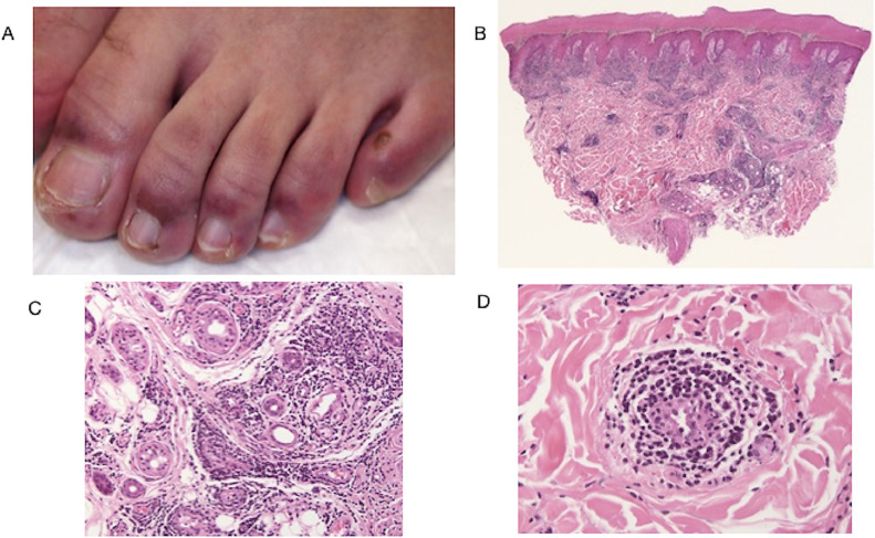
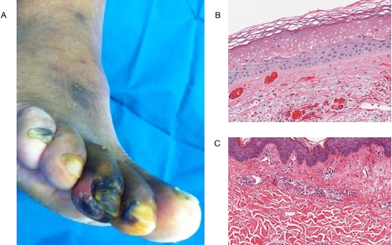
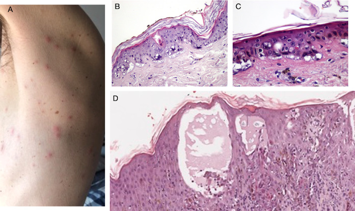
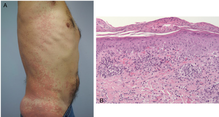
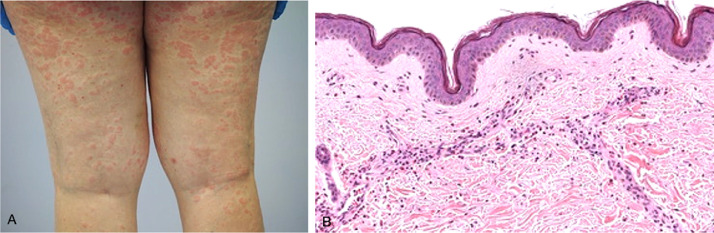
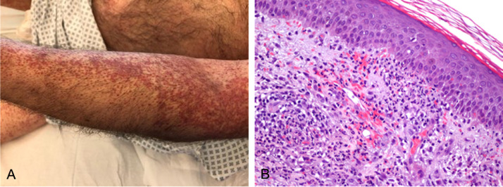
Skin is one of target organs affected by the novel coronavirus SARS-CoV-2, and in response to the current COVID-19 pandemic, a fast body of literature has emerged on related cutaneous manifestations. Current perspective is that the skin is not only a bystander of the general cytokines storm with thrombophilic multiorgan injury, but it is directly affected by the epithelial tropism of the virus, as confirmed by the detection of SARS-CoV-2 in endothelial cells and epithelial cells of epidermis and eccrine glands. In contrast with the abundance of epidemiologic and clinical reports, histopathologic characterization of skin manifestations is limited. Without an adequate clinicopathologic correlation, nosology of clinically similar conditions is confusing, and effective association with COVID-19 remains presumptive. Several patients with different types of skin lesions, including the most specific acral chilblains-like lesions, showed negative results at SARS-CoV-2 nasopharyngeal and serologic sampling. The aim of this review is to provide an overview of what has currently been reported worldwide, with a particular emphasis on microscopic patterns of the skin manifestations in patients exposed to or affected by COVID-19. Substantial breakthroughs may occur in the near future from more skin biopsies, improvement of immunohistochemistry studies, RNA detection of SARS-CoV-2 strain by real-time polymerase chain reaction-based assay, and electron microscopic studies.
Copyright © 2020 Elsevier Ltd. All rights reserved.
Figures
Similar articles
-
SARS-CoV-2 endothelial infection causes COVID-19 chilblains: histopathological, immunohistochemical and ultrastructural study of seven paediatric cases.Br J Dermatol. 2020 Oct;183(4):729-737. doi: 10.1111/bjd.19327. Epub 2020 Aug 5. Br J Dermatol. 2020. PMID: 32562567 Free PMC article.
-
Skin manifestations of COVID-19 in children: Part 2.Clin Exp Dermatol. 2021 Apr;46(3):451-461. doi: 10.1111/ced.14482. Epub 2020 Nov 9. Clin Exp Dermatol. 2021. PMID: 33166429 Free PMC article. Review.
-
COVID-19: The experience from Italy.Clin Dermatol. 2021 Jan-Feb;39(1):12-22. doi: 10.1016/j.clindermatol.2020.12.008. Epub 2020 Dec 17. Clin Dermatol. 2021. PMID: 33972039 Free PMC article.
-
Skin Manifestations Associated with COVID-19: Current Knowledge and Future Perspectives.Dermatology. 2021;237(1):1-12. doi: 10.1159/000512932. Epub 2020 Nov 24. Dermatology. 2021. PMID: 33232965 Free PMC article. Review.
-
Identification, Mechanism, and Treatment of Skin Lesions in COVID-19: A Review.Viruses. 2021 Sep 24;13(10):1916. doi: 10.3390/v13101916. Viruses. 2021. PMID: 34696346 Free PMC article. Review.
Cited by
-
Cutaneous manifestations of COVID-19 in a tertiary COVID-19 referral hospital in the Philippines.JAAD Int. 2022 Jun;7:44-51. doi: 10.1016/j.jdin.2022.01.007. Epub 2022 Feb 2. JAAD Int. 2022. PMID: 35128486 Free PMC article.
-
SARS-CoV-2 and Skin: The Pathologist's Point of View.Biomolecules. 2021 Jun 4;11(6):838. doi: 10.3390/biom11060838. Biomolecules. 2021. PMID: 34200112 Free PMC article.
-
Complanatuside alleviates inflammatory cell damage induced by pro-inflammatory cytokines in skin keratinocytes.Front Chem. 2022 Aug 10;10:909651. doi: 10.3389/fchem.2022.909651. eCollection 2022. Front Chem. 2022. PMID: 36034662 Free PMC article.
-
Chilblain-like lesions onset during SARS-CoV-2 infection in a COVID-19-vaccinated adolescent: case report and review of literature.Ital J Pediatr. 2022 Jun 13;48(1):93. doi: 10.1186/s13052-022-01296-5. Ital J Pediatr. 2022. PMID: 35698236 Free PMC article. Review.
-
Multisystem Inflammatory Syndrome Associated with SARS-CoV-2 Infection in an Adult: A Case Report from the Maldives.Trop Med Infect Dis. 2021 Oct 19;6(4):187. doi: 10.3390/tropicalmed6040187. Trop Med Infect Dis. 2021. PMID: 34698279 Free PMC article.
References
Publication types
MeSH terms
Supplementary concepts
LinkOut - more resources
Full Text Sources
Other Literature Sources
Medical
Miscellaneous
