Review
. 2021 Jun;48(2):247-266.
doi: 10.1016/j.ogc.2021.02.001.

Placental Function and the Development of Fetal Overgrowth and Fetal Growth Restriction

Affiliations
Review

Placental Function and the Development of Fetal Overgrowth and Fetal Growth Restriction

Jerad H Dumolt et al. Obstet Gynecol Clin North Am. 2021 Jun.

Abstract

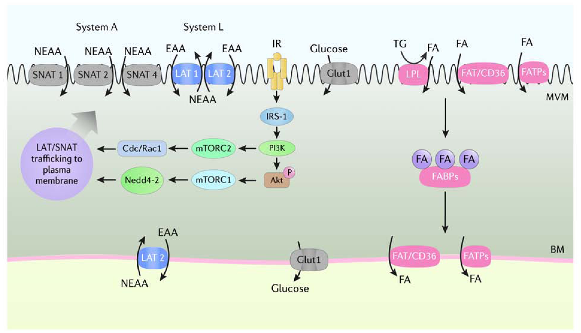
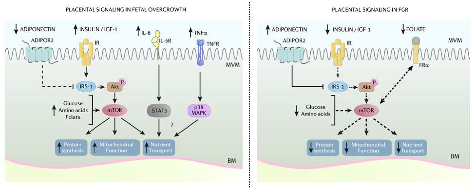
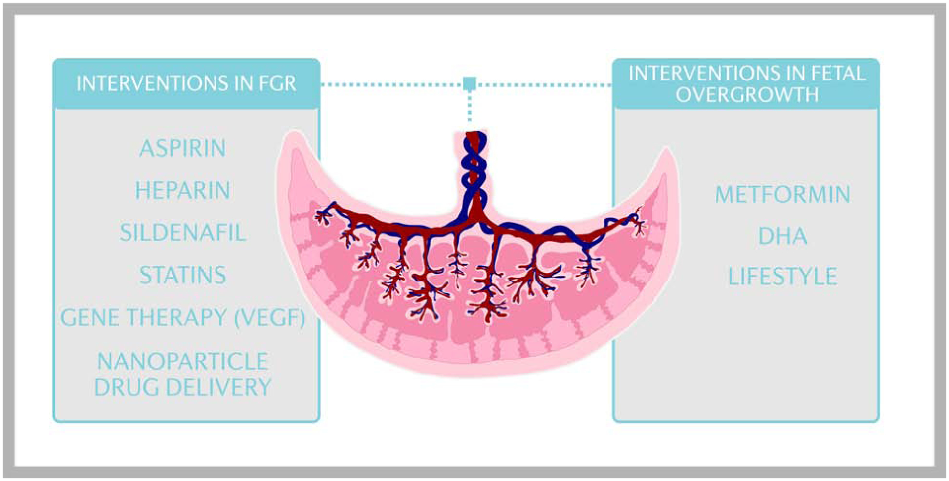
Placental regulation of fetal growth involves the integration of multiple signaling pathways that modulate an array of placental functions, including nutrient transport. As a result, the flux of oxygen and nutrients to the fetus is altered, leading to changes in placental and fetal growth. Placental insulin/insulinlike growth factor-1 and mechanistic target of rapamycin signaling and amino acid transport capacity are inhibited in fetal growth restriction and activated in fetal overgrowth, implicating these placental functions in driving fetal growth. With novel approaches to specifically target the placenta, clinical interventions to modulate placental function in high-risk pregnancies can be developed.

Keywords: Fetal development; Fetal growth restriction; Fetal overgrowth; Maternal-fetal exchange; Placental transport; Syncytiotrophoblast.


Conflict of interest statement

Conflict of Interests: The authors declare that there are no competing interests associated with the manuscript.

Figures

Figure 1.
Figure 1.. Some key placental signaling pathways and nutrient transporters
The syncytiotrophoblast consists of two polarized plasma membranes, microvillous plasma membrane (MVM) and basal membrane (BM), that express an array of transport proteins that mediate maternal-to-fetal transfer of amino acids, glucose, and fatty acids. The uptake of nonessential and essential amino acids from maternal circulation across the MVM is mediated by System A (SNAT1, 2, 4) and System L (LAT1, 2) transport systems that are trafficked to the plasma membrane as a result of activation of insulin/IGF-1 and mTOR signaling. GLUT-1 is highly expressed in the MVM and BM of the syncytiotrophoblast and is considered the primary glucose transporter in the human placenta at term. Maternal triglycerides are hydrolyzed into FFA by membrane-bound lipases and transferred across the MVM by FAT/CD36 and FATPs. Internalized FFA are transferred to the BM by FABPs for export into fetal circulation. Akt, Protein Kinase B; Cdc/Rac1, cell division control protein/ras-related C3 botulinum toxin substrate 1; EAA, essential amino acids; FA, fatty acids; FABPs, fatty acid binding proteins; FAT/CD36, fatty acid translocase/cluster of differentiation 36; FATPs, fatty acid transport proteins; GLUT1, glucose transporter 1; IGF-1, insulin-like growth factor; IR, insulin receptor; IRS-1, insulin receptor substrate 1; LAT 1, L-amino acid transporter 1, 2; LPL, lipoprotein lipase, mTORC1, mechanistic target of rapamycin complex 1; mTORC2, mechanistic target of rapamycin complex 2; Nedd4–2, neuronal precursor cell-expressed, developmentally down-regulated gene 4 isoform 2; NEAA, non-essential amino acids; SNAT 1, Sodium-coupled neutral amino acid transporter 1, 2, 4; TG, triglycerides. Courtesy of KIMEN Design4Research, with permission.
Figure 2.
Figure 2.. Placental signaling in fetal overgrowth and FGR
The coordinated actions of placental insulin/IGF-1, mTOR, adiponectin, and inflammatory cytokine signaling pathways act to regulate mitochondrial function, protein synthesis and the flux of glucose, amino acids, lipids, and folate across the placental barrier. Placental insulin/IGF-1 and mTOR signaling is activated in women with obesity delivering LGA infants likely due to elevated circulating maternal insulin/IGF-1 and increased availably of nutrients. Moreover, low maternal levels of circulating adiponectin in pregnancies complicated by maternal obesity contribute to enhance placental insulin signaling due to decreased inhibition of IRS-1. Increased maternal circulating proinflammatory cytokines IL-6 and TNF-α are increased in pregnancies with obesity and may contribute to fetal overgrowth by activating STAT3 and p38 MAPK signaling pathways. Conversely, lower maternal circulating insulin/IGF-1, folate, and increased adiponectin contribute to reduced placental insulin/IGF-1 and mTOR signaling in FGR pregnancies. Thus, differential regulation of placental signaling pathways and subsequent impact on placental function and nutrient transfer likely contribute to fetal overgrowth and FGR. ADIPOR2, adiponectin receptor 2; Akt, Protein Kinase B; BM, basal membrane; FRα, folate receptor-α; IGF-1, insulin-like growth factor 1; IR, insulin receptor; IRS-1, insulin receptor substrate 1; IL-6, interleukin-6; IL-6R, interleukin-6 receptor; FGR, fetal growth restriction; mTORC, mechanistic target of rapamycin complex; MVM, microvillous membrane; p38 MAPK, p38 mitogen-activated protein kinases; STAT3, Signal transducer and activator of transcription 3; TNF-α, tumor necrosis factor-α; TNFR, tumor necrosis factor-α receptor. Courtesy of KIMEN Design4Research, with permission.
Figure 3.
Figure 3.. The influence of small extracellular vesicles on placental function.
Small extracellular vesicles (sEV) are membrane-bound particles containing bioactive proteins, lipids, DNA, mRNA, and miR that are secreted from most cells and participate in cell to cell communication. The functions of maternal and placental sEVs remain to be fully established but are believed to be involved in immune response, angiogenesis, placentation, and the transfer of nucleic acids and proteins important in normal and complicated pregnancies that may influence fetal growth. miR, microRNA; MVB, multivesicular bodies. Courtesy of KIMEN Design4Research, with permission.
Figure 4.
Figure 4.. Clinical interventions targeting placental function to restore fetal growth
Numerous studies have tested the hypothesis that systemic administration of vasodilators such as aspirin, low molecular weight heparin, and sildenafil increases utero-placental blood flow and promote fetal growth in FGR, however the results have not been encouraging. Clinical trials to improve fetal growth in FGR pregnancies using statins have been initiated. In addition, gene therapy and nanoparticle drug delivery designed to alter the expression of genes in the uteroplacental circulation and the placenta, including VEGF, is an area of active research. Limited clinical approaches to prevent fetal overgrowth currently exist, however, treatment of GDM and/or maternal obesity with metformin, DHA supplementation, or lifestyle interventions may mitigate fetal overgrowth in high risk pregnancies. DHA, docosahexaenoic acid; GDM, gestational diabetes mullites; FGR, fetal growth restriction; VEGF, vascular endothelial growth factor. Courtesy of KIMEN Design4Research, with permission.

References

    1. Goldstein RF, Abell SK, Ranasinha S, et al. Association of Gestational Weight Gain With Maternal and Infant Outcomes: A Systematic Review and Meta-analysis. JAMA. 2017;317(21):2207–2225. - PMC - PubMed
    1. Gluckman PD, Hanson MA, Cooper C, Thornburg KL. Effect of in utero and early-life conditions on adult health and disease. N Engl J Med. 2008;359(1):61–73. - PMC - PubMed
    1. Reynolds RM, Allan KM, Raja EA, et al. Maternal obesity during pregnancy and premature mortality from cardiovascular event in adult offspring: follow-up of 1 323 275 person years. BMJ. 2013;347:f4539. - PMC - PubMed
    1. Catalano PM, Ehrenberg HM. The short- and long-term implications of maternal obesity on the mother and her offspring. BJOG. 2006;113(10):1126–1133. - PubMed
    1. Gluckman PD, Hanson MA, Pinal C. The developmental origins of adult disease. Matern Child Nutr. 2005;1(3):130–141. - PMC - PubMed