Review
. 2021 Jul;44(7):550-563.
doi: 10.1016/j.tins.2021.03.006. Epub 2021 May 7.

Circuit organization of the rodent medial prefrontal cortex

Affiliations
Review

Circuit organization of the rodent medial prefrontal cortex

Paul G Anastasiades et al. Trends Neurosci. 2021 Jul.

Abstract

The prefrontal cortex (PFC) orchestrates higher brain function and becomes disrupted in many mental health disorders. The rodent medial PFC (mPFC) possesses an enormous variety of projection neurons and interneurons. These cells are engaged by long-range inputs from other brain regions involved in cognition, motivation, and emotion. They also communicate in the local network via specific connections between excitatory and inhibitory cells. In this review, we describe the cellular diversity of the rodent mPFC, the impact of long-range afferents, and the specificity of local microcircuits. We highlight similarities with and differences between other cortical areas, illustrating how the circuit organization of the mPFC may give rise to its unique functional roles.

Keywords: cell types; circuits; interneuron; prefrontal cortex; projection neuron; synapses.


Conflict of interest statement

Declaration of interests The authors declare no interests in relation to this work.

Figures

Figure 1.
Figure 1.. General organization of the rodent mPFC
A) Key brain regions that communicate with the mPFC by providing input to it, receiving output projections from the mPFC, or making reciprocal connections with the mPFC. BLA = basolateral amygdala, CLA = claustrum, PAG = periaqueductal gray, STR = striatum, THAL = thalamus, vHPC = ventral hippocampus, VTA = ventral tegmental area. Note that many connections are reciprocal. B) Distribution of the subdivisions of the mPFC along the dorso-ventral axis. ACC = anterior cingulate cortex, PL = prelimbic, IL = infralimbic.
Figure 2.
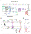
Figure 2.. mPFC projection neuron diversity
A) Laminar distributions of projection neurons in the mPFC, identified by injection of retrograde tracers into target brain regions. BLA = basolateral amygdala, cPFC = contralateral PFC, iSTR = ipsilateral striatum, cSTR = contralateral striatum, cCLA = contralateral claustrum, PT = pyramidal tract, PT / CT = PT cells that send collaterals to the thalamus, CT = corticothalamic, non-PT cells. PIA = pial surface, WM = white matter, L5b up = upper L5b, L5b low = lower L5b. Note that projection neurons are found from L2 to L6. B) Dendritic morphologies of mPFC projection neurons. From left to right: L2/3 intratelencephalic (IT) cell, L5 IT cell, L5 pyramidal tract (PT) cell, L6 IT cell, and L6 corticothalamic (CT) cell. Note that L5 PT cells have more elaborate dendrites than adjacent IT cells, and that L6 CT cells have apical dendrites that often extend to L1, unlike L6 IT cells that can display inverted pyramidal morphologies. C) Intrinsic firing properties of mPFC projection neurons. From top to bottom: L5 IT cells, L5 PT cells and L6 CT cells. Note that L5 IT cells lack Ih-mediated voltage sag, L5 PT cells display strong Ih-mediated voltage sag, and L6 CT cells display highly non-adapting firing patterns. Panel A adapted from references & . Data in panels B & C used with permission from references , & ,
Figure 3.
Figure 3.. Long-range excitatory inputs to the mPFC
A) Laminar distribution of axons to the prelimbic mPFC from different brain regions. From left to right: BLA = basolateral amygdala, cPFC = contralateral PFC, MD = mediodorsal thalamus, VM = ventromedial thalamus, vHPC = ventral hippocampus. Note that inputs arrive in characteristic patterns across L1 to L6. B) Summary of the main projection neuron(s) contacted by long-range excitatory inputs. BLA activates reciprocally-projecting L2 cortico-amygdala (CA) neurons. cPFC innervates more broadly, including L5 PT cells and a range of IT cells. MD strongly activates L3 IT cells, while VM engages the dendrites of L5 PT cells. vHPC primarily targets L5 IT cells. Note that the most strongly driven targets are shown for simplicity, but most inputs provide some excitation to all cells in the network. C) Biased long-range excitatory inputs onto neighboring projection neurons. Top, BLA preferentially contacts L2 CA neurons over L2 cortico-cortical (CC) neurons, whereas cPFC shows no bias. Middle, BLA and cPFC preferentially contact L5 PT cells over L5 IT cells, with vHPC showing opposite bias. Bottom, MD preferentially contacts L5 IT cells over L5 PT cells, but make stronger contacts onto dendrites of L5 PT cells. VM inputs make highly enriched inputs to L5 PT apical dendrites, where they can evoke dendritic Ca2+ spikes. Images in panel A adapted with permission from reference and unpublished data from the Carter lab,
Figure 4.
Figure 4.. Local excitatory connections in the mPFC
A) Simplified wiring diagram of the local excitatory circuit in sensory cortex (left) and mPFC (right). Individual arrows represent major excitatory connections, with the weight of the arrow indicating connection strength. B) Biased local excitatory connectivity within and across layers of the mPFC. Note that IT cells, PT cells and CT cells make within-class connections, and that IT cells also make between-class connections onto PT and CT cells, which are the major outputs to subcortical brain regions.
Figure 5.
Figure 5.. GABAergic interneuron subtypes in the mPFC
A) Segregation of different subpopulations of interneurons based on gene expression and morphological properties. PV = parvalbumin, SOM = somatostatin, 5HT3aR = serotonin receptor 3a, CCK = cholecystokinin, VIP = vasoactive intestinal peptide, NDNF = neuron derived neurotrophic factor. B) Laminar distributions of interneurons based on labelling with transgenic mouse lines. PIA = pial surface, WM = white matter, L5b up = upper L5b, L5b low = lower L5b. C) Characteristic firing properties of different interneurons. Grey traces show peri-threshold spikes and colored traces show sustained firing in response to larger current steps. Data in panel B adapted from references , , & . Traces in panel C adapted with permission from reference and unpublished data from the Carter lab,
Figure 6:
Figure 6:. Feed-forward inhibitory circuits in the mPFC
A) Targeting of pyramidal cells by PV+ basket cells, PV+ chandelier cells, SOM+ cells, CCK+ cells and NDNF+ cells, along with targeting of SOM+ cells by VIP+ cells and PV+ cells by SOM+ cells and NDNF+ cells. B) Biased inhibitory inputs onto neighboring projection neurons. PV+ (basket and chandelier cells) and SOM+ cells preferentially contact L2 CA neurons over L2 CC neurons. PV+, SOM+, CCK+ and NDNF+ preferentially contact L5 PT cells over L5 IT cells. C) Summary of the currently known interneurons contacted by long-range excitatory inputs. BLA activates L2 PV+ and SOM+ cells. cPFC innervates L2 Chandelier cells, in addition to L5 PV+ and SOM+ cells. MD engages both L3 PV+ and SOM+ cells, as well as L1 VIP+ cells. VM drives L1 NDNF+ cells. vHPC activates L2/3 VIP+ cells and L5 PV+, SOM+ and CCK+ cells. Note that the distinct presynaptic release properties of inputs onto these cells mean they also fire during different phases of activity.

Similar articles

Cited by

References

    1. Miller EK and Cohen JD (2001) An integrative theory of prefrontal cortex function. Annu Rev Neurosci 24, 167–202 - PubMed
    1. Robbins TW and Arnsten AFT (2009) The Neuropsychopharmacology of Fronto-Executive Function: Monoaminergic Modulation. Annu Rev Neurosci 32, 267–287 - PMC - PubMed
    1. Hare BD and Duman RS (2020) Prefrontal cortex circuits in depression and anxiety: contribution of discrete neuronal populations and target regions. Molecular Psychiatry DOI: 10.1038/s41380-020-0685-9 - DOI - PMC - PubMed
    1. Arnsten AFT and Rubia K (2012) Neurobiological Circuits Regulating Attention, Cognitive Control, Motivation, and Emotion: Disruptions in Neurodevelopmental Psychiatric Disorders. Journal of the American Academy of Child & Adolescent Psychiatry 51, 356–367 - PubMed
    1. Harris KD and Shepherd GMG (2015) The neocortical circuit: themes and variations. Nature Neuroscience 18, 170–181 - PMC - PubMed

Publication types