. 2022 Feb;106(2):288-296.
doi: 10.1136/bjophthalmol-2020-318529. Epub 2021 May 10.

Evaluation of intravitreal topotecan dose levels, toxicity and efficacy for retinoblastoma vitreous seeds: a preclinical and clinical study

Affiliations

Evaluation of intravitreal topotecan dose levels, toxicity and efficacy for retinoblastoma vitreous seeds: a preclinical and clinical study

Carley M Bogan et al. Br J Ophthalmol. 2022 Feb.

Abstract

Background: Current melphalan-based intravitreal regimens for retinoblastoma (RB) vitreous seeds cause retinal toxicity. We assessed the efficacy and toxicity of topotecan monotherapy compared with melphalan in our rabbit model and patient cohort.

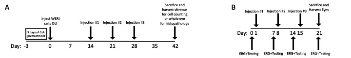
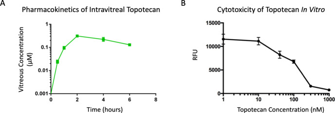
Methods: Rabbit experiments: empiric pharmacokinetics were determined following topotecan injection. For topotecan (15 μg or 30 µg), melphalan (12.5 µg) or saline, toxicity was evaluated by serial electroretinography (ERG) and histopathology, and efficacy against vitreous seed xenografts was measured by tumour cell reduction and apoptosis induction.

Patients: retrospective cohort study of 235 patients receiving 990 intravitreal injections of topotecan or melphalan.

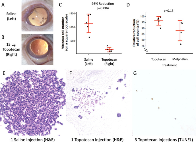
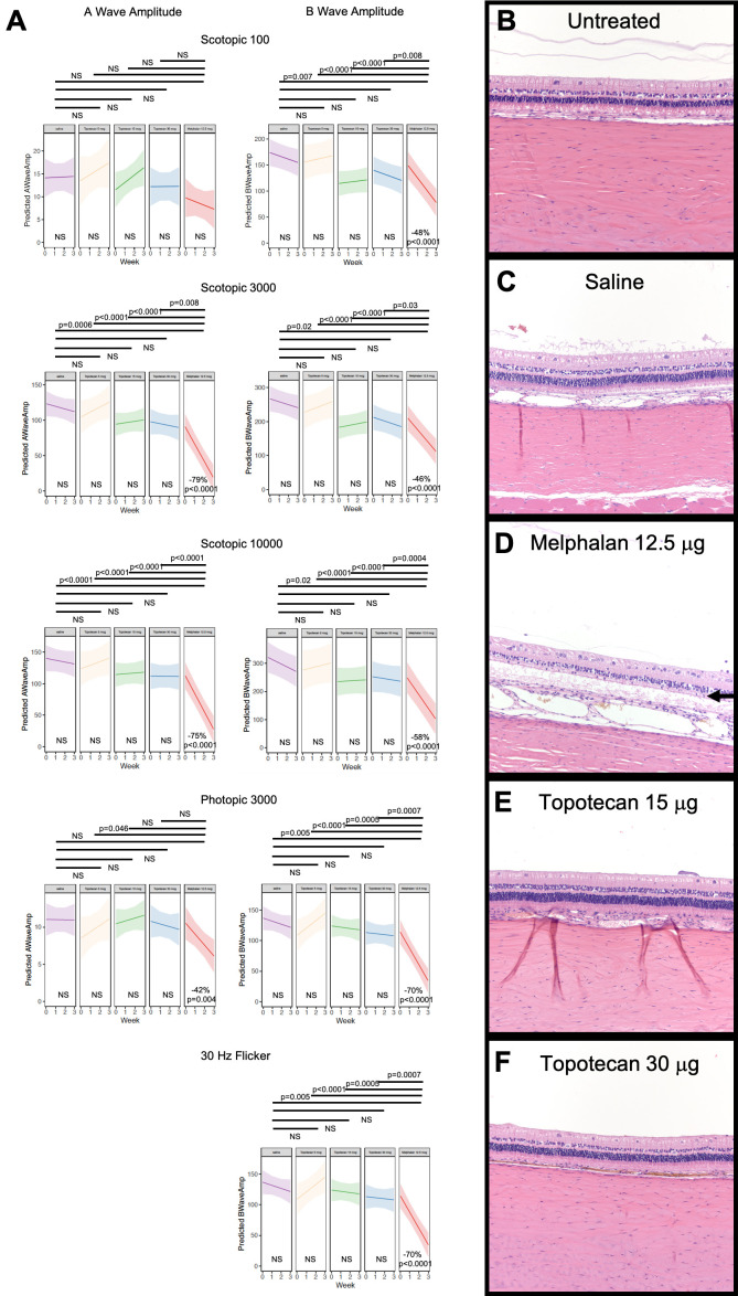
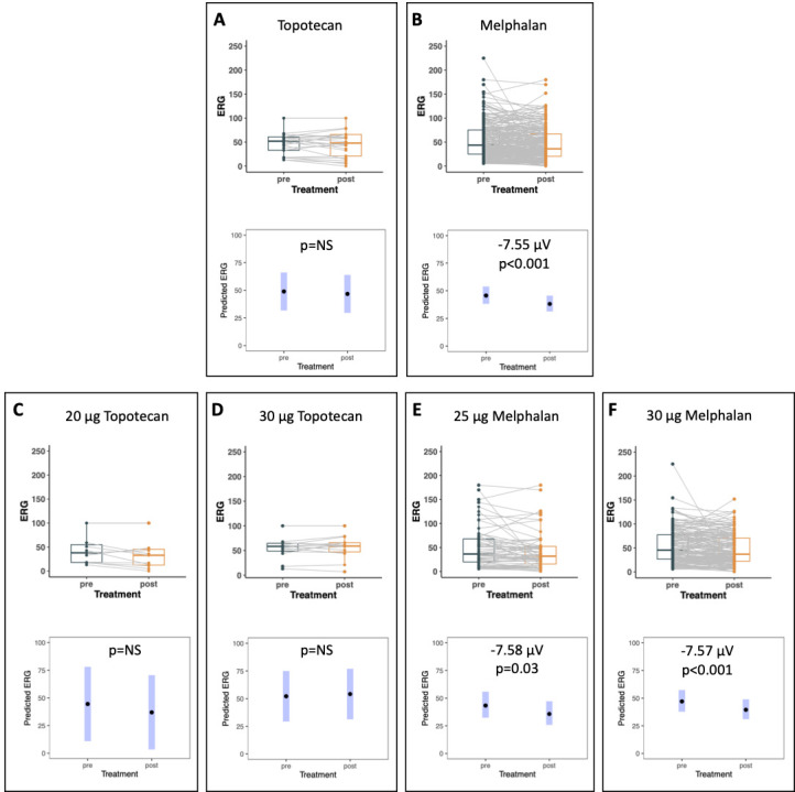
Results: Intravitreal topotecan 30 µg (equals 60 µg in humans) achieved the IC90 across the rabbit vitreous. Three weekly topotecan injections (either 15 µg or 30 µg) caused no retinal toxicity in rabbits, whereas melphalan 12.5 µg (equals 25 µg in humans) reduced ERG amplitudes 42%-79%. Intravitreal topotecan 15 µg was equally effective to melphalan to treat WERI-Rb1 cell xenografts in rabbits (96% reduction for topotecan vs saline (p=0.004), 88% reduction for melphalan vs saline (p=0.004), topotecan vs melphalan, p=0.15). In our clinical study, patients received 881 monotherapy injections (48 topotecan, 833 melphalan). Patients receiving 20 µg or 30 µg topotecan demonstrated no significant ERG reductions; melphalan caused ERG reductions of 7.6 μV for every injection of 25 µg (p=0.03) or 30 µg (p<0.001). Most patients treated with intravitreal topotecan also received intravitreal melphalan at some point during their treatment course. Among those eyes treated exclusively with topotecan monotherapy, all eyes were salvaged.

Conclusions: Taken together, these experiments suggest that intravitreal topotecan monotherapy for the treatment of RB vitreous seeds is non-toxic and effective.

Keywords: animal models; efficacy; intravitreal chemotherapy; pharmacokinetics; retinoblastoma; topotecan; toxicity; vitreous seeds.


Conflict of interest statement

Competing interests: ABD and DLF have a patent with Vanderbilt University Medical Center. ABD has received research funding from Spectrum Pharmaceuticals (now Acrotech Biopharma) through an investigator-initiated study separate from the data presented in this manuscript. None of the other authors has any conflicts of interest or financial disclosures.

Figures

Figure 1
Figure 1
Experimental design of efficacy and toxicity experiments in the rabbit model. (A) Efficacy experiments. Following 3 days of cyclosporine immunosuppression (which also continued throughout the course of the experiment), WERI-Rb1 cells were injected into the vitreous of both eyes of New Zealand white rabbits. After 2 weeks of growth, once-per-week injections of drug (topotecan 15 µg or melphalan 12.5 µg) were given into the right eye (OD), and saline into the left eye (OS). Imaging was obtained and rabbits were sacrificed 2 weeks after the third injection. (B) Toxicity experiments. Drug (topotecan, melphalan or saline, depending on the cohort) was injected into the right eye (OD) of New Zealand white rabbits once per week for three consecutive weeks. One day prior to each injection, functional testing was obtained (see the Methods section). One week following the final (third) injection, testing was again performed, and then the rabbits were sacrificed and globes harvested for histopathological evaluation. ERG, electroretinography. CsA=cyclosporine A.
Figure 2
Figure 2
Pharmacokinetics of intravitreal topotecan in the rabbit eye and dose-dependent survival curves of retinoblastoma cells to transient exposure to topotecan in vitro. (A) Pharmacokinetic curve in the rabbit vitreous following intravitreal injection of 1 µg of topotecan hydrochloride. Sampling was performed through a valved vitrectomy cannula inserted on the contralateral side of the eye relative to the injection site, and all sampling was performed on the far side of the vitreous relative to the injection site. The use of a valved vitrectomy cannula helped to maintain eye stability and prevent leakage throughout the experiment. (B) CellTiter Blue survival curve in human WERI-Rb1 cells exposed in vitro to various doses of topotecan hydrochloride for 16 hours (equal to five vitreous half-lives). Surviving cells were measured at the 7-day time-point. The IC90 can be found to correspond to 300 nM topotecan hydrochloride for this length of exposure. RFU=relative fluorescence units.
Figure 3
Figure 3
Efficacy of intravitreal topotecan against retinoblastoma vitreous seeds in a rabbit xenograft model. One million WERI-Rb1 human retinoblastoma cells were injected into the vitreous of both eyes of cyclosporine-immunosuppressed rabbits. Following 2 weeks of growth, the eyes were given three weekly injections of the intravitreal drug (right eye) or intravitreal saline (left eye). (A–B) Residual vitreous seeds 2 weeks following the final (third) injection, showing (A) significant large seeds in a representative left eye, and (B) complete clinical resolution of the vitreous seeds in a representative topotecan-treated right eye. (C) Quantification of residual live vitreous seeds 2 weeks after the third injection of topotecan (right eyes) or saline (left eyes), showing reduction in live tumour cells with topotecan 15 µg. Note the scale is a square root scale and is not linear. (D) Comparison of relative reduction with topotecan versus melphalan (compared with their respective saline-treated contralateral eyes), demonstrating no statistically significant difference between the two treatments. Note the abbreviated Y-axis only extends from 75% to 100%. (E) H&E stain of a vitreous seed 2 weeks after a single saline injection in the control eye. (F) H&E stain of a vitreous seed 2 weeks after one single injection of 15 µg topotecan, showing significant cell death in most cells. (G) An eye treated with 3 weekly injections of 15 µg topotecan. TUNEL staining shows that all remaining cells are in the process of dying. Taken together with our quantitative cell counting experiments, which showed a 96% reduction in tumour cells with three weekly injections of topotecan, this indicates that even the ‘surviving’ 4% of cells were actually in the process of dying as well. H&E=hematoxylin and eosin.
Figure 4
Figure 4
Absence of retinal toxicity with various doses of intravitreal topotecan, compared with melphalan. (A) Retinal function. Electroretinography was performed weekly, 1 day prior to each of the three planned injections, as well as 1 week after the final injection (immediately prior to euthanising the rabbit). Retinal responses to scotopic 100 mcd flashes, scotopic 3000 mcd flashes, scotopic 10 000 mcd flashes, photopic 3000 mcd flashes and 30 Hz flicker flashes were recorded. A-wave and B-wave amplitudes, and A-wave and B-wave implicit times were recorded (except for the 30 Hz flicker, for which there is only a B wave). Shaded areas on the graphs represent 95% CIs. No toxicity (see the Methods section for toxicity criteria) was observed for any parameter in the saline-treated control eyes, as well as in the cohorts treated with either 15 µg or 30 µg of topotecan. However, significant toxicity was seen in the cohort of rabbits treated with 12.5 µg of melphalan. Graphs of amplitudes are shown, but similar results were seen for implicit times, as well. For those particular tests where significant toxicity was seen, per cent change and p values for estimates of trend are shown alongside the particular graph. P values of the difference between groups are shown at the top of each graph. (B–F) Histopathology of treated eyes demonstrating (B) normal retinal architecture in untreated eyes and in (C) the saline-treated eyes. (D) In contrast, eyes treated with 12.5 µg melphalan showed significant retinal atrophy on histopathology (arrow shows the location of loss of outer retinal architecture). Eyes treated with intravitreal injections of (E) 15 µg topotecan, or (F) 30 µg topotecan were histologically indistinguishable from saline-treated (or untreated) eyes. Retinal detachments are artefactual. NS=not significant.
Figure 5
Figure 5
Changes in retinal function in topotecan-treated versus melphalan-treated eyes of patients with retinoblastoma vitreous seeds. Within each cohort or subcohort (delineated within a box), the top panel represents the univariate analysis, with each ‘string’ representing the electroretinography (ERG) changes with a single intravitreal injection. The bottom panel within each pair represents the results of the mixed-effect modelling, accounting for inter-eye/intra-patient and intra-eye correlations, with the appropriate statistical analysis results labelled on the panel. (A–B) ERG amplitude changes per injection for patients treated with topotecan (A), or melphalan (B). Topotecan caused no significant reduction in ERG parameters, whereas significant reductions in ERG amplitudes were seen with each injection of melphalan (7.55 μV per injection, p<0.001). (C–F) ERG amplitude changes by drug and dose. (C–D) represent sub-cohorts of the full topotecan-treated cohort presented in (A), and (E–F) represent subcohorts of the full melphalan-treated cohort presented in (B). Topotecan caused no reduction in ERG parameters at either 20 µg (C) or 30 µg (D), whereas significant reductions in ERG amplitudes were seen with each injection of either 25 µg melphalan (E; 7.58 μV per injection, p=0.03) or 30 µg melphalan (F; 7.57 μV per injection, p<0.001). NS=not significant.

Similar articles

Cited by

References

    1. Shields CL, Mashayekhi A, Au AK, et al. . The International classification of retinoblastoma predicts chemoreduction success. Ophthalmology 2006;113:2276–80. 10.1016/j.ophtha.2006.06.018 - DOI - PubMed
    1. Shields CL, Honavar SG, Meadows AT, et al. . Chemoreduction plus focal therapy for retinoblastoma: factors predictive of need for treatment with external beam radiotherapy or enucleation. Am J Ophthalmol 2002;133:657–64. 10.1016/s0002-9394(02)01348-x - DOI - PubMed
    1. Abramson DH, Beaverson KL, Chang ST, et al. . Outcome following initial external beam radiotherapy in patients with Reese-Ellsworth group Vb retinoblastoma. Arch Ophthalmol 2004;122:1316–23. 10.1001/archopht.122.9.1316 - DOI - PubMed
    1. Murphree AL, Villablanca JG, Deegan WF, et al. . Chemotherapy plus local treatment in the management of intraocular retinoblastoma. Arch Ophthalmol 1996;114:1348–56. 10.1001/archopht.1996.01100140548005 - DOI - PubMed
    1. Daniels AB, Patel SN, Milam RW, et al. . Effect of intravenous chemotherapy regimen on globe salvage success rates for retinoblastoma based on disease Class—A meta-analysis. Cancers 2021;13:2216. 10.3390/cancers13092216 - DOI - PMC - PubMed

Publication types