. 2021 Jul 16;65(8):e0030021.
doi: 10.1128/AAC.00300-21. Epub 2021 Jul 16.

RNase P Inhibitors Identified as Aggregators

Affiliations

RNase P Inhibitors Identified as Aggregators

Isabell Schencking et al. Antimicrob Agents Chemother. .

Abstract

RNase P is an essential enzyme responsible for tRNA 5'-end maturation. In most bacteria, the enzyme is a ribonucleoprotein consisting of a catalytic RNA subunit and a small protein cofactor termed RnpA. Several studies have reported small-molecule inhibitors directed against bacterial RNase P that were identified by high-throughput screenings. Using the bacterial RNase P enzymes from Thermotoga maritima, Bacillus subtilis, and Staphylococcus aureus as model systems, we found that such compounds, including RNPA2000 (and its derivatives), iriginol hexaacetate, and purpurin, induce the formation of insoluble aggregates of RnpA rather than acting as specific inhibitors. In the case of RNPA2000, aggregation was induced by Mg2+ ions. These findings were deduced from solubility analyses by microscopy and high-performance liquid chromatography (HPLC), RnpA-inhibitor co-pulldown experiments, detergent addition, and RnpA titrations in enzyme activity assays. Finally, we used a B. subtilis RNase P depletion strain, whose lethal phenotype could be rescued by a protein-only RNase P of plant origin, for inhibition zone analyses on agar plates. These cell-based experiments argued against RNase P-specific inhibition of bacterial growth by RNPA2000. We were also unable to confirm the previously reported nonspecific RNase activity of S. aureus RnpA itself. Our results indicate that high-throughput screenings searching for bacterial RNase P inhibitors are prone to the identification of "false positives" that are also termed pan-assay interference compounds (PAINS).

Keywords: RNase P inhibitors; RnpA protein subunit; bacterial RNase P; protein aggregators.


Figures

FIG 1
FIG 1
Inhibition of T. maritima RNase P by RNPA2000. (A) T. maritima RNase P holoenzyme activity in the presence of 140 μM RNPA2000 (green). Kinetics were performed with 50 nM holoenzyme and 500 nM pre-tRNAGly substrate in kinetic (KIN) buffer with 10 mM MgCl2. Neomycin (Neo; pink) was used for all experiments (A to D) as a control at its 50% inhibitory concentration (IC50) of ∼40 μM (see Fig. S2). Mean values (± standard error of the mean [SEM]), normalized to the 2% dimethyl sulfoxide (DMSO)-only control, are based on 6 biological replicates. (B) Effect of 140 μM RNPA2000 on the T. maritima P RNA-alone reaction, using KIN buffer with 100 mM MgCl2, 50 nM P RNA, and 500 nM pre-tRNAGly. Mean values (±SEM), normalized to the 2% DMSO-only control, are based on 3 biological replicates. (C) Effect of addition of 0.01% Triton X-100. Relative activity of the T. maritima RNase P holoenzyme assayed in KIN buffer with 10 mM MgCl2, 0.01% Triton X-100, and 2% DMSO. Experiments were performed with 50 nM holoenzyme (equal amounts of P RNA and RnpA) and 500 nM pre-tRNAGly. Mean values (±SEM), normalized to the 2% DMSO-only control, are based on 4 biological replicates. (D) Effect of a 10-fold increase in RnpA concentration. Activity of the T. maritima holoenzyme was assayed in KIN buffer with 10 mM MgCl2 and containing 50 nM P RNA, 500 nM RnpA protein, and 500 nM pre-tRNAGly. Mean values (±SEM), normalized to the 2% DMSO-only control, are based on 3 biological replicates.
FIG 2
FIG 2
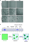
Protein aggregation. (A) Microscopy of RNPA2000 in different buffers with various Mg2+ concentrations. Three different buffers were chosen, as follows: (i) KIN buffer, used for enzyme kinetics in the present study, (ii) 50 mM Tris buffer, previously used for kinetic experiments (20), and (iii) phosphate-buffered saline (PBS), used for solubility determinations (31). Solutions (200 μl) with 2% DMSO and 200 μM RNPA2000 were prepared either without or with Mg2+ at the indicated concentration. After incubation for 15 min at room temperature, solutions were applied to microscopy. (B) SDS-PAGE of a cosedimentation experiment with RNPA2000; s, supernatant; p, pellet. Lane 1, a 15-kDa marker (M); lane 2, 1 μg of T. maritima RnpA; lanes 3 and 4, 5 μg T. maritima RnpA only; lanes 5 and 6, 140 μM RNPA2000 only; lanes 7 and 8, 5 μg T. maritima RnpA and 140 μM RNPA2000. Proteins in lanes 1 and 2 were in double-distilled water (ddH2O), samples in lanes 3 to 8 were in KIN buffer containing 10 mM MgCl2 and 2% DMSO, and lanes 9 and 10 were derived from samples containing 5 μg T. maritima RnpA and 140 μM RNPA2000 in KIN buffer with 2% DMSO but lacking MgCl2 (indicated by an asterisk [*]). (C) Model sketches of protein aggregation induced by RNPA2000. (Left) RNPA2000 is soluble in the absence of Mg2+ (depicted by the homogeneous greenish color); (middle) addition of Mg2+ induces the formation of insoluble compound aggregates (green spheres); (right) protein (in blue) adsorbs to green RNPA2000 aggregates.
FIG 3
FIG 3
Model of aggregator effects on RNase P. (Left) After aggregate formation of the small molecule (green spheres), RnpA protein (blue) adsorbs to such aggregates, leading to the depletion of soluble protein that is available for enzyme function. (Middle) Increasing the RnpA concentration (10-fold in the sketch) saturates the contact sites on the aggregates, leaving a fraction of soluble protein, which manifests as attenuated enzyme inhibition. (Right) Triton X-100 interferes with aggregate formation or disrupts aggregates, thereby restoring protein solubility.
FIG 4
FIG 4
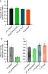
Effect of RNPA2000 and its derivatives NL20 and NL48 on RNase P cleavage. (A) Relative activity of the T. maritima RNase P holoenzyme in the presence of the three compounds (chemical structures shown in Fig. S1). Experiments were performed with 50 nM T. maritima holoenzyme and 500 nM pre-tRNAGly substrate using KIN buffer, 10 mM MgCl2, and 10% DMSO. DMSO (10%) was chosen to maximize compound solubility while maintaining substantial enzyme activity (see Fig. S2). The compound concentrations used were selected based on the solubility data shown in Table 1; for NL20 and NL48, compound concentrations were above the IC50 values reported for inhibition of S. aureus RNase P (75 μM for NL20 [31] and 1 μM for NL48 [21]). Mean cleavage rate constants (± SEM), derived from 3 biological replicates each, were normalized to reaction mixtures containing 10% DMSO without added compound. (B) Relative S. aureus RNase P holoenzyme activity in the absence or presence of RNPA2000 assayed in KIN buffer, 10 mM MgCl2, and 2% DMSO. P RNA (50 nM) and substrate (500 nM) were used at the same concentrations as for kinetic assays with T. maritima RNase P (Fig. 1A). Only the S. aureus RnpA concentration was increased to 100 nM for optimal enzyme activity. The RNPA2000 concentration (140 μM) corresponds to the previously determined IC50 value for the same enzyme reaction (20, 31). Mean values (±SEM), normalized to the 2% DMSO control, are based on 4 biological replicates. (C) S. aureus RNase P activity assayed in KIN buffer, 10 mM MgCl2, and 10% DMSO, using the same enzyme and substrate concentrations as in panel B. Compound concentrations were identical to those used in panel A for the T. maritima enzyme. Mean values (±SEM), normalized to 10% DMSO, are based on 4 biological replicates.
FIG 5
FIG 5
Assay for the detection of S. aureus RnpA-mediated RNA degradation. Incubation of 5′-[32P]-end-labeled T. thermophilus pre-tRNAGly (<5 nM) with 50 pmol S. aureus RnpA for 60 min. Buffer (storage buffer of S. aureus RnpA; 50 mM Tris-HCl [pH 7.0], 100 mM NaCl, and 10% glycerin) only and bovine serum albumin (BSA) were used as negative controls and incubation with 1 U RNase T1 was used as a positive control. Samples were analyzed by 8% denaturing PAGE, and radioactive bands were visualized by phosphorimaging.
FIG 6
FIG 6
Iriginol hexaacetate (Ir6Ac) testing. (A to D) Single exponential fittings (see the supplemental material) for pre-tRNAGly processing by the B. subtilis holoenzyme or P RNA alone in the presence of 10 μM Ir6Ac (red curves). DMSO (1%) was used as a control (black dashed curves). Experiments were performed in KIN buffer with 1% DMSO and 5 mM (A to C) or 100 mM (D) MgCl2. Concentrations of P RNA, RnpA, and pre-tRNAGly substrate were varied. All data are based on at least three biological replicates each. (A) Holoenzyme (1 nM; equal amounts of P RNA and RnpA protein) and 10 nM pre-tRNAGly substrate; (B) 1 nM P RNA, 10 nM RnpA, and 10 nM pre-tRNAGly substrate; (C) 10 nM holoenzyme (equal amounts of P RNA and RnpA) and 100 nM pre-tRNAGly substrate. (D) RNA-alone activity using 1 nM P RNA and 10 nM substrate and assayed at 100 mM Mg2+.

References

    1. Hartmann RK, Gößringer M, Späth B, Fischer S, Marchfelder A. 2009. The making of tRNAs and more—RNase P and tRNase Z. Prog Mol Biol Transl Sci 85:319–368. 10.1016/S0079-6603(08)00808-8. - DOI - PubMed
    1. Gopalan V, Jarrous N, Krasilnikov AS. 2018. Chance and necessity in the evolution of RNase P RNA 24:1–5. 10.1261/rna.063107.117. - DOI - PMC - PubMed
    1. Guerrier-Takada C, Gardiner K, Marsh T, Pace N, Altman S. 1983. The RNA moiety of ribonuclease P is the catalytic subunit of the enzyme. Cell 35:849–857. 10.1016/0092-8674(83)90117-4. - DOI - PubMed
    1. Gössringer M, Kretschmer-Kazemi Far R, Hartmann RK. 2006. Analysis of RNase P protein (rnpA) expression in Bacillus subtilis utilizing strains with suppressible rnpA expression. J Bacteriol 188:6816–6823. 10.1128/JB.00756-06. - DOI - PMC - PubMed
    1. Waugh DS, Pace NR. 1990. Complementation of an RNase P RNA (rnpB) gene deletion in Escherichia coli by homologous genes from distantly related eubacteria. J Bacteriol 172:6316–6322. 10.1128/jb.172.11.6316-6322.1990. - DOI - PMC - PubMed

Publication types

LinkOut - more resources