. 2021 May 10;4(6):e202000958.
doi: 10.26508/lsa.202000958. Print 2021 Jun.

Cell-free expression of the outer membrane protein OprF of Pseudomonas aeruginosa for vaccine purposes

Affiliations

Cell-free expression of the outer membrane protein OprF of Pseudomonas aeruginosa for vaccine purposes

Géraldine Mayeux et al. Life Sci Alliance. .

Abstract

Pseudomonas aeruginosa is the second-leading cause of nosocomial infections and pneumonia in hospitals. Because of its extraordinary capacity for developing resistance to antibiotics, treating infections by Pseudomonas is becoming a challenge, lengthening hospital stays, and increasing medical costs and mortality. The outer membrane protein OprF is a well-conserved and immunogenic porin playing an important role in quorum sensing and in biofilm formation. Here, we used a bacterial cell-free expression system to reconstitute OprF under its native forms in liposomes and we demonstrated that the resulting OprF proteoliposomes can be used as a fully functional recombinant vaccine against P. aeruginosa Remarkably, we showed that our system promotes the folding of OprF into its active open oligomerized state as well as the formation of mega-pores. Our approach thus represents an easy and efficient way for producing bacterial membrane antigens exposing native epitopes for vaccine purposes.


Conflict of interest statement

The authors declare that the research was conducted in the absence of any commercial or financial relationships that could be construed as a potential conflict of interest.

Figures

Figure 1.
Figure 1.. Production and purification of recombinant OprF proteoliposomes.
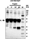
(A) Comparison, using anti-His Western blot analysis, of cell-free OprF expression in the presence of liposomes of six different lipid compositions (LC 1 to LC 3′) 3 and 5 μl of each cell-free reaction mixture were analyzed. Anti-His antibody was conjugated to HRP and was detected using chemiluminescence. (B) Comparison, using anti-His Western blot analysis, of the influence of liposome concentration on OprF expression for each LC. (C) Purification of OprF proteoliposomes (LC 1) by sucrose gradient. Anti-His Western blot analysis of each 1 ml layer of the sucrose gradient (10 lanes from 0 to 40% sucrose), with OprF monomeric and oligomeric forms indicated (only dimeric, trimeric, and tetrameric forms are visible). (D) Evaluation, using Coomassie gel, of the purity of OprF proteoliposomes after purification by sucrose gradient and subsequent pellet washing with NaCl. (E) Trypsin digestion of OprF proteoliposomes after purification. The digested bands were revealed by Western blotting using an anti-his antibody.
Figure 2.
Figure 2.. Determination of OprF orientation in proteoliposomes by atomic force microscopy.
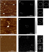
(A, B, C) Atomic force microscopy topographic images of an OprF proteoliposomes’ sample absorbed on mica surface, acquired using a tip functionalized with Tris–NTA, without (A, B) and with Triton X-100 (C). The circles in white dotted lines correspond to proteoliposomes. (A′, B′, C′) Corresponding adhesion maps of specific adhesion forces between the Tris–NTA modified tip and OprF His-tag, without (A′, B′) and with Triton X-100 (C′). The white to grey squares represent values of adhesion forces between 80 and 150 pN, as illustrated by the scale bar on the right. The circles in white dotted lines corresponding to proteoliposomes are zoomed on the right. The white arrows indicate specific adhesion points validated by the force distance curves and correspond to 16 transmembrane passages based on the theoretical model Worm-Like Chain.
Figure 3.
Figure 3.. Determination of the membrane topology of OprF in liposomes by trypsin digestion.
The result of the trypsin digestion of OprF at different incubation times is visualized by anti-His Western blot (FL, full length). Anti-His antibody was conjugated to HRP and was detected using chemiluminescence.
Figure 4.
Figure 4.. Detection of two transmembrane topologies adopted by OprF in proteoliposomes by Atomic Force Microscopy.
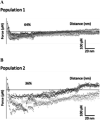
(A, B) Force distance curves, obtained with a tip functionalized with Tris–NTA in Triton X-100 solution, detecting 64% of specific adhesion events corresponding to eight transmembrane domains (A) and 36% of specific adhesion events corresponding to 16 transmembrane domains (B), based on the theoretical model Worm-Like Chain.
Figure 5.
Figure 5.. Superimposition of circular dichroism spectra of OprF proteoliposomes before (black curve) and after addition of IFN-γ (red curve).
Figure 6.
Figure 6.. Visualization of liposomal surface by negative-staining (NS) EM. Scale bar 100 nm.
(A) EM image of empty liposomes (incubated using the cell-free cell lysate and reaction mixture = negative control). (B, C, D, E) EM images of OprF proteoliposomes (LC1). The membrane was perforated with a series of holes with an average size of 9.5 ± 4 nm corresponding to OprF pores.
Figure 7.
Figure 7.. Visualization of OprF pore-forming activity by Atomic Force Microscopy.
(A) Topographic image of the surface of an OprF proteoliposome absorbed onto a freshly cleaved mica support, acquired using a tip functionalized with Tris–NTA, in presence of Triton X-100. (B) Corresponding adhesion map of specific adhesion forces between the tris–NTA modified tip and OprF His-tag. The white to grey squares represent adhesion forces values between 80 and 150 pN, as illustrated by the scale bar on the right. The circles in white dotted lines highlight pores. (C) Force distance curves detecting specific adhesion events (indicated by arrows).
Figure S1.
Figure S1.. Measurements of OprF channel activity using impedance spectrometry and effect of IFN-γ.
(A) Conductance of the t-BLM after the insertion of OprF proteoliposomes (blue squares) and empty liposomes (green triangles) for different KCl concentrations (150, 200, 300, 400, 500, 750 mM, and 1M), in 50 mM Tris pH 7.5. (B) Effects of increasing concentrations of IFN-γ (0–1.5 μM) on the conductance of the tBLM after incorporation of OprF (blue diamonds) and empty liposomes (orange squares) in 50 mM Tris, pH 7.5, 150 mM KCl. The conductance values correspond to the mean (±SD) of three values measured during 10 min. (A) and (B) are two separated experiments.
Figure 8.
Figure 8.. Immunization with recombinant OprF proteoliposomes protects mice against a bacterial challenge.
(A) C57Bl/6 mice were immunized subcutaneously with 25, 50 or 75 μg of OprF in proteoliposome. 1 wk after the fourth immunization, OprF-immunized groups (n = 10 or 11) or control group (n = 11) were subcutaneously challenged with a lethal dosage of Pseudomonas aeruginosa CHA strain (2 × 10⁷ CFU/mouse) and monitored for survival and disease symptoms for up to 80 h. (B) Naïve mice were subcutaneously challenged with a lethal dose of the CHA strain and an intraperitoneal injection with sera containing polyclonal antibodies induced by the OprF proteoliposomes was performed 1 h after the bacterial challenge. Mice were monitored for survival and disease symptoms for up to 80 h.
Figure S2.
Figure S2.. Additional immunization with recombinant OprF proteoliposomes protects mice against a bacterial challenge.
C57Bl/6 mice were immunized subcutaneously with 25, 50, or 75 μg of OprF in proteoliposome. 1 wk after the fourth immunization, OprF-immunized groups (n = 5 or 6) or control group (n = 6) were subcutaneously challenged with lethal dosage of Pseudomonas aeruginosa CHA strain (2 × 10⁷ CFU/mouse) and monitored for survival and disease symptoms for up to 60 h.
Figure 9.
Figure 9.. Anti-OprF antibody titration curves of sera from immunized mice.
Antibody titration curves of sera from mice immunized four times with 25, 50, or 75 μg of recombinant OprF proteoliposome. Control mice were immunized with liposomes previously incubated with cell-free lysate. Values are means of three mice per group determined by indirect ELISA using recombinant OprF proteoliposome as coated antigen.
Figure 10.
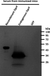
Figure 10.. Western blot illustrating the binding of both endogenous and recombinant OprF by polyclonal antibodies found in sera from immunized mice.
GFP was used as a negative control. The secondary antibody binding polyclonal antibodies was an HRP-conjugated rabbit anti-mouse IgG and it was detected using chemiluminescence.
Figure 11.
Figure 11.
Analysis of the OprF proteoliposomes at different temperatures. (A, B) Evaluation of OprF stability in liposomes before (clt, control) and after 14 d storage at RT, 4°C, −20°C, and −80°C using Coomassie gel—SDS–PAGE (A) and anti-HIS Western blot analysis (B). The anti-His antibody was conjugated to HRP and was detected using chemiluminescence.

References

    1. Adlbrecht C, Wurm R, Depuydt P, Spapen H, Lorente JA, Staudinger T, Creteur J, Zauner C, Meier-Hellmann A, Eller P, et al. (2020) Efficacy, immunogenicity, and safety of IC43 recombinant Pseudomonas aeruginosa vaccine in mechanically ventilated intensive care patients-a randomized clinical trial. Crit Care 24: 74. 10.1186/s13054-020-2792-z - DOI - PMC - PubMed
    1. Alhede M, Bjarnsholt T, Givskov M, Alhede M (2014) Pseudomonas aeruginosa biofilms: Mechanisms of immune evasion. Adv Appl Microbiol 86: 1–40. 10.1016/B978-0-12-800262-9.00001-9 - DOI - PubMed
    1. Azghani AO, Idell S, Bains M, Hancock REW (2002) Pseudomonas aeruginosa outer membrane protein F is an adhesin in bacterial binding to lung epithelial cells in culture. Microb Pathog 33: 109–114. 10.1006/mpat.2002.0514 - DOI - PubMed
    1. Bahey-El-Din M, Mohamed SA, Sheweita SA, Haroun M, Zaghloul TI (2020) Recombinant N-terminal outer membrane porin (OprF) of Pseudomonas aeruginosa is a promising vaccine candidate against both P. aeruginosa and some strains of Acinetobacter baumannii. Int J Med Microbiol 310: 151415. 10.1016/j.ijmm.2020.151415 - DOI - PubMed
    1. Baker SM, McLachlan JB, Morici LA (2020) Immunological considerations in the development of Pseudomonas aeruginosa vaccines. Hum Vaccin Immunother 16: 412–418. 10.1080/21645515.2019.1650999 - DOI - PMC - PubMed

Publication types

MeSH terms

LinkOut - more resources