. 2021 Jun 23;41(7):e0066220.
doi: 10.1128/MCB.00662-20. Epub 2021 Jun 23.

Vps34 and TOR Kinases Coordinate HAC1 mRNA Translation in the Presence or Absence of Ire1-Dependent Splicing

Affiliations

Vps34 and TOR Kinases Coordinate HAC1 mRNA Translation in the Presence or Absence of Ire1-Dependent Splicing

Jagadeesh Kumar Uppala et al. Mol Cell Biol. .

Abstract

In the budding yeast Saccharomyces cerevisiae, an mRNA, called HAC1, exists in a translationally repressed form in the cytoplasm. Under conditions of cellular stress, such as when unfolded proteins accumulate inside the endoplasmic reticulum (ER), an RNase Ire1 removes an intervening sequence (intron) from the HAC1 mRNA by nonconventional cytosolic splicing. Removal of the intron results in translational derepression of HAC1 mRNA and production of a transcription factor that activates expression of many enzymes and chaperones to increase the protein-folding capacity of the cell. Here, we show that Ire1-mediated RNA cleavage requires Watson-Crick base pairs in two RNA hairpins, which are located at the HAC1 mRNA exon-intron junctions. Then, we show that the translational derepression of HAC1 mRNA can occur independent of cytosolic splicing. These results are obtained from HAC1 variants that translated an active Hac1 protein from the unspliced mRNA. Additionally, we show that the phosphatidylinositol-3-kinase Vps34 and the nutrient-sensing kinases TOR and GCN2 are key regulators of HAC1 mRNA translation and consequently the ER stress responses. Collectively, our data suggest that the cytosolic splicing and the translational derepression of HAC1 mRNA are coordinated by unique and parallel networks of signaling pathways.

Keywords: ER; Gcn2; Hac1; Ire1; TOR; UPR; Vps34; translation.


Figures

FIG 1
FIG 1
Translational fate of HAC1 mRNA. The m7G cap, 5′-UTR (68 nucleotides, [nts]) and 3′-UTR (416 nucleotides), two exons (black boxes, 661 and 56 nucleotides), intron, and polyadenylated tail (An) of HAC1 mRNA are shown. The intron interacts with the 5′-UTR to form an RNA duplex (RD). Under ER stress conditions, Ire1 cleaves two phosphodiester bonds at the nucleotide positions G661 and G913 (shown by scissors). Both positions are present in the two RNA hairpins, referred to here as 5′-hairpin (5′-HP) and 3′-hairpin (3′-HP). The nucleotide compositions of the RD, 5′-HP, and 3′-HP are shown. The unspliced HAC1 mRNA can translate an Hac1u protein (230 amino acids) by leaky scanning (16), which contains a bZIP domain, a transcription activation domain (TAD), and a short tail of 10 amino acids (green box). The spliced HAC1 mRNA yields Hac1i protein (238 amino acids), containing a bZIP domain, a TAD, and an altered C-terminal tail of 18 amino acids (purple).
FIG 2
FIG 2
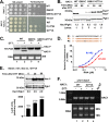
Ire1-mediated HAC1 mRNA cleavage requires Watson-Crick base pairs in the cleavage RNA hairpins. (A) Growth of yeast cells on tunicamycin medium requires Ire1 and Hac1. WT and its isogenic hac1Δ or ire1Δ yeast strains containing a URA3 plasmid (vector) or the same plasmid bearing an HAC1 or IRE1 gene were grown overnight, serially diluted, and spotted on the synthetic complete (SC) medium without uracil and the same medium containing tunicamycin. (B) ER stress activates HAC1 mRNA splicing. Total RNA was extracted from the indicated yeast strains grown for 2 h in a liquid YEPD medium containing tunicamycin. RT-PCR was used to analyze the unspliced (HAC1u) and spliced (HAC1s) forms of HAC1 mRNA. (C) Reciprocal exchange of nucleotides C658 and G666 restores HAC1 mRNA splicing. (Top) An hac1Δ strain containing a URA3 plasmid (vector) or the same plasmid bearing the indicated HAC1 mutant was tested for growth on the SC and tunicamycin media. (Bottom) RT-PCR was used to analyze the unspliced (HAC1u) and spliced (HAC1s) isoforms of HAC1 mRNA in the above strains grown in the presence of tunicamycin. (D) Reciprocal exchange of nucleotides U910 and G918 restores HAC1 mRNA splicing. An hac1Δ strain containing a URA3 plasmid (vector) or the same plasmid bearing the indicated HAC1 mutant was tested for growth on the SC and tunicamycin media (top). RT-PCR was used to analyze the unspliced (HAC1u) and spliced (HAC1s) forms of HAC1 mRNA in the above strains grown in the presence of tunicamycin (bottom).
FIG 3
FIG 3
Translational derepression of HAC1-G771A and HAC1-G661C,G771A mRNA. (A) The HAC1-G661C,G771A mutant grows on the tunicamycin medium. The hac1Δ or ire1Δ hac1Δ strain containing the indicated HAC1 mutants in a URA3 plasmid was tested for growth on the SC medium without uracil and the same medium containing tunicamycin. (B) Hac1u expression from the HAC1-G661C,G771A mutant is enhanced under ER stress conditions. (Top) the hac1Δ strain containing the indicated HAC1 mutants (WT, G661A, or G661A,G771A) were grown in the SC-uracil medium until OD600 reached ∼0.6 to 0.8; 5 mM DTT was then added to cells. After indicated times (0, 30, and 60 min), cells were harvested, whole-cell extracts (WCEs) were prepared and subjected to Western blot analysis using anti-Hac1 and Pgk1 antibodies. The intensities of Hac1 and Pgk1 protein bands were measured by ImageJ software (bottom). The ratios of the Hac1 and Pgk1 protein band intensities are shown. Experiments were repeated twice. A representative result is shown. (C) The HAC1-G661C,G771A mutant is deficient in splicing. Total RNA was prepared from the hac1Δ strain containing the indicated HAC1 mutants in a URA3 plasmid. RT-PCR was used to analyze the unspliced (HAC1u) and spliced (HAC1s) forms of HAC1 mRNA. (D) In vitro reconstitution of the 5′-UTR and intron interaction. As indicated, three RNA oligonucleotides corresponding to the 5′-UTR (R1), the intron (R2), and the intron with a G771A mutation (R3) were synthesized from Sigma (USA). Mixtures of RNA oligonucleotides R1 and R2 and R1 and R3 were heated to 95°C for 5 min and then annealed at room temperature slowly. The RNA mixture was diluted, and SYBR green was added to the diluted samples. Samples were then read in a spectrophotometer. The relative fluorescence units were averaged and then plotted against the RNA concentrations. The calculated Kd values were 0.692 ± 0.18 for R1+R2 and 1.755 ± 0.02 for R1+R3. (E) Hac1u expression from the HAC1-G771A mutant is enhanced during ER stress. WCEs were prepared from the ire1Δ hac1Δ strain containing the HAC1-G771A mutant and subjected to Western blot analysis using Hac1 and Pgk1 antibodies. The ratios of the Hac1 and Pgk1 protein band intensities are shown (top). The ire1Δ hac1Δ strain containing a HAC1-G771A allele in a URA3 vector and an UPRE-driven LacZ reporter plasmid in a LEU2 vector was grown as described in Fig. 3B. WCEs were prepared and subjected to β-galactosidase assay. The average values of three experiments are shown with standard errors (bottom). (F) ER stress does not induce the transcript levels of WT or HAC1-G771A mutant carried in plasmid. Total RNA was isolated from the ire1Δ hac1Δ strain containing WT HAC1 or HAC1-G771A mutant in a URA3 plasmid (bottom). Total RNA was reverse transcribed into cDNA. The synthesized cDNA was amplified by PCR using the exon1-specific primers of the HAC1 gene (top). The RNA sample obtained from the WT cells was used as a PCR template without the reverse transcriptase reaction (no RT).
FIG 4
FIG 4
ER stress provokes Hac1u expression from the HAC1-G771A mutant. The ire1Δ strain containing the HAC1-G771A mutant was grown in the SC-uracil medium in the presence of DTT (5 mM), 3-AT (30 mM), or H2O2 (0.5 mM) for 1 h. WCEs were prepared and subjected to Western blot analysis using anti-Hac1 and Pgk1 antibodies (top). The relative intensity of Hac1 protein band was measured by ImageJ software. The ratios of the Hac1 and Pgk1 protein band intensities are shown (bottom).
FIG 5
FIG 5
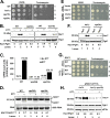
The PI3-kinase Vps34 contributes to Hac1i expression. (A) The vps34Δ strain grows slowly on the tunicamycin medium. The indicated WT and its isogenic hac1Δ, vps15Δ, or vps34Δ yeast strains were tested for their growth on rich YEPD medium and the same medium containing tunicamycin. (B) Expression of Hac1i protein is reduced in the vps34Δ strain. WCEs were prepared from the indicated yeast strain in the presence and absence of DTT and subjected to Western blot analysis using anti-Hac1 and Pgk1 antibodies. The intensities of Hac1 and Pgk1 protein bands were measured by ImageJ software, and the ratios of the Hac1 and Pgk1 bands are shown. (C) Reduced expression of UPRE-driven LacZ in both vps34Δ and vps15Δ strains. WCEs were prepared from the indicated yeast strains grown in the presence and absence of DTT and subjected to β-galactosidase assay. The average values of three experiments are shown with standard errors. (D) Deletion of Vps34 protein had no effect on HAC1 mRNA splicing. Total RNA was prepared from the indicated WT, vps34Δ, and vps15Δ strains grown in a liquid YEPD medium containing tunicamycin. RT-PCR was used to analyze the unspliced (HAC1u) and spliced (HAC1s) forms of HAC1 mRNA. (E) Reduced growth of the vps34Δ strain expressing an intronless HAC1 variant. The indicated ire1Δ and vps34Δ strains containing a vector plasmid or the same vector plasmid harboring the intronless HAC1 variant (HAC1c) were tested for their growth on rich YEPD medium and the same medium containing tunicamycin. (F) Reduced expression of Hac1c in the vps34Δ strain. WCEs were prepared from the indicated ire1Δ or vps34Δ strains expressing an Hac1c derivative and subjected to Western blot analysis using Hac1 and Pgk1 antibodies. The intensities of Hac1 and Pgk1 protein bands were measured by ImageJ software, and the ratios of the Hac1c and Pgk1 band intensities are shown. (G) The ire1Δ vps15Δ strain expressing HAC1-G771A allele grows slowly on the tunicamycin medium. The indicated ire1Δ strain and its isogenic ire1Δ vps15Δ strain expressing HAC1-G771A allele were tested for their growth on the medium containing tunicamycin. (H) Reduced expression of Hac1u in the ire1Δ vps15Δ strain. WCEs were prepared from the strains shown in Fig. 4G and subjected to Western blot analysis using Hac1, Pgk1, and eIF2α antibodies.
FIG 6
FIG 6
Translational activation of HAC1-G771A mRNA requires Gcn2 activation. (A) Analysis of yeast growth under ER stress condition. The ire1Δ, ire1Δ gcn2Δ, and hac1Δ gcn2Δ strains containing indicated WT HAC1 and HAC1-G771A or HAC1-G661C,G771A mutant were tested for growth on SC and tunicamycin media. (B) Reduced expression of Hac1u from HAC1-G771A mutant in the gcn2Δ strain. The hac1Δ or hac1Δ gcn2Δ strain containing the HAC1-G661C,G771A mutant was grown in the presence and absence of DTT. WCEs were prepared and subjected to Western blot analysis using Hac1 and Pgk1 antibodies. The ratios of the Hac1u and Pgk1 band intensities are shown. (C) Increased phosphorylation of eIF2α upon DTT treatment. WT yeast cells were grown in the presence of 3-AT (30 mM), rapamycin (0.5 mM), or DTT (5 mM) for 1 h. WCEs were prepared and subjected to Western blot analysis using Ser-51 phospho-specific antibody against eIF2α (top). The membrane was stripped and reprobed with total eIF2α antibody (bottom). The ratios of the eIF2α-P and eIF2α band intensities are shown. (D) Modest reduction of Hac1i expression in the gcn2Δ strain. WT and gcn2Δ strains were grown in the presence (+) and absence (−) of DTT. WCEs were prepared and subjected to Western blot analysis using Hac1, Pgk1, and eIF2α antibodies. The ratios of the Hac1 and Pgk1 protein band intensities are shown.
FIG 7
FIG 7
Translational activations of HAC1 mRNA requires TOR kinase function. (A) The tor1Δ tor2ts strain does not grow at 37°C. The tor1Δ tor2ts strains expressing WT Tor2 and Tor2-D2298E mutant were tested for growth at 25°C and 37°C. (B) The ire1Δ tor1Δ tor2ts strain containing the HAC1-G771A mutant grows under the ER stress condition. The indicated ire1Δ or ire1Δ tor1Δ tor2ts strain containing WT HAC1 or HAC1-G771A mutant was tested for growth on SC and tunicamycin media at 25°C and 37°C. (C) Reduced expression of Hac1u from the HAC1-G771A mutant in the ire1Δ tor1Δ tor2ts strain. The indicated yeast strains were grown in the presence of DTT at 37°C for 2 or 4 h. WCEs were prepared and subjected to Western blot analysis using Hac1u, Pgk1, and eIF2α antibodies. The ratios of the Hac1u and Pgk1 protein band intensities are shown. (D) Reduced expression of Hac1i in the tor1Δ tor2ts strain. The indicated yeast strains were grown in the presence (+) and absence (−) of DTT. WCEs were prepared and subjected to Western blot analysis using Hac1 and Pgk1 antibodies. The ratios of the Hac1i and Pgk1 protein band intensities are shown. (E) Reduced expression of UPRE-driven LacZ in the tor1Δ tor2ts strain. WCEs were prepared from the indicated yeast strains grown at 37°C for 4 h in the presence (+) and absence (−) of 5 mM DTT and subjected to β-galactosidase assay. The average values of three experiments are shown with standard errors.
FIG 8
FIG 8
Proposed model for translational derepression of HAC1 mRNA. The color scheme of HAC1 mRNA is the same as in Fig. 1. The intron interacts with the 5′-UTR to form an RNA duplex (RD), thus inhibiting ribosome or helicase recruitment. The diagram shows the core components of the (m7G) cap complex (eIF4E, eIF4G, and eIF4A) with its RD bound to a suppressor protein (blue box). The diagram also shows the proposed assembly of eIF4G, eIF4E, and eIF4A on the (m7G) cap and their regulation by kinases Vps34, TOR, and Gcn2 as well as the proposed role of Kin kinases (65) in splicing of HAC1 mRNA.

Similar articles

Cited by

References

    1. Luo Y, Na Z, Slavoff SA. 2018. P-bodies: composition, properties, and functions. Biochemistry 57:2424–2431. 10.1021/acs.biochem.7b01162. - DOI - PMC - PubMed
    1. Standart N, Weil D. 2018. P-bodies: cytosolic droplets for coordinated mRNA storage. Trends Genet 34:612–626. 10.1016/j.tig.2018.05.005. - DOI - PubMed
    1. Baker KE, Coller J. 2006. The many routes to regulating mRNA translation. Genome Biol 7:332. 10.1186/gb-2006-7-12-332. - DOI - PMC - PubMed
    1. Nissan T, Parker R. 2008. Analyzing P-bodies in Saccharomyces cerevisiae. Methods Enzymol 448:507–520. 10.1016/S0076-6879(08)02625-6. - DOI - PMC - PubMed
    1. Aragon T, van Anken E, Pincus D, Serafimova IM, Korennykh AV, Rubio CA, Walter P. 2009. Messenger RNA targeting to endoplasmic reticulum stress signalling sites. Nature 457:736–740. 10.1038/nature07641. - DOI - PMC - PubMed

Publication types

MeSH terms

Substances

LinkOut - more resources