. 2021 Jun 16;41(24):5274-5286.
doi: 10.1523/JNEUROSCI.1605-20.2021. Epub 2021 May 10.

Associative Learning Requires Neurofibromin to Modulate GABAergic Inputs to Drosophila Mushroom Bodies

Affiliations

Associative Learning Requires Neurofibromin to Modulate GABAergic Inputs to Drosophila Mushroom Bodies

Eirini-Maria Georganta et al. J Neurosci. .

Abstract

Cognitive dysfunction is among the hallmark symptoms of Neurofibromatosis 1, and accordingly, loss of the Drosophila melanogaster ortholog of Neurofibromin 1 (dNf1) precipitates associative learning deficits. However, the affected circuitry in the adult CNS remained unclear and the compromised mechanisms debatable. Although the main evolutionarily conserved function attributed to Nf1 is to inactivate Ras, decreased cAMP signaling on its loss has been thought to underlie impaired learning. Using mixed sex populations, we determine that dNf1 loss results in excess GABAergic signaling to the central for associative learning mushroom body (MB) neurons, apparently suppressing learning. dNf1 is necessary and sufficient for learning within these non-MB neurons, as a dAlk and Ras1-dependent, but PKA-independent modulator of GABAergic neurotransmission. Surprisingly, we also uncovered and discuss a postsynaptic Ras1-dependent, but dNf1-independnet signaling within the MBs that apparently responds to presynaptic GABA levels and contributes to the learning deficit of the mutants.

Keywords: Drosophila; GABA; Ras1; cAMP; learning; neurofibromatosis 1.


Conflict of interest statement

The authors declare no competing financial interests.

Figures

Figure 1.
Figure 1.
Adult-specific requirement of dNf1 in OK72 neurons for olfactory associative learning. Performance assessment 3 min after conditioning in control (white bars), mutant (black bars), experimental (dark gray bars), and transgene (UAS-dNf1)-expressing (light gray bars) animals. The genotypes are indicated below each bar. Data are mean ± SEM. *Statistically significant difference of the experimental from the mutant flies. #Statistically significant difference of the experimental from the control flies. G80ts indicates the ubiquitously expressed temperature-sensitive Gal4 repressor Tub-Gal80ts. A, Adult-specific expression of an Nf1 transgene throughout the MBs under Leo-Gal4 does not restore learning in dNf1E2 homozygotes. B, RNAi-mediated (UASdNf1Ri) dNf1 attenuation in MBs (gray bar) does not affect learning. C, Restoration of dNf1 expression within cholinergic neurons under Cha-Gal4 (gray bar) does not reverse the learning deficit of dNf1E2 homozygotes (black bar), which remained significantly different from that of controls (white bar). The UAS-Nf1 transgene-expressing flies (light gray bar) presented the same learning performance with control flies. D, UAS-Nf1 expression in dorsal paired medial neurons under c316-Gal4 does not restore learning to dNf1E2 homozygotes. E, UAS-Nf1 expression in dorsal anterior lateral neurons under the G0431-Gal4 driver does not restore associative learning to mutant homozygotes. F, UAS-Nf1 expression in antennal lobe projection neurons, including the APL, marked with the NP5288-Gal4 driver does not restore associative learning to mutant homozygotes. G, Adult-specific UAS-Nf1 expression in neurons marked with the OK72-Gal4 driver (gray bar) reverses the associative learning deficit of dNf1E2 nulls. H, RNAi-mediated dNf1 abrogation (UASdNf1Ri) in OK72 neurons in control flies and dNf1E2 heterozygotes (gray bars) induces significant learning deficits.
Figure 2.
Figure 2.
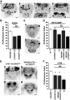
MB-independent requirement of dNf1 in OK72 neurons for olfactory associative learning. A, Grayscale inverted confocal images of adult brains stained with anti-GFP driven by the OK72-Gal4 driver, arranged from anterior to posterior (A1–A4): A1, AL glomeruli (narrow arrowhead) and MB lobes (wide arrowhead). Note the constellation of positive neurons between the vertical MB lobes in the center of the brain. A2, A single confocal section at the anterior of the brain clearly indicating expression in the α/β MB lobes (wide arrowhead) and AL glomeruli (narrow arrowhead). A3, A mid-frontal view of a brain revealing the constellation of positive neurons at the SMP, as well as symmetrical clusters of lateral neurons (open arrow pointing to the right cluster). The fan-shaped body (fs) is indicated with the staining pattern flanked by the MB pedunculi, better revealed in the single confocal section in the insert (arrows). A4, A posterior view of the brain reveals the MB dendrites/calyces (ca), the posterior SMP neurons (open triangle), and the lateral clusters (open arrow). The single confocal plain insert focuses on the posterior SMP neurons (open triangle). B, UAS-Nf1 expression in olfactory receptor neurons and antennal lobe glomeruli marked with the Or83b-Gal4 driver do not restore associative learning to dNf1E2 homozygotes. C, Anti-GFP stained grayscale inverted confocal images of adult brains of OK72-Gal4/MBG80 driving UAS-mcD8GFP in an anterior (C1) and mid-posterior (C2) view. Although staining remains in the fs, antennal lobe, and SMP neurons, the area occupied by the MBs does not express GFP (large arrowheads). D, Adult-specific dNf1 expression within the OK72 marked neurons, excluding the MBs, restores associative learning in dNf1E2homozygotes. MBG80 encodes a Gal80 repressor expressed constitutively within MB neurons. The performance of OK72-Gal4/MBG80; UAS-Nf1,dNf1E2/TubG80ts,dNf1E2 (second gray bar) was significantly different from that of animals not expressing the transgene (black bars), but not from controls (white bar). *Statistically significant difference of the experimental from the mutant flies. E, Confocal images of adult brains stained with anti-GFP driven by the c739-Gal4 driver in anterior (E1) and posterior (E2) views, illustrating strong MB expression as well as a constellation of non-MB GFP-positive neurons, especially in the posterior of the brain (E2). These non-MB neurons appear prominently in the anterior (E3) and mostly in the posterior (E4) of c739-Gal4/MBG80 brains where GFP expression in the MBs is eliminated. F, c739-Gal4;UAS-Nf1,dNf1E2/dNf1E2 animals (first gray bar) express transgenic dNf1 in c739 neurons, and this partially restores learning performance in dNf1E2nulls equally well as in animals expressing the transgene under c739 at the exclusion of the MBs (second gray bar). *Statistically significant difference of the experimental from the mutant flies.
Figure 3.
Figure 3.
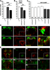
Olfactory associative learning requires dNf1 in GABAergic OK72-marked neurons. A–C, Performance was assessed within 3 min after conditioning in control (white bars), mutant (black bars), and experimental (gray bars) animals. The genotypes are indicated below each bar, representing the mean ± SEM. *Statistically significant difference of the experimental from mutant flies. G80ts indicates the ubiquitously expressed temperature-sensitive Gal4 repressor Tub-Gal80ts. A, Expression of the UAS-Nf1 transgene within GABAergic neurons restores normal learning to dNf1E2 nulls. B, Adult-specific Gad abrogation (UASGadRi) within OK72 neurons reverses the learning deficit of dNf1E2 homozygotes to levels not significantly different from that of controls. C, RNAi-mediated abrogation of Gad (UASGadRi) in OK72 neurons of adult WT flies does not alter learning at the restrictive (18°C, UN) or the permissive temperature (30°C, IN), respectively (gray bars). White bars represent the control genotypes, raised at both temperatures. D, All images are at 40× magnification. Size bar indicates relative size. Anti-GFP staining (green), anti-GABA staining (red), and merging of the two (yellow) in representative optical sections from fly brains where GFP expression is driven by OK72-Gal4. The insets in the GABA panels are maximum projections to demonstrate ample penetration of the anti-GABA antibody not obvious in the optical sections used for colocalization. Colocalization of GFP and GABA immunofluorescence is detected laterally (top) and the SMP (bottom). Right panels (both top and bottom), Magnifications of the respective marked boxes, showing colocalization in single cells. E, In flies bearing the trans-Tango components, driving ligand and myrGFP expression under OK72-Gal4 (green) results in HA-mtdTomato expression in postsynaptic MB dendrites (red). Anterior (E1) and posterior (E3) view of maximum projections of presynaptic and postsynaptic neurons under OK72Gal4-driven TANGO. E2a–E2c, Representative optical sections of presynaptic (OK72; green) and postsynaptic (red) neurons at the dendritic level, where yellow represents their interaction. Insets, Higher magnification of confocal images from different brains at the level of MB dendrites. E4a, Posterior view maximum projection of OK72 presynaptic neurons (green) and GABA-positive neurons (red). White box represents the area magnified (80×) in E4b with the white arrow indicating colocalization. E5a, Posterior view maximum projection of postsynaptic to OK72-marked neurons (green) and GABAergic ones (red), clearly showing no colocalization. E5b, Area in the posterior of a different brain than in E5a verifying independently the lack of GABA (red) colocalization with postsynaptic to OK72-marked neurons (green).
Figure 4.
Figure 4.
The cAMP/PKA pathway is not implicated in Nf1-related learning deficit within OK72 neurons. Performance assessment after conditioning of control (white bars), mutant (black bars), experimental (dark gray bars), and transgene-overexpressing (light gray bars) animals. The genotypes of all animals are indicated below each bar. Data are mean ± SEM. G80ts indicates the ubiquitously expressed temperature-sensitive Gal4 repressor Tub-Gal80ts. A, Expression of a constitutively active Drosophila PKA transgene in OK72 neurons (dark gray bar) does not rescue the dNf1E2 learning deficit (black bar) relative to controls (white bar). The second and third bars (light gray) represent learning of WT animals expressing the UAS-PKAC1 transgene in OK72 neurons, including or excluding the MBs, respectively, and are indistinguishable from the control. B, Expression of a constitutively active murine PKA* transgene with 2 (first gray bar) or 4 (second gray bar) UAS elements in OK72 neurons does not reverse the learning deficit of dNf1E2 mutant homozygotes (black bars). The performance of both null mutants (black bars) and PKA* transgene-expressing mutant animals (gray bars) is significantly different from that of control flies (white bar). C, Pan-neuronal expression, under the Alk-Gal4 driver, of a constitutively active murine PKA* transgene with 2 UAS elements (gray bar) fails to reverse the learning deficiency of dNf1E2 homozygotes (black bar). D, Expression of a constitutively active murine PKA* transgene with 2 (first gray bar) or 4 (second gray bar) UAS elements in adult MBs does not rescue the learning deficit of dNf1E2 homozygotes. *Statistically significant difference of the experimental from mutant flies. §Statistical difference of the respective experimental group from the mutant flies (p = 0.0277). E, Rolipram administration (1 mm) to dNf1E2 null homozygotes (gray bar) does not restore learning, which remains indistinguishable from that of untreated mutant flies (black bar). Treated and untreated control flies (white bars) perform significantly different from the mutant groups. Control flies are OK72-Gal4 homozygotes, whereas the genotype of E2 mutants is OK72-Gal4;E2 homozygotes.
Figure 5.
Figure 5.
dNf1 engages Alk and Ras1 in OK72-GABAergic neurons to mediate learning. Performance assessment after conditioning of control (white bars), mutant (black bars), experimental (dark gray bars), and transgene-overexpressing (light gray bars) animals. The genotypes of all animals are indicated below each bar. Data are mean ± SEM. *Statistically significant difference in the performance of the experimental from mutant flies. G80ts indicates the ubiquitously expressed temperature-sensitive Gal4 repressor Tub-Gal80ts. A, Adult-induced dAlk abrogation in OK72 neurons ameliorates the Nf1E2 learning deficit. The performance of UASAlkRi-expressing mutants (gray bar) is significantly different from that of the nulls (black bars). B, Adult-induced dAlk inhibition in OK72 neurons rescues Nf1E2 learning defects. ANOVA indicated significant effects on UASAlkDN expression (dark gray bar), compared with Nf1E2 homozygotes (black bars), but not compared with controls (white bars). UASAlkDN expressing WT flies (light gray bar) perform equally to control animals. C, dAlk suppression specifically in cholinergic neurons (gray bar) does not alter the learning deficit of dNf1E2homozygotes (black bars). The performance of UASAlkRi-expressing mutant flies (gray bar) is significantly different from that of control animals (white bar). D, RNAi-mediated dAlk abrogation in GABAergic neurons (gray bar) improves the learning deficiency of Nf1E2 null flies. E, Adult-specific dRas1 abrogation in OK72 neurons of null mutants ameliorates their learning deficiency. F, Adult-specific abrogation of dRas2 within OK72 neurons does not reverse the learning deficit of dNf1E2 homozygotes. G, RNAi-mediated adult-specific dRas1 attenuation in GABAergic neurons fully rescues the dNf1E2 learning deficiency. H, Adult-specific RNAi-mediated abrogation of MEK (UASDsorRi) in OK72 neurons fails to rescue the learning deficit of dNf1E2 homozygotes (dark gray bar). I, Adult specific RNAi-mediated abrogation of MAPK (UASrlRi) in OK72 neurons does not restore the learning deficits of dNf1E2 homozygotes. J, Overexpression of a constitutively active MAPK transgene (UASrlsem) in OK72 neurons does not precipitate learning deficits in WT flies.
Figure 6.
Figure 6.
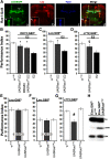
Ras1 hyperactivation in the MBs contributes to the learning deficiency of dNf1 nulls. A, Confocal images illustrate the distribution of Ras1 in the MBs of adult flies. Ras1-Gal4 drives the expression of UAS-mCD8GFP, especially in the α/β MB lobes (Aa–Ad), the γ lobe (Aa, arrowhead), and at lower levels in the α/′β′ lobes, where it colocalizes with Leo that marks all MB lobes (Aa,Ab,Ad, arrow in the marked boxes). Marked boxes represent higher magnification of the α/α′ lobes. B–G, Performance assessment after conditioning in control (white bars), mutant (black bars), and experimental (gray bars) animals. The genotypes of all animals are indicated below each bar. Data are mean ± SEM. G80ts indicates the ubiquitously expressed temperature-sensitive Gal4 repressor Tub-Gal80ts. B, Adult-specific dRas1 abrogation within the OK72 marked neurons at the exclusion of MB expression (second gray bar) partially restores learning in dNf1E2 homozygotes. *Statistically significant difference of the experimental from mutant flies. C, dRas1 abrogation in MB neurons (Leo-Gal4 driver) during adulthood rescues the dNf1E2 learning deficits. *Statistically significant difference of the experimental from mutant flies. D, dRas1 abrogation in MB neurons (c772-Gal4 driver) during adulthood improves the dNf1E2 learning deficit. §Statistical difference of the experimental from the mutant flies (p = 0.0312). E, F, Adult-specific abrogation of Ras1 (UASRas1Ri) in MB neurons does not yield learning deficits in control flies. The performance of animals with reduced Ras1 within MBs was not significantly different from that of controls both after 6 (E) and 3 (F) CS-US pairings. G, Overexpression of the constitutively active Ras1 (UASRas1v12) in the MBs of WT adult flies yields learning deficits. #Statistically significant difference of the experimental from the control groups. H, Representative Western blot of head lysates from adult flies expressing pan-neuronally a constitutively active form of Ras1 (UASRas1v12) under Elav-Gal4;Gal80ts using an antibody against phosphorylated ERK (p-ERK), revealing elevated active kinase compared with control animals. The amount of total-ERK protein is not affected. The genotypes of animals used are indicated above the immunoblot. Tubulin levels serve as loading control.

Similar articles

Cited by

References

    1. Acebes A, Ferrús A (2001) Increasing the number of synapses modifies olfactory perception in Drosophila. J Neurosci 21:6264–6273. 10.1523/JNEUROSCI.21-16-06264.2001 - DOI - PMC - PubMed
    1. Anastasaki C, Gutmann DH (2014) Neuronal NF1/RAS regulation of cyclic AMP requires atypical PKC activation. Hum Mol Genet 23:6712–6721. 10.1093/hmg/ddu389 - DOI - PMC - PubMed
    1. Bazigou E, Apitz H, Johansson J, Loren CE, Hirst EM, Chen PL, Palmer RH, Salecker I (2007) Anterograde Jelly belly and Alk receptor tyrosine kinase signaling mediates retinal axon targeting in Drosophila. Cell 128:961–975. 10.1016/j.cell.2007.02.024 - DOI - PubMed
    1. Brown JA, Gianino SM, Gutmann DH (2010) Defective cAMP generation underlies the sensitivity of CNS neurons to neurofibromatosis-1 heterozygosity. J Neurosci 30:5579–5589. 10.1523/JNEUROSCI.3994-09.2010 - DOI - PMC - PubMed
    1. Buchanan ME, Davis RL (2010) A distinct set of Drosophila brain neurons required for neurofibromatosis type 1-dependent learning and memory. J Neurosci 30:10135–10143. 10.1523/JNEUROSCI.0283-10.2010 - DOI - PMC - PubMed

Publication types

MeSH terms

LinkOut - more resources