. 2021 May 25;118(21):e2100170118.
doi: 10.1073/pnas.2100170118.

2'-O methylation of RNA cap in SARS-CoV-2 captured by serial crystallography

Affiliations

2'-O methylation of RNA cap in SARS-CoV-2 captured by serial crystallography

Mateusz Wilamowski et al. Proc Natl Acad Sci U S A. .

Abstract

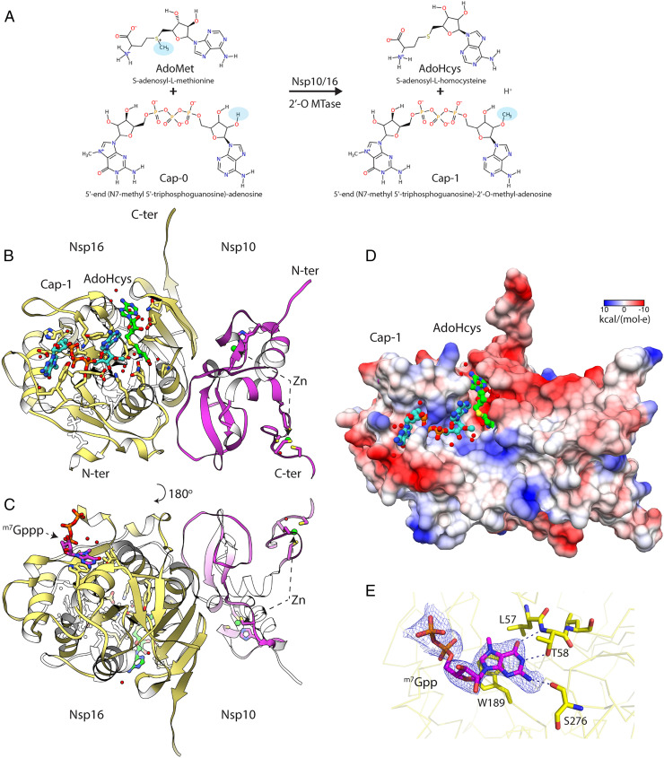
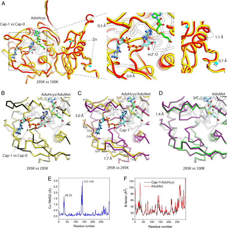
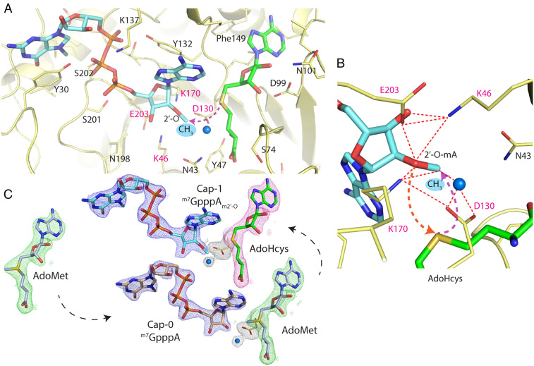
The genome of the severe acute respiratory syndrome coronavirus 2 (SARS-CoV-2) coronavirus has a capping modification at the 5'-untranslated region (UTR) to prevent its degradation by host nucleases. These modifications are performed by the Nsp10/14 and Nsp10/16 heterodimers using S-adenosylmethionine as the methyl donor. Nsp10/16 heterodimer is responsible for the methylation at the ribose 2'-O position of the first nucleotide. To investigate the conformational changes of the complex during 2'-O methyltransferase activity, we used a fixed-target serial synchrotron crystallography method at room temperature. We determined crystal structures of Nsp10/16 with substrates and products that revealed the states before and after methylation, occurring within the crystals during the experiments. Here we report the crystal structure of Nsp10/16 in complex with Cap-1 analog (m7GpppAm2'-O). Inhibition of Nsp16 activity may reduce viral proliferation, making this protein an attractive drug target.

Keywords: CAP-1; Nsp10/16; SARS-CoV-2; mRNA; serial crystallography.


Conflict of interest statement

The authors declare no competing interest.

Figures

Fig. 1.
Fig. 1.
Fixed-target serial crystallography data collection on Nsp10/16 crystals from SARS-CoV-2. System for SSX with moving crystal holder developed at SBC beamline 19ID.
Fig. 2.
Fig. 2.
Crystal structure of the Nsp10/16 in a complex with m7GpppAm2′-O (Cap-1) and AdoHcys determined using fixed-target SSX at 295 K. (A) Diagram of the 2′-O methyl transfer reaction performed by Nsp10/16; methyl groups are highlighted with blue ovals. (B) The assembly of the Nsp10/16 heterodimer; Nsp10 (pink), Nsp16 (yellow) in complex with Cap-1 (aquamarine), and AdoHcys (green) (PDB entry 7JHE). (C) The m7Gppp at the potential allosteric site of Nsp16. (D) The Coulombic electrostatic potential mapped on the surface of the Nsp10/16 heterodimer in complex with Cap-1/AdoHcys at 295 K calculated using default settings in UCSF Chimera. (E) 2mFo-DFc composite omit map calculated for m7Gpp and contoured at 1.2 σ (PDB entry 7JPE). The Nsp16 is shown as yellow ribbon, selected residues (yellow), and m7Gpp are depicted as pink sticks.
Fig. 3.
Fig. 3.
Comparison of crystal structures of the Nsp10/16 determined at 295 K and at 100 K. (A) Crystal structure of the Nsp10/16 with Cap-1 and AdoHcys (yellow, 7JHE) determined using SSX superimposed with Nsp10/16 with Cap-0 and AdoHcys (red, 6WQ3) determined at 100 K. (B) Comparison of 295 K crystal structures of Nsp10/16/Cap-1/AdoHcys (yellow, 7JHE) superimposed to the structure of Nsp10/16/Cap-0/AdoMet (black, 7JPE). (C) Comparison of 295 K crystal structures of the Nsp10/16/Cap-1/AdoHcys (yellow, 7JHE) superimposed to the structure of Nsp10/16/AdoMet (pink, 6XKM). (D) Comparison of the Nsp10/16 crystal structures with AdoMet determined respectively using SSX (pink, 6XKM) and standard data collection at 100 K (green, 6W4H). (E and F) Binding of Cap-1 to Nsp16. Plots of rmsd (E) and B factor of structures with AdoMet and Cap-1/AdoHcys (F).
Fig. 4.
Fig. 4.
Methylation of the 2′-O-ribose of the m7GpppA catalyzed by Nsp10/16 from SARS-CoV-2. (A) Active site of Nsp16 2′-O methyltransferase (yellow) in complex with m7GpppAm2′-O (Cap-1) as aquamarine sticks and AdoHcys as green sticks. (B) Magnification of the active site depicts catalytic tetrad Lys46-Asp130-Lys170-Glu203 essential for the 2′-O MTase activity shown as yellow sticks. Red dashed lines show close distances between residues and arrows depict simplified nucleophilic attack and subsequent movement of methyl group to the 2′-O position. The water molecule is represented as a blue sphere. (C) The 2mFo-DFc maps contoured at 1.2 σ around ligands of the structures determined by SSX; cap analogs are in blue, AdoHcys in pink, and AdoMet in green.

Similar articles

Cited by

References

    1. Stadler K., et al. ., SARS–Beginning to understand a new virus. Nat. Rev. Microbiol. 1, 209–218 (2003). - PMC - PubMed
    1. Huang C., et al. ., Clinical features of patients infected with 2019 novel coronavirus in Wuhan, China. Lancet 395, 497–506 (2020). - PMC - PubMed
    1. Dong E., Du H., Gardner L., An interactive web-based dashboard to track COVID-19 in real time. Lancet Infect. Dis. 20, 533–534 (2020). - PMC - PubMed
    1. Wu A., et al. ., Genome composition and divergence of the novel coronavirus (2019-nCoV) originating in China. Cell Host Microbe 27, 325–328 (2020). - PMC - PubMed
    1. Muthukrishnan S., Moss B., Cooper J. A., Maxwell E. S., Influence of 5′-terminal cap structure on the initiation of translation of vaccinia virus mRNA. J. Biol. Chem. 253, 1710–1715 (1978). - PubMed

Publication types

MeSH terms

LinkOut - more resources