. 2021 May 18;118(20):e2026104118.
doi: 10.1073/pnas.2026104118.

AGO2 promotes tumor progression in KRAS-driven mouse models of non-small cell lung cancer

Affiliations

AGO2 promotes tumor progression in KRAS-driven mouse models of non-small cell lung cancer

Jean Ching-Yi Tien et al. Proc Natl Acad Sci U S A. .

Abstract

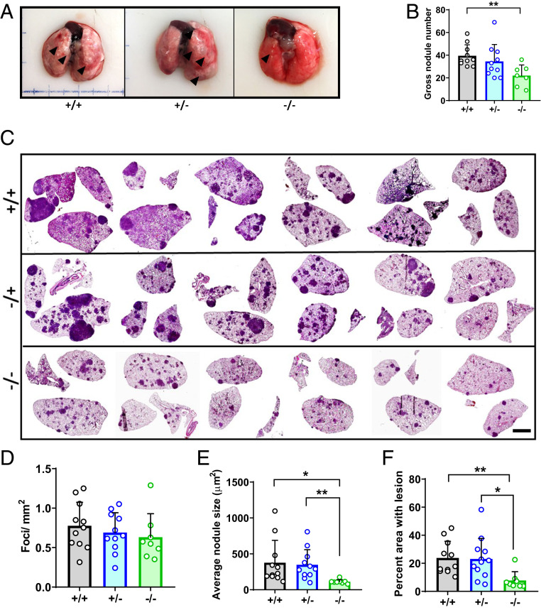
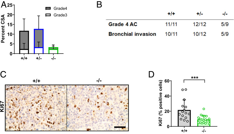
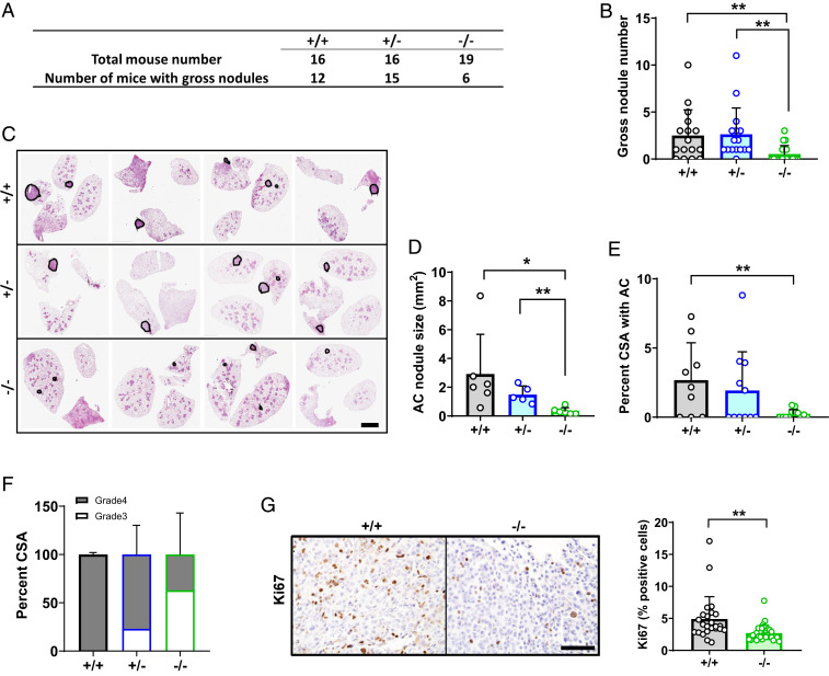
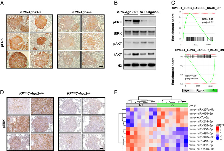
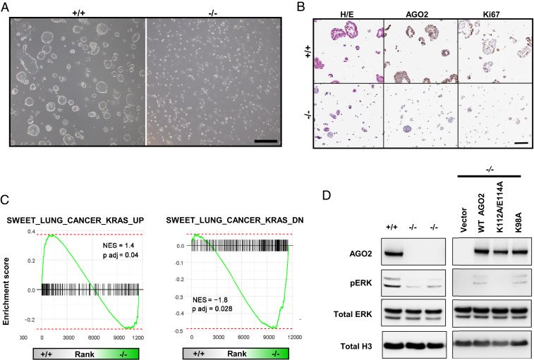
Lung cancer is the deadliest malignancy in the United States. Non-small cell lung cancer (NSCLC) accounts for 85% of cases and is frequently driven by activating mutations in the gene encoding the KRAS GTPase (e.g., KRASG12D). Our previous work demonstrated that Argonaute 2 (AGO2)-a component of the RNA-induced silencing complex (RISC)-physically interacts with RAS and promotes its downstream signaling. We therefore hypothesized that AGO2 could promote KRASG12D-dependent NSCLC in vivo. To test the hypothesis, we evaluated the impact of Ago2 knockout in the KPC (LSL-KrasG12D/+;p53f/f;Cre) mouse model of NSCLC. In KPC mice, intratracheal delivery of adenoviral Cre drives lung-specific expression of a stop-floxed KRASG12D allele and biallelic ablation of p53 Simultaneous biallelic ablation of floxed Ago2 inhibited KPC lung nodule growth while reducing proliferative index and improving pathological grade. We next applied the KPHetC model, in which the Clara cell-specific CCSP-driven Cre activates KRASG12D and ablates a single p53 allele. In these mice, Ago2 ablation also reduced tumor size and grade. In both models, Ago2 knockout inhibited ERK phosphorylation (pERK) in tumor cells, indicating impaired KRAS signaling. RNA sequencing (RNA-seq) of KPC nodules and nodule-derived organoids demonstrated impaired canonical KRAS signaling with Ago2 ablation. Strikingly, accumulation of pERK in KPC organoids depended on physical interaction of AGO2 and KRAS. Taken together, our data demonstrate a pathogenic role for AGO2 in KRAS-dependent NSCLC. Given the prevalence of this malignancy and current difficulties in therapeutically targeting KRAS signaling, our work may have future translational relevance.

Keywords: AGO2; KRAS; nonsmall cell lung cancer.


Conflict of interest statement

The authors declare no competing interest.

Figures

Fig. 1.
Fig. 1.
Ago2 ablation impairs tumor growth in KPC lung cancer. (A) Gross lung samples from KPC-Ago2+/+ (+/+), KPC-Ago2+/− (+/−), and KPC-Ago2−/− (−/−) mice. Representative images from n = 11, n = 11, and n = 8 animals per group, respectively. Arrowheads indicate grossly visible tumor nodules. (B) Quantification of grossly visible nodules from aforementioned animals expressed as number of total nodules per pair of lungs. (C) Representative H&E-stained image series of lung cross-sections from +/+, +/−, and −/− mice. (Scale bar, 2.5 mm.) (D) Quantification of tumor foci per mm2 cross-sectional area. Average of three nonconsecutive sections from each of 11 +/+, 11 +/−, and 8 −/− animals. (E) Average surface area of individual nodules described in D. (F) Percentage of total lung cross-sectional area occupied by tumor lesion in the same animals as described in CE. *P < 0.05, **P < 0.01.
Fig. 2.
Fig. 2.
Ago2 facilitates progression of KPC lung adenocarcinoma. (A) Percentage of lung cross-sectional area occupied by adenocarcinoma (subdivided into grade 3 and grade 4) in KPC-Ago2+/+ (+/+) (n = 11), KPC-Ago2+/− (+/−) (n = 12), and KPC-Ago2−/− (−/−) (n = 9) mice. Analyses based on pathological assessment of H&E staining of whole lung cross-sections. (B) Number of mice per group (represented as a fraction of total animals) with any evidence of grade 4 adenocarcinoma (AC) or bronchial invasion. (C and D) Ki67 staining in adenocarcinoma lesions from +/+ and f/f animals with quantification (n = 3 sections/animal, 6 animals/group). (Scale bar, 50 μm.) ***P < 0.001.
Fig. 3.
Fig. 3.
Ago2 ablation impairs tumor growth in KPHet-C lung cancer. (A and B) Quantification of grossly visible nodules from lungs of KPHetC-Ago2+/+ (+/+), KPHetC-Ago2+/− (+/−), and KPHetC-Ago2−/− (−/−) mice. (C) Representative H&E-stained image series of lung cross-sections from +/+, f/+, and f/f mice. (Scale bar, 2.5 mm.) (D) Quantification of adenocarcinoma (AC) foci per mm2 cross-sectional area. (E) Average lung cross-sectional surface area (CSA) occupied by adenocarcinoma. (F) Percent grade 3 and grade 4 tissue within adenocarcinoma lesions of mice of each genotype. (G) Ki67 staining (Left images) and proliferative index (Right) in +/+ and −/− mice. (Scale bar, 50 μm.) n = 16 to 19 mice/group. *P < 0.05, **P < 0.01.
Fig. 4.
Fig. 4.
Ago2 ablation reduces Kras signaling in vivo. (A) pERK immunohistochemistry of lung cross-sections from KPC-Ago2+/+ versus KPC-Ago2−/− mice. (Scale bar, 50 μm.) (B) Immunoblot analysis of protein lysates from isolated lung nodules of KPC-Ago2+/+ and KPC-Ago2−/− mice. Phosphorylated ERK (pERK), total ERK (tERK), phosphorylated AKT (Ser-473) (pAKT), total AKT (tAKT), and histone H3 (H3). (C) GSEA assessing genes associated with increased KRAS signaling (SWEET_LUNG_CANCER_KRAS_UP and SWEET_LUNG_CANCER_KRAS_DOWN). Comparison of genes differentially expressed in KPC-Ago2+/+ versus KPC-Ago2−/− (isolated nodules). NES, normalized enrichment score. (D) pERK immunohistochemistry of lung cross-sections from KPHetC-Ago2+/+ and KPHetC-Ago2−/− mice. (Scale bar, 100 μm.) (E) Heat map of differentially expressed (false discovery rate [FDR] < 0.05) species from miRNA QPCR performed on nodules from KPC mice with normal Ago2 (+/+) or with biallelic Ago2 knockout (−/−).
Fig. 5.
Fig. 5.
AGO2 promotes MAPK signaling through direct interaction. (A) Organoids isolated from lung nodules of KPC-Ago2+/+ versus KPC-Ago2−/− mice. Bright field image. (Scale bar, 500 μm.) (B) Histological sections of KPC-Ago2+/+ and KPC-Ago2−/− organoids stained with H&E and antibodies directed against AGO2 and Ki67. (Scale bar, 150 μm.) (C) GSEA assessing genes associated with increased KRAS signaling (SWEET_LUNG_CANCER_KRAS_UP and SWEET_LUNG_CANCER_KRAS_DOWN). Comparison of genes differentially expressed in KPC-Ago2+/+ versus KPC-Ago2−/− organoids. NES, normalized enrichment score. (D, Left) Immunoblot analysis of protein lysates from KPC-Ago2+/+ versus KPC-Ago2−/− organoid-derived cells. Phosphorylated ERK (pERK), total ERK (tERK), and histone H3 (H3). (D, Right) Immunoblot analysis of KPC-Ago2−/− organoid-derived cells transfected with constructs encoding wild-type AGO2 (WT AGO2), AGO2K112A/E114A (K112A/E114A), and AGO2K98A (K98A).

Similar articles

Cited by

References

    1. Siegel R. L., Miller K. D., Jemal A., Cancer statistics, 2017. CA Cancer J. Clin. 67, 7–30 (2017). - PubMed
    1. Ettinger D. S.et al. .; NCCN (National Comprehensive Cancer Network) , Non-small cell lung cancer. J. Natl. Compr. Canc. Netw. 10, 1236–1271 (2012). - PubMed
    1. Schubbert S., Shannon K., Bollag G., Hyperactive Ras in developmental disorders and cancer. Nat. Rev. Cancer 7, 295–308 (2007). - PubMed
    1. Karnoub A. E., Weinberg R. A., Ras oncogenes: Split personalities. Nat. Rev. Mol. Cell Biol. 9, 517–531 (2008). - PMC - PubMed
    1. Trahey M., McCormick F., A cytoplasmic protein stimulates normal N-ras p21 GTPase, but does not affect oncogenic mutants. Science 238, 542–545 (1987). - PubMed

Publication types

MeSH terms