. 2021 May 18;118(20):e2100466118.
doi: 10.1073/pnas.2100466118.

Comprehensive pregnancy monitoring with a network of wireless, soft, and flexible sensors in high- and low-resource health settings

Affiliations

Comprehensive pregnancy monitoring with a network of wireless, soft, and flexible sensors in high- and low-resource health settings

Dennis Ryu et al. Proc Natl Acad Sci U S A. .

Abstract

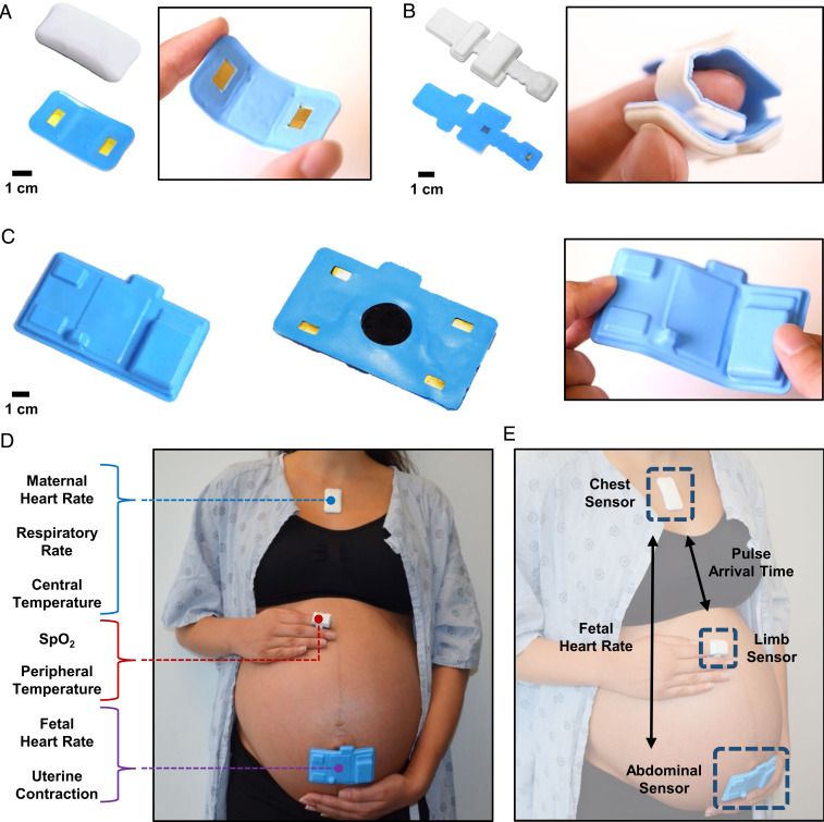
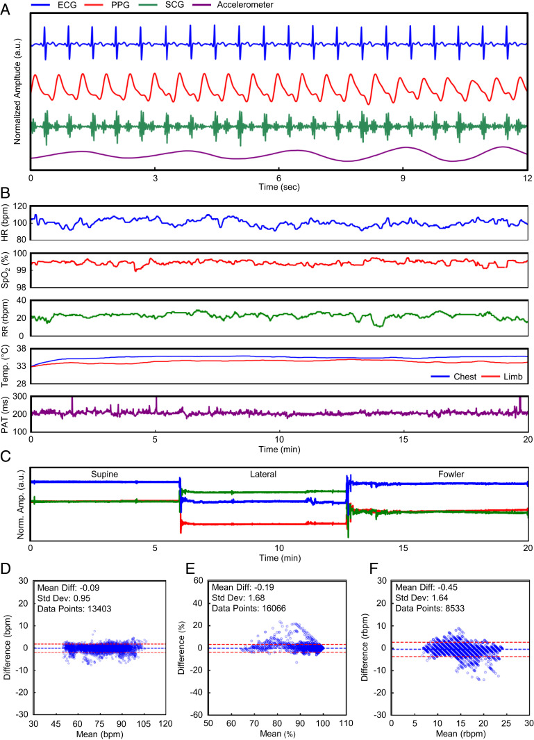
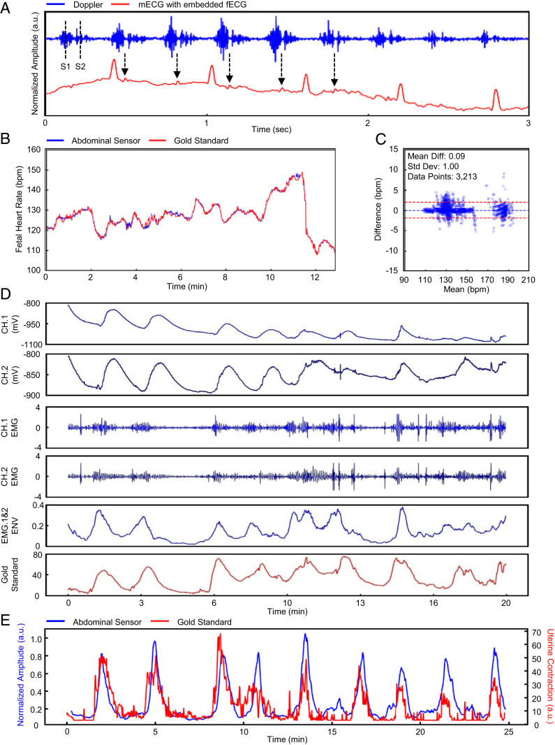
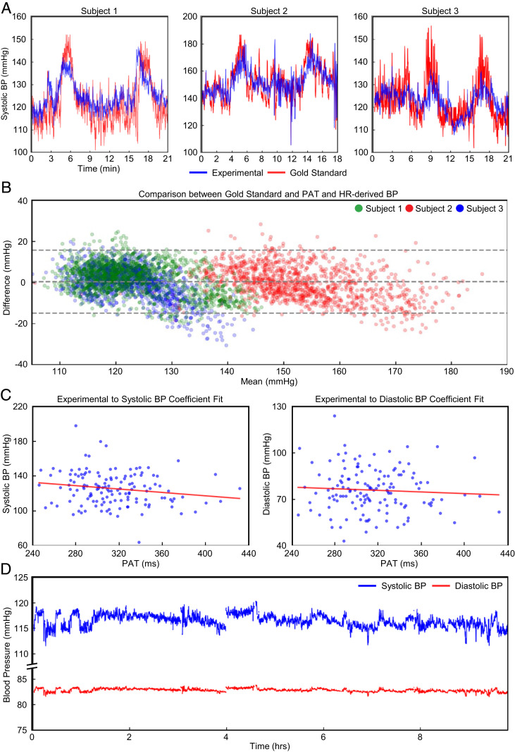
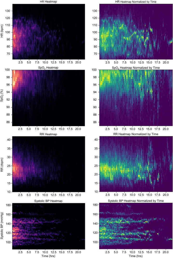
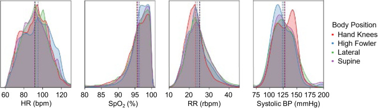
Vital signs monitoring is a fundamental component of ensuring the health and safety of women and newborns during pregnancy, labor, and childbirth. This monitoring is often the first step in early detection of pregnancy abnormalities, providing an opportunity for prompt, effective intervention to prevent maternal and neonatal morbidity and mortality. Contemporary pregnancy monitoring systems require numerous devices wired to large base units; at least five separate devices with distinct user interfaces are commonly used to detect uterine contractility, maternal blood oxygenation, temperature, heart rate, blood pressure, and fetal heart rate. Current monitoring technologies are expensive and complex with implementation challenges in low-resource settings where maternal morbidity and mortality is the greatest. We present an integrated monitoring platform leveraging advanced flexible electronics, wireless connectivity, and compatibility with a wide range of low-cost mobile devices. Three flexible, soft, and low-profile sensors offer comprehensive vital signs monitoring for both women and fetuses with time-synchronized operation, including advanced parameters such as continuous cuffless blood pressure, electrohysterography-derived uterine monitoring, and automated body position classification. Successful field trials of pregnant women between 25 and 41 wk of gestation in both high-resource settings (n = 91) and low-resource settings (n = 485) demonstrate the system's performance, usability, and safety.

Keywords: biosensors; pregnancy; vital signs.


Conflict of interest statement

Competing interest statement: Coauthors with a Sibel, Inc. affiliation are working with the Bill & Melinda Gates Foundation to deploy these technologies into low- and middle-income countries.

Figures

Fig. 1.
Fig. 1.
Overview of the maternal fetal monitoring system. The on-body network of the maternal–fetal sensor system is visualized. (A and B) Photographs of the front and back side of the chest and limb sensors. The silicone encapsulation allows the sensors to be soft and stretchable while still operating wirelessly. (C) The abdominal sensor is designed to conform around the abdomen of the patient without the need for an external strap. (D) Individually, each sensor captures unique signals from the patient. (E) Together, the sensors constitute an on-body network capable of acquiring more advanced metrics.
Fig. 2.
Fig. 2.
Maternal vital signs. The end-to-end data analytics of the sensor system is outlined. (A) Waveforms from the patient are obtained by the chest, limb, and abdominal sensors. (B) The raw signals are then processed to yield the representative vital signs of a clinical setting. (C) The patient’s body orientation is also identifiable through the use of an embedded IMU. (D–F) Our calculated metrics are statistically comparable to the gold standard used in modern hospitals.
Fig. 3.
Fig. 3.
Doppler-derived FHR and EMG-derived uterine contraction. Data analytics of the FHR and maternal uterine contraction are outlined. (A) The raw US Doppler signal is obtained by the abdominal sensor. We can identify the S1 and S2 waves and see the signal aligned with peaks indicative of fetal ECG. (B and C) Our calculated FHR is statistically comparable to the gold standard. (D) The raw biosignal is obtained by the abdominal sensor. We acquire two channels for sequential processing of the EMG signal. (E) Our calculated uterine contraction output is overlaid onto the gold standard.
Fig. 4.
Fig. 4.
Continuous blood pressure correlation. The derivation of continuous BP from PAT and HR measurements. (A) In a cold-pressor test, we use the first 50 s of sampled data to derive linear coefficients for PAT. For each sample, we overlay the gold-standard SBP. (B) A Bland–Altman plot comparing the calibrated PAT to the gold-standard SBP from the cold-pressor test is presented. (C) An inverse relationship across all participants is derived between PAT and sampled BP cuff measurements. (D) n = 1 is shown for the converted systolic and diastolic BPs from PAT.
Fig. 5.
Fig. 5.
Heat-map analysis of maternal vital signs for laboring women in low-resource settings. A heat map of each vital signal is generated for all 485 participants. (Left) Plot (in red–black) for each vital is a heat map without normalization as the majority of labors last less than 5 h. (Right) Plot (in yellow–green) is normalized by frequency with a 4-h window to illustrate average vital signs over longer labors.
Fig. 6.
Fig. 6.
Body position and vital signs. Small, but clear, differences are seen in maternal vital signs based on body position for HR, SpO2, RR, and SBP derived from PAT. A vertical line is depicted in each of the vital sign distribution plots to denote the mean value for the specific body position.

References

    1. Sedgh G., Singh S., Hussain R., Intended and unintended pregnancies worldwide in 2012 and recent trends. Stud. Fam. Plann. 45, 301–314 (2014). - PMC - PubMed
    1. MacDorman M. F., Declercq E., Cabral H., Morton C., Recent increases in the U.S. maternal mortality rate: Disentangling trends from measurement issues. Obstet. Gynecol. 128, 447–455 (2016). - PMC - PubMed
    1. Organization World Health, Trends in Maternal Mortality 2000 to 2017: Estimates by WHO, UNICEF, UNFPA (World Bank Group and the United Nations Population Division, 2019).
    1. Rochat R. W., Koonin L. M., Atrash H. K., Jewett J. F., Maternal mortality in the United States: Report from the maternal mortality collaborative. Obstet. Gynecol. 72, 91–97 (1988). - PubMed
    1. Kodio B., et al. ., Levels and causes of maternal mortality in Senegal. Trop. Med. Int. Health 7, 499–505 (2002). - PubMed

Publication types