. 2021 May 11;13(9):12817-12832.
doi: 10.18632/aging.202949. Epub 2021 May 11.

Hydroxyurea-induced membrane fluidity decreasing as a characterization of neuronal membrane aging in Alzheimer's disease

Affiliations

Hydroxyurea-induced membrane fluidity decreasing as a characterization of neuronal membrane aging in Alzheimer's disease

Qiujian Yu et al. Aging (Albany NY). .

Abstract

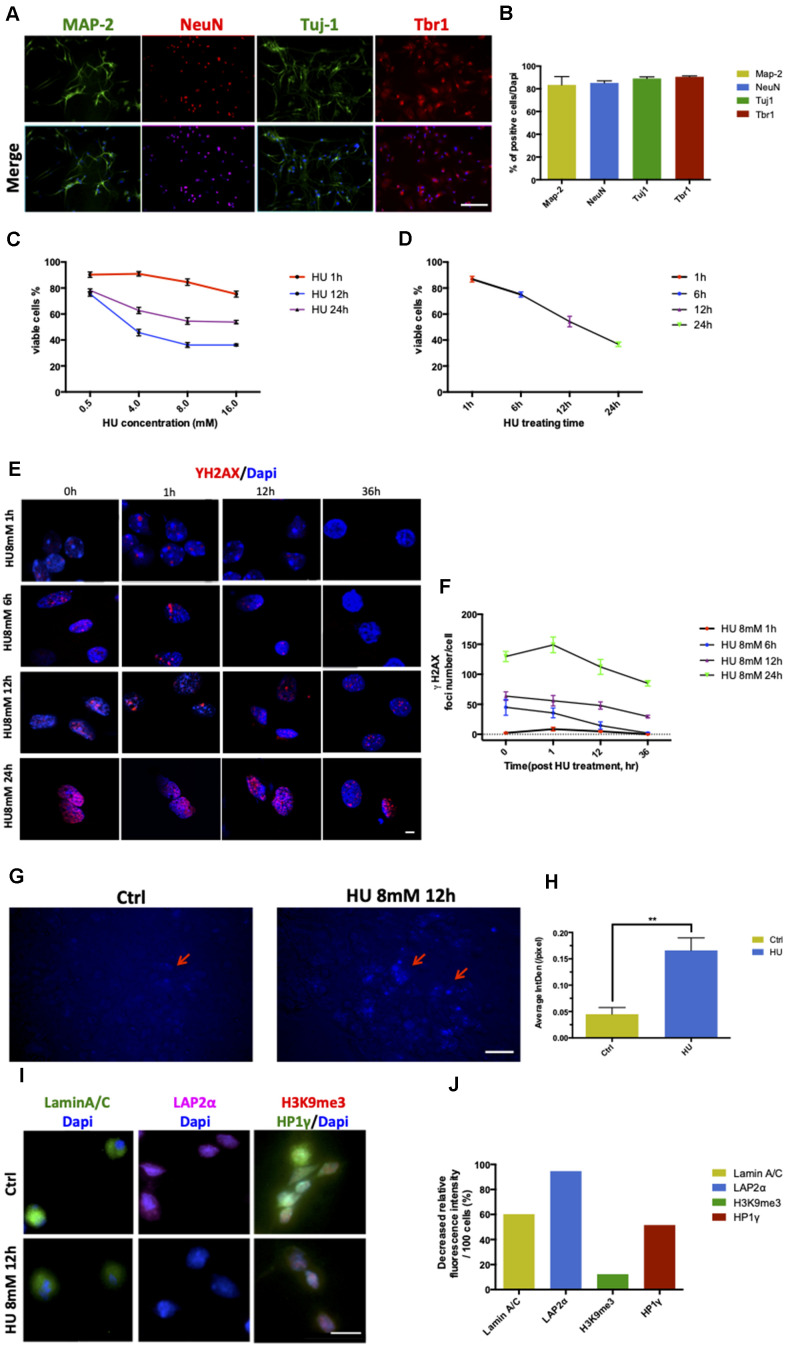
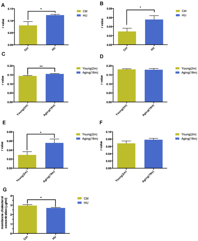
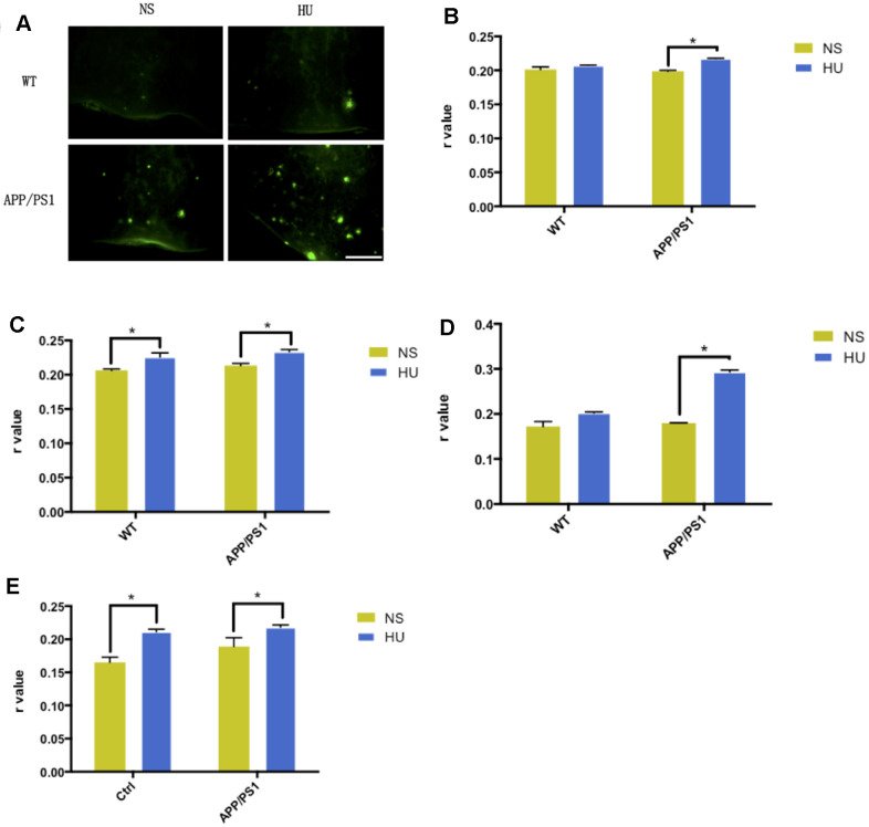
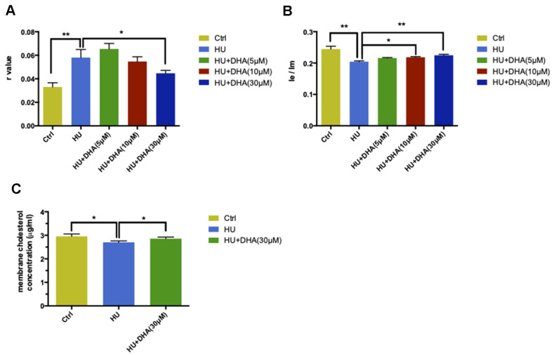
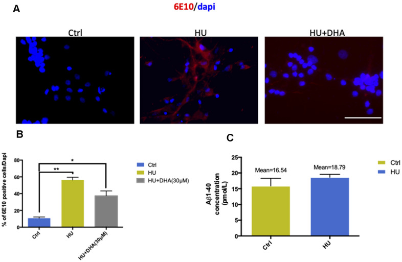
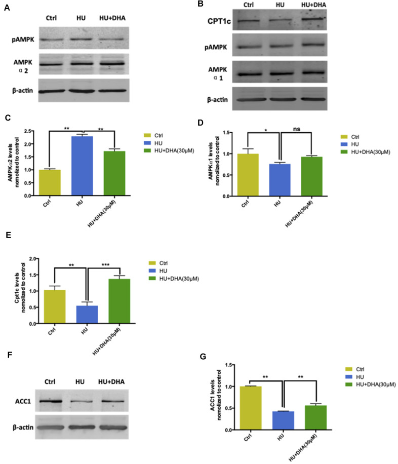
Aging is one of the significant risk factors for Alzheimer's disease (AD). Therefore, this study aimed to propose a new hypothesis "membrane aging" as a critical pathogenesis of AD. The concept of "membrane aging" was reviewed, and the possible mechanisms of membrane aging as the primary culprit of AD were clarified. To further prove this hypothesis, a hydroxyurea-induced "membrane aging" model was established in vitro and in vivo. First, neuronal aging was validated by immunocytochemistry with age-related markers, and membrane aging phenotypes were confirmed. The alterations of membrane fluidity within APP/PS1 mice were re-proved by intracerebroventricular injection of hydroxyurea. Decreased membrane fluidity was found in vitro and in vivo, accompanied by increased total cholesterol concentration in neurons but decreased cholesterol levels within membrane fractions. The Aβ level increased considerably after hydroxyurea treatment both in vitro and in vivo. DHA co-treatment ameliorated membrane aging phenotypes and Aβ aggregation. The study revealed the AMP-activated protein kinase/acetyl CoA carboxylase/carnitine palmitoyl transferase 1 pathway involved in membrane aging processes. These results strongly supported the idea that membrane aging was a pathogenesis of AD and might serve as a new therapeutic target for AD.

Keywords: Alzheimer's disease; DHA; hydroxyurea; membrane aging; β-amyloid.


Conflict of interest statement

CONFLICTS OF INTEREST: The authors declare that they have no conflicts of interest.

Figures

Figure 1
Figure 1
Establishment and characterization of neuronal senescence. More than 80% of rat primary cortical neurons (cultured till day 5) were positively stained with neuronal markers of MAP-2 (green), NeuN (red), Tju1 (green), and Tbr1 (red). Scale bar: 200 μm (A, B). Dose-dependent and time-dependent curves represented neuronal viability using CCK8 test detection (C, D). Formation of DNA double-strand breaks in rat primary cortical neurons (5 days) after HU treatment. Neurons were treated with 8mM HU for 1, 6, 12, and 24 h and allowed to recover until 36 h after treatment. The cells were fixed and stained for γH2AX foci (red) at the indicated time points after HU treatment. Scale bar: 5 μm (E). Graphical quantification of the number of γH2AX foci in neurons treated with 8mM HU for different times over 36 h (F). In vitro staining of total cholesterol (blue) in the 12-h 8mM HU treatment group compared with the control neurons. Scale bar: 500 μm (G). Average fluorescent intensity of the cholesterol-positive cells among control and HU-treated cells. As shown earlier, HU treatment significantly increased the level of blue-staining intensity (P < 0.01) (H). Immunocytochemistry for markers identifying the nuclear lamina (Lamin A/C), a lamina-associated protein (LAP2α), and peripheral heterochromatin (3K9me3, HP1γ) in HU-treated neurons compared with the control ones. Scale bar: 25 μm (I). Quantification of the markers depicted in (I) demonstrated the decreased relative fluorescence intensity per 100 cells in the HU group compared with the control group (J). All the data are expressed as mean ± SD from three independent experiments (N = 3). *P < 0.05, **P < 0.01, ***P < 0.001. The Student t test was used to determine the statistical significance of the differences.
Figure 2
Figure 2
Validation of “membrane aging” in vivo and in vitro. Changes in membrane fluidity and cholesterol levels in membrane pellets are two main characteristics of membrane aging. Fluorescent probe polarization is one of the most direct measurements representing the alterations in membrane fluidity. HU treatment significantly increased the r value compared with the control in vitro via applying the DPH fluorescent probe (P < 0.05) (A). The same trend was observed while probing with TMA-DPH (P < 0.01) (B). The results demonstrated that HU treatment decreased neuronal membrane fluidity dramatically because the r value was inversely proportional to membrane mobility. In vivo, native membrane pellets were carefully extracted, followed by hippocampus and cortex isolation in 18-month-old male SD rats (n = 6) and 2-month-old male SD rats (n = 6). When probing with DPH in cortical membrane pellets, the r value increased significantly in the aging group compared with the young group (P < 0.01) (C). In hippocampal regions, no difference was observed between two groups (D). Similar results were obtained by applying a TMA-DPH fluorescent probe in cortical and hippocampal native membrane pellets in vivo. The r value considerably increased in elderly rats only in the cortical regions (P < 0.05) but without apparent changes in the hippocampus (E, F). Regarding the alterations in membrane lipid composition, HU-treated neurons showed a decreased cholesterol level in neuronal membrane pellets (P < 0.05) (G). All the data are expressed as mean ± SD from three independent experiments (N = 3). *P < 0.05, **P < 0.01, ***P < 0.001. The Student t test was used to determine the statistical significance of the differences.
Figure 3
Figure 3
Validation of membrane aging in APP/PS1-mutated transgenic mice. In the dissected cortical region, APP/PS1-mutated transgenic mice showed accelerated Aβ deposition after HU intracerebroventricular injection compared with mice with NS injection and wild-type controls. Scale bar: 1 mm (A). For the membrane fluidity determination probed with TMA-DPH and DPH, 32μM HU injection dramatically increased r values in the hippocampal and cortical membranes of APP/PS1 mice compared with NS-injected ones (n = 3, P < 0.05). Same trends were found only in hippocampal membranes in wild-type controls (n = 3, P < 0.05) (BE). All the data are expressed as mean ± SD from three independent experiments (N = 3). *P < 0.05. Two-way ANOVA was used to determine the statistical significance of the differences.
Figure 4
Figure 4
DHA intervention ameliorated membrane aging phenotypes. TMA-DPH and DPP were used as two fluorescent probes to detect membrane mobility in vitro. In the TMA-DPH probing experiment, HU treatment considerably increased the r value compared with the control (P < 0.01). However, co-treatment with different concentrations of DHA gradually decreased the r value. Especially after 30μM DHA treatment, the r value showed a dramatic decline compared with HU treatment (P < 0.05) (A). Similar results were confirmed by the DPP probing test. HU treatment decreased the Ie/Im ratio noticeably (P < 0.01). The ratio gradually increased after co-treatment with the increasing concentrations of DHA. Ie/Im was considerably augmented after treatment with 10μM and 30μM DHA compared with HU treatment (P < 0.05 and P < 0.01) (B). The r value is inversely proportional to membrane fluidity, while the ratio of Ie/Im is directly proportional to membrane mobility. Regarding membrane lipid composition in vitro, the cholesterol level in membrane pellets significantly increased in the 30μM DHA co-treatment group compared with HU treatment group (C). All the data are expressed as mean ± SD from three independent experiments (N = 3). *P < 0.05, **P < 0.01. Student t test and one-way ANOVA were used to determine the statistical significance of the differences.
Figure 5
Figure 5
Membrane aging increased Aβ aggregation in vitro. The results of immunostaining with 6E10 showed that membrane aging significantly increased the number of positively stained neurons compared with the control (P < 0.01). After co-treatment with DNA, Aβ aggregation decreased (A, B). Further quantification of Aβ in vitro was done by ELISA detection. The average concentration of Aβ1-40 was at least 2 pmol/L higher in the membrane aging group compared with the control group (C). However, Aβ1-42 was under the detection threshold, which was hard to quantify. All the data are expressed as mean ± SD from three independent experiments (N = 3). *P < 0.05, **P < 0.01. The Student t test was used to determine the statistical significance of the differences.
Figure 6
Figure 6
AMPK/ACC/CPT1 pathway was involved in membrane aging processes. Membrane aging activated protein expression of AMPK-α2 through Ser485/491 residue phosphorylation by DHA treatment. However, AMPK-α1 showed no significant change in the protein expression level (A, B). Membrane aging also increased AMPK-α2 mRNA levels (P < 0.01), and DHA co-treatment partly decreased mRNA levels of AMPK-α2 (P < 0.01) (C). AMPK-α1 mRNA levels decreased in the membrane aging group and slightly increased in the DHA treatment group (D). CPT1c protein and mRNA levels were also inhibited by membrane aging (P < 0.01) and eased by DHA intervention (P < 0.01) (B, E). The protein and mRNA expression of two downstream factors of AMPK were detected. Membrane aging inhibited the expression of ACC1 protein; DHA co-treatment comparatively increased the protein level (F). The ACC1 mRNA level followed the same trend, which was reduced by membrane aging (P < 0.05) and then ameliorated by DHA administration (P < 0.01) (G). All the data are expressed as mean ± SD from three independent experiments (N = 3). *P < 0.05, **P < 0.01, ***P < 0.001. One-way ANOVA was used to determine the statistical significance of the differences.

Similar articles

Cited by

References

    1. Brookmeyer R, Gray S, Kawas C. Projections of Alzheimer’s disease in the United States and the public health impact of delaying disease onset. Am J Public Health. 1998; 88:1337–42. 10.2105/AJPH.88.9.1337 - DOI - PMC - PubMed
    1. Chan KY, Wang W, Wu JJ, Liu L, Theodoratou E, Car J, Middleton L, Russ TC, Deary IJ, Campbell H, Wang W, Rudan I, and Global Health Epidemiology Reference Group (GHERG). Epidemiology of Alzheimer’s disease and other forms of dementia in China, 1990-2010: a systematic review and analysis. Lancet. 2013; 381:2016–23. 10.1016/S0140-6736(13)60221-4 - DOI - PubMed
    1. Hirsch HR. The waste-product theory of aging: cell division rate as a function of waste volume. Mech Ageing Dev. 1986; 36:95–107. 10.1016/0047-6374(86)90142-9 - DOI - PubMed
    1. Strehler BL. Genetic instability as the primary cause of human aging. Exp Gerontol. 1986; 21:283–319. 10.1016/0531-5565(86)90038-0 - DOI - PubMed
    1. Freitas AA, de Magalhães JP. A review and appraisal of the DNA damage theory of ageing. Mutat Res. 2011; 728:12–22. 10.1016/j.mrrev.2011.05.001 - DOI - PubMed

Publication types

MeSH terms