Review
. 2021 May 31;44(5):342-355.
doi: 10.14348/molcells.2021.0067.

MiT Family Transcriptional Factors in Immune Cell Functions

Affiliations
Review

MiT Family Transcriptional Factors in Immune Cell Functions

Seongryong Kim et al. Mol Cells. .

Abstract

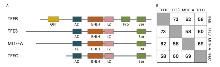
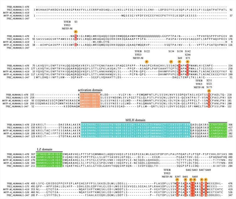
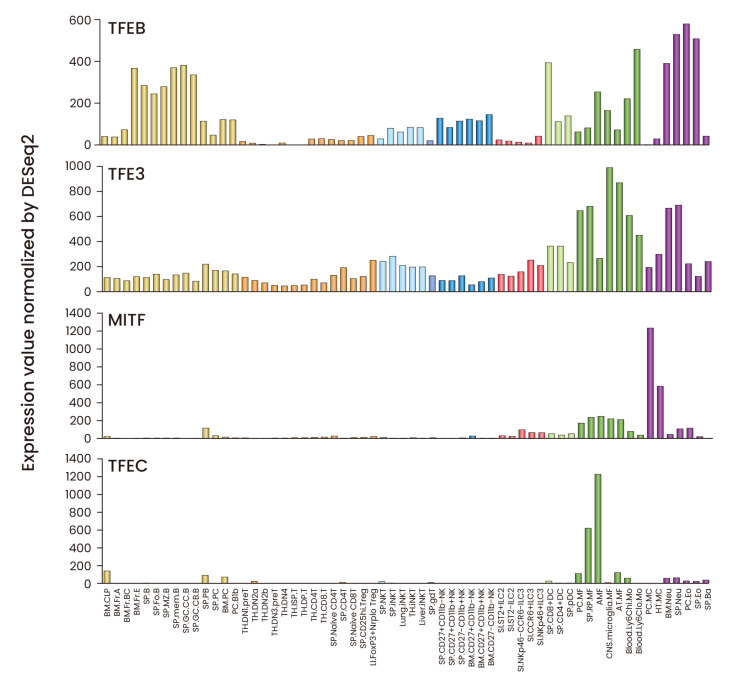
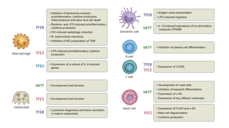
The microphthalmia-associated transcription factor family (MiT family) proteins are evolutionarily conserved transcription factors that perform many essential biological functions. In mammals, the MiT family consists of MITF (microphthalmia-associated transcription factor or melanocyte-inducing transcription factor), TFEB (transcription factor EB), TFE3 (transcription factor E3), and TFEC (transcription factor EC). These transcriptional factors belong to the basic helix-loop-helix-leucine zipper (bHLH-LZ) transcription factor family and bind the E-box DNA motifs in the promoter regions of target genes to enhance transcription. The best studied functions of MiT proteins include lysosome biogenesis and autophagy induction. In addition, they modulate cellular metabolism, mitochondria dynamics, and various stress responses. The control of nuclear localization via phosphorylation and dephosphorylation serves as the primary regulatory mechanism for MiT family proteins, and several kinases and phosphatases have been identified to directly determine the transcriptional activities of MiT proteins. In different immune cell types, each MiT family member is shown to play distinct or redundant roles and we expect that there is far more to learn about their functions and regulatory mechanisms in host defense and inflammatory responses.

Keywords: MiT family transcription factors; autophagy; immune cells; lysosome; metabolism; microphthalmia-associated transcription factor (MITF); mitochondria; stress response; transcription factor E3 (TFE3); transcription factor EB (TFEB); transcription factor EC.


Conflict of interest statement

CONFLICT OF INTEREST

The authors have no potential conflicts of interest to disclose.

Figures

Fig. 1
Fig. 1. Protein domain structures and amino acid sequence similarities of human MiT family proteins.
(A) The four mammalian MiT family members share highly conserved basic helix-loop-helix, leucine zipper (bHLH-ZIP) domain, which serves as a DNA binding and dimerization domain. They also contain an activation domain (or acidic domain, AD) required for transcriptional activation and a serine-rich domain (Ser). In addition, TFEB has a glutamine-rich domain (Gln) and a proline-rich domain (Pro). (B) The amino acid sequence similarities among human MiT family proteins were analyzed using NCBI protein blast. Aligned amino acid sequence ranges are: MITF-A (53-425) vs TFEB (1-349); MITF-A (4-521) vs TFE3 (46-573); MITF-A (221-509) vs TFEC (59-331); TFEB (62-330) vs TFE3 (167-441); TFEB (119-476) vs TFEC (20-347); TFE3 (229-561) vs TFEC (24-331).
Fig. 2
Fig. 2. Amino acid sequence alignment of human MiT family proteins.
A multiple sequence alignment of TFEB (P19484-1), TFE3 (P19532-1), MITF-M (O75030-9), MITF-A (O75030-1), and TFEC (O14948-1) was performed using clustal Omega (ver. 1.2.4). Major phosphorylation sites are denoted as P, and the conserved phosphorylation sites are in red. The activation domain, bHLH (basic-helix-loop-helix) domain, and LZ (leucine zipper) domain are also indicated.
Fig. 3
Fig. 3. Expression patterns of MiT family proteins in mouse immune cells.
The mRNA expression data were downloaded from the ImmGen (Immunological Genome Project) database (https://www.immgen.org) and displayed in bar graphs. The mRNA expression values and the full names of individual cell types can be found in Supplementary Table S1.
Fig. 4
Fig. 4. Diverse functions of MiT family proteins in immune cells.
MiT family transcription factors play diverse roles in various immune cell types. The known roles of each transcription factor in macrophages, osteoclasts, dendritic cells, B cells, T cells, and mast cells are summarized.

References

    1. Aksan I., Goding C.R. Targeting the microphthalmia basic helix-loop-helix-leucine zipper transcription factor to a subset of E-box elements in vitro and in vivo. Mol. Cell. Biol. 1998;18:6930–6938. doi: 10.1128/MCB.18.12.6930. - DOI - PMC - PubMed
    1. Argani P., Reuter V.E., Zhang L., Sung Y.S., Ning Y., Epstein J.I., Netto G.J., Antonescu C.R. TFEB-amplified renal cell carcinomas: an aggressive molecular subset demonstrating variable melanocytic marker expression and morphologic heterogeneity. Am. J. Surg. Pathol. 2016;40:1484–1495. doi: 10.1097/PAS.0000000000000720. - DOI - PMC - PubMed
    1. Beckmann H., Su L.K., Kadesch T. TFE3: a helix-loop-helix protein that activates transcription through the immunoglobulin enhancer muE3 motif. Genes Dev. 1990;4:167–179. doi: 10.1101/gad.4.2.167. - DOI - PubMed
    1. Betschinger J., Nichols J., Dietmann S., Corrin P.D., Paddison P.J., Smith A. Exit from pluripotency is gated by intracellular redistribution of the bHLH transcription factor Tfe3. Cell. 2013;153:335–347. doi: 10.1016/j.cell.2013.03.012. - DOI - PMC - PubMed
    1. Brady O.A., Jeong E., Martina J.A., Pirooznia M., Tunc I., Puertollano R. The transcription factors TFE3 and TFEB amplify p53 dependent transcriptional programs in response to DNA damage. Elife. 2018;7:e40856. doi: 10.7554/eLife.40856. - DOI - PMC - PubMed

MeSH terms

Substances

LinkOut - more resources