. 2021 May 10;12(1):2614.
doi: 10.1038/s41467-021-22758-0.

Deep learning-based predictive identification of neural stem cell differentiation

Affiliations

Deep learning-based predictive identification of neural stem cell differentiation

Yanjing Zhu et al. Nat Commun. .

Abstract

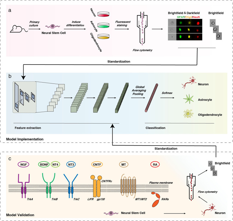
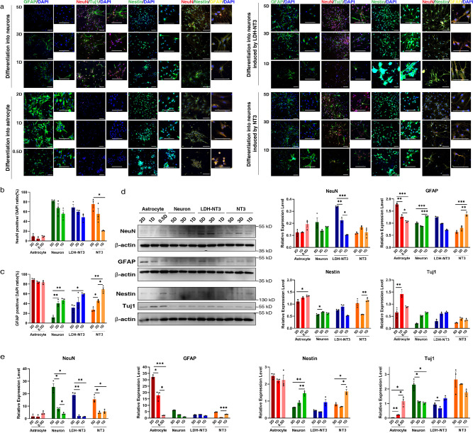
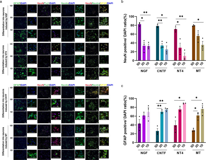
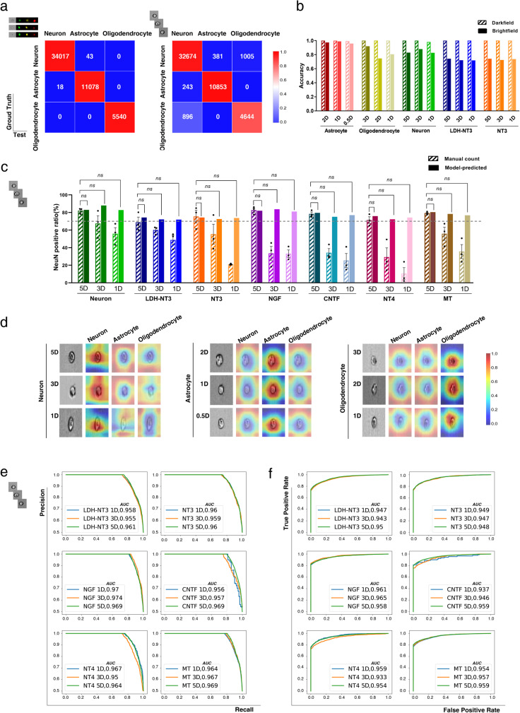
The differentiation of neural stem cells (NSCs) into neurons is proposed to be critical in devising potential cell-based therapeutic strategies for central nervous system (CNS) diseases, however, the determination and prediction of differentiation is complex and not yet clearly established, especially at the early stage. We hypothesize that deep learning could extract minutiae from large-scale datasets, and present a deep neural network model for predictable reliable identification of NSCs fate. Remarkably, using only bright field images without artificial labelling, our model is surprisingly effective at identifying the differentiated cell types, even as early as 1 day of culture. Moreover, our approach showcases superior precision and robustness in designed independent test scenarios involving various inducers, including neurotrophins, hormones, small molecule compounds and even nanoparticles, suggesting excellent generalizability and applicability. We anticipate that our accurate and robust deep learning-based platform for NSCs differentiation identification will accelerate the progress of NSCs applications.


Conflict of interest statement

The authors declare no competing interests.

Figures

Fig. 1
Fig. 1. Overview of workflow.
a NSCs were induced to differentiate into neurons/astrocytes/oligodendrocytes, and the cells were stained with NeuN (red)/GFAP (green)/Olig2 (yellow), collected and subjected to image flow cytometry. b Brightfield and darkfield (labelled) single-cell images were used as training data for the screening system; a schematic of the convolutional neural network (CNN) is presented. c Various inducers in different forms that act on different pathways were used to guide NSCs to differentiate into neurons, and brightfield (unlabelled) single-cell image patches were obtained by flow cytometry. These independent test sets were evaluated with the deep network model to show its generalizability.
Fig. 2
Fig. 2. Differentiation efficiency of training and testing sets identified by immunofluorescence, western blot and RT-qPCR.
a Images of immunofluorescent staining using NeuN, GFAP, Tuj1 and Nestin as characteristic markers of neuron and astrocyte differentiation induction at 5D (5 days), 3D (3 days), 2D (2 days), 1D (1 day) and 0.5D (0.5 days). Differentiated cells treated with NT3 and LDH-NT3 were stained following the same protocols. Quantification of immunostaining data. The y-axis shows the number of: b NeuN- and c GFAP-positive cells, n = 3 imaging field repeats. d Western blot analysis of NeuN, GFAP, Nestin and Tuj1 protein in NSCs in different states of differentiation, n = 3 biological repeats. e Quantitative real-time PCR detection of NeuN, GFAP, Nestin and Tuj1 gene expression in cells in different states of differentiation, n = 3 biological repeats. Data are shown as mean ± SEM. Statistical significance was determined by two-sided Welch’s ANOVA. *p < 0.05, **p < 0.01, ***p < 0.001. Scale bar = 100 μm. LDH: layered double hydroxide.
Fig. 3
Fig. 3. Differentiation efficiency of various neuron inducers, identified by immunofluorescence.
a Images of immunofluorescence using NeuN, GFAP, Tuj1 and Nestin as characteristic markers of NGF/CNTF/NT4/MT-treated cells at 5D (5 days), 3D (3 days) and 1D (1 day). Quantification of immunostaining data. The y-axis shows the number of: b NeuN- and c GFAP-positive cells, n = 3 imaging field repeats. Data are shown as mean ± SEM. Statistical significance was determined by two-sided Welch’s ANOVA. *p < 0.05, **p < 0.01, ***p < 0.001. Scale bar = 100 μm.
Fig. 4
Fig. 4. Deep learning prospectively identifies the differentiation of NSCs at 5D (5 days), 3D (3 days), 2D (2 days), 1D (1 day) and 0.5D (0.5 days).
a Confusion matrices for the darkfield (left) and brightfield (right) models for the classification of each differentiated cell type. b Accuracy of each training set and the independent LDH-NT3 test set in both darkfield and brightfield models, the size of each testing dataset is available in Source Data file. c The proportion of neurons calculated by immunofluorescence and brightfield-based model benchmark, the size of each testing dataset is available in Source Data file. Data are shown as mean ± SEM, statistical significance was determined by two-sided single population t-test. ns: not significant. d The CAM highlights the class-specific discriminative regions of cells, blue represents low attention while red represents high attention. Benchmarking differentiated cell type predictions on independent testing data with: e ROC (receiver operating characteristic) and f PR (precision-recall) curves. LDH: layered double hydroxide.
Fig. 5
Fig. 5. Performance comparison of models established with different structures.
Accuracy comparison of models with: a different architectures and b Xception models with different size and input resolutions. Loss curves comparison of models with: c different architectures and d Xception models with different size and input resolutions. The size of each testing dataset is available in Source Data file. MLP, multi-layer perceptron.

References

    1. Lane SW, Williams DA, Watt FM. Modulating the stem cell niche for tissue regeneration. Nat. Biotechnol. 2014;32:795–803. doi: 10.1038/nbt.2978. - DOI - PMC - PubMed
    1. Madl CM, Heilshorn SC, Blau HM. Bioengineering strategies to accelerate stem cell therapeutics. Nature. 2018;557:335–342. doi: 10.1038/s41586-018-0089-z. - DOI - PMC - PubMed
    1. Mizuno H, Tobita M, Uysal AC. Concise review: adipose-derived stem cells as a novel tool for future regenerative medicine. Stem Cells. 2012;30:804–810. doi: 10.1002/stem.1076. - DOI - PubMed
    1. Blurton-Jones M, et al. Neural stem cells improve cognition via BDNF in a transgenic model of Alzheimer disease. Proc. Natl Acad. Sci. USA. 2009;106:13594–13599. doi: 10.1073/pnas.0901402106. - DOI - PMC - PubMed
    1. Lindvall O, Kokaia Z. Stem cells in human neurodegenerative disorders - time for clinical translation? J. Clin. Invest. 2010;120:29–40. doi: 10.1172/JCI40543. - DOI - PMC - PubMed

Publication types

MeSH terms