. 2021 May 10;11(1):9856.
doi: 10.1038/s41598-021-89053-2.

Dermal bacterial LPS-stimulation reduces susceptibility to intradermal Trypanosoma brucei infection

Affiliations

Dermal bacterial LPS-stimulation reduces susceptibility to intradermal Trypanosoma brucei infection

Omar A Alfituri et al. Sci Rep. .

Abstract

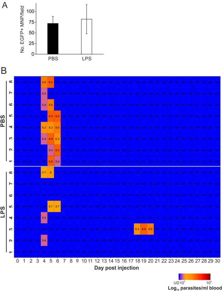
Infections with Trypanosoma brucei sp. are established after the injection of metacyclic trypomastigotes into the skin dermis by the tsetse fly vector. The parasites then gain access to the local lymphatic vessels to infect the local draining lymph nodes and disseminate systemically via the bloodstream. Macrophages are considered to play an important role in host protection during the early stage of systemic trypanosome infections. Macrophages are abundant in the skin dermis, but relatively little is known of their impact on susceptibility to intradermal (ID) trypanosome infections. We show that although dermal injection of colony stimulating factor 1 (CSF1) increased the local abundance of macrophages in the skin, this did not affect susceptibility to ID T. brucei infection. However, bacterial LPS-stimulation in the dermis prior to ID trypanosome infection significantly reduced disease susceptibility. In vitro assays showed that LPS-stimulated macrophage-like RAW264.7 cells had enhanced cytotoxicity towards T. brucei, implying that dermal LPS-treatment may similarly enhance the ability of dermal macrophages to eliminate ID injected T. brucei parasites in the skin. A thorough understanding of the factors that reduce susceptibility to ID injected T. brucei infections may lead to the development of novel strategies to help reduce the transmission of African trypanosomes.


Conflict of interest statement

The authors declare no competing interests.

Figures

Figure 1
Figure 1
Enhanced abundance of Csf1r-EGFP + mononuclear phagocytes (MNP) in the dermis of Csf1r-EGFP+ mice after intradermal (ID) injection with CSF1-Fc. (A) Csf1r-EGFP reporter mice were injected ID with CSF1-Fc, or PBS as a control, for three days and 24 h after the final injection the ears were removed and viewed using a 2-photon microscope. Images show EGFP + MNP (green, arrowheads) and background collagen signal (purple) in the dermis. Arrows, auto-fluorescent hair follicles. (B) The numbers of EGFP + MNP in the dermis local to the injection site were significantly increased after CSF1-Fc-treatment when compared to PBS-treated controls (P = 0.001, Student’s t-test; CSF1-Fc, n = 4; PBS, n = 3).
Figure 2
Figure 2
Intradermal CSF1-Fc treatment does not affect susceptibility to ID T. brucei infection. Groups of C57BL/6J mice were injected ID with CSF1-Fc, or PBS as a control, for three consecutive days and 24 h after the final injection the mice were injected ID with a 1 × 105 dose of T. brucei STIB 247 parasites (n = 8 mice/group). Heatmap shows the log10 number of trypanosomes/mL of blood on each day after ID infection. Each row represents an individual mouse. UD = below detection limit (~ 5.4 log10 parasites/mL blood).
Figure 3
Figure 3
ID LPS treatment decreases susceptibility to ID T. brucei infection. (A) Csf1r-EGFP reporter mice were injected ID with LPS, or PBS as a control, and 24 h later the abundance of EGFP + MNP cells in the dermis local to the injection site determined as in Fig. 1. Similar numbers of EGFP+ MNP were observed in the ear following ID LPS or PBS treatment (n = 2–3 mice/group). (B) Groups of C57BL/6 J mice were injected ID with LPS, or PBS as a control, and 24 h later injected with ID with a 1 × 105 dose of T. brucei STIB 247 parasites (n = 8 mice/group). Heatmap shows the log10 number of trypanosomes/mL of blood on each day after ID infection. Each row represents an individual mouse. UD = below detection limit of 5.4 log10 parasites/mL.
Figure 4
Figure 4
Enhanced ability of LPS-stimulated RAW264.7 cells to kill T. brucei. (A) Macrophage-like RAW264.7 cells were stimulated with 10 µg LPS (or PBS as a control), co-cultivated with T. brucei STIB 247 parasites, and the number of viable parasites determined 24 h later. (B,C) Macrophage-like RAW264.7 cells were stimulated with 10 µg LPS and/or 1 mM indolepyruvate, and then co-cultivated with T. brucei STIB 247 parasites. Twenty four hours later (B) nitrite levels in the culture supernatant, and (C) the number of viable parasites were determined. Data are shown as the mean ± SD for 3 wells/group from an individual experiment, and are representative of data from 3 independent experiments. *, P < 0.05; **, P < 0.01; ***, P < 0.001; # below the limit of detection.

Similar articles

Cited by

References

    1. Alfituri OA, et al. Effects of host-derived chemokines on the motility and viability of Trypanosoma brucei. Parasite Immunol. 2019;41:e12609. doi: 10.1111/pim.12609. - DOI - PMC - PubMed
    1. Caljon G, et al. The dermis as a delivery site of Trypanosoma brucei for tsetse flies. PLoS Pathog. 2016;12:e1005744. doi: 10.1371/journal.ppat.1005744. - DOI - PMC - PubMed
    1. Adams ARD. Trypanosiomiasis of stock in Mauritius III. The diagnosis and course of untreated T. vivax infections in domestic animals. Ann. Trop. Med. Parasitol. 1936;30:521–531. doi: 10.1080/00034983.1936.11684957. - DOI
    1. Emery DL, Barry JD, Moloo SK. The appearance of Trypanosoma (Duttonella) vivax in lymph following challenge of goats with infected Glossina moritans moritans. Acta Trop. 1980;37:375–379. - PubMed
    1. Tabel H, Wei G, Bull HJ. Immunosuppression: Cause for failures of vaccines against African trypanosomiases. PlOS Negl. Trop. Dis. 2013;7:e2090. doi: 10.1371/journal.pntd.0002090. - DOI - PMC - PubMed

Publication types

MeSH terms

Substances