

Identification of a peptide motif that potently inhibits two functionally distinct subunits of Shiga toxin
- PMID: 33972673
- PMCID: PMC8111002
- DOI: 10.1038/s42003-021-02068-3
Identification of a peptide motif that potently inhibits two functionally distinct subunits of Shiga toxin
Abstract
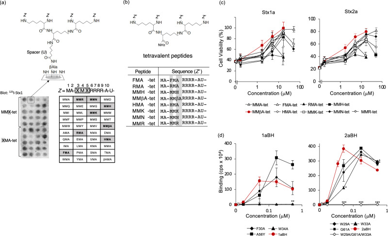
Shiga toxin (Stx) is a major virulence factor of enterohemorrhagic Escherichia coli, which causes fatal systemic complications. Here, we identified a tetravalent peptide that inhibited Stx by targeting its receptor-binding, B-subunit pentamer through a multivalent interaction. A monomeric peptide with the same motif, however, did not bind to the B-subunit pentamer. Instead, the monomer inhibited cytotoxicity with remarkable potency by binding to the catalytic A-subunit. An X-ray crystal structure analysis to 1.6 Å resolution revealed that the monomeric peptide fully occupied the catalytic cavity, interacting with Glu167 and Arg170, both of which are essential for catalytic activity. Thus, the peptide motif demonstrated potent inhibition of two functionally distinct subunits of Stx.
Conflict of interest statement
The authors declare no competing interests.
Figures


Similar articles
-
Identification of a wide range of motifs inhibitory to shiga toxin by affinity-driven screening of customized divalent peptides synthesized on a membrane.Appl Environ Microbiol. 2015 Feb;81(3):1092-100. doi: 10.1128/AEM.03517-14. Epub 2014 Dec 1. Appl Environ Microbiol. 2015. PMID: 25452283 Free PMC article.
-
A multivalent peptide library approach identifies a novel Shiga toxin inhibitor that induces aberrant cellular transport of the toxin.FASEB J. 2006 Dec;20(14):2597-9. doi: 10.1096/fj.06-6572fje. Epub 2006 Oct 25. FASEB J. 2006. PMID: 17065223
-
Identification of a peptide-based neutralizer that potently inhibits both Shiga toxins 1 and 2 by targeting specific receptor-binding regions.Infect Immun. 2013 Jun;81(6):2133-8. doi: 10.1128/IAI.01256-12. Epub 2013 Apr 1. Infect Immun. 2013. PMID: 23545297 Free PMC article.
-
[New drugs that prevent cytotoxicity of Shiga toxins].Nihon Rinsho. 2002 Jun;60(6):1131-7. Nihon Rinsho. 2002. PMID: 12078085 Review. Japanese.
-
[Novel Shiga toxin inhibitor that induces aberrant cellular transport of the toxin].Tanpakushitsu Kakusan Koso. 2008 Jan;53(1):44-51. Tanpakushitsu Kakusan Koso. 2008. PMID: 18186302 Review. Japanese. No abstract available.
Cited by
-
Therapeutic Uses of Bacterial Subunit Toxins.Toxins (Basel). 2021 May 26;13(6):378. doi: 10.3390/toxins13060378. Toxins (Basel). 2021. PMID: 34073185 Free PMC article. Review.
-
Clustered peptide regulating the multivalent interaction between RANK and TRAF6 inhibits osteoclastogenesis by fine-tuning signals.Commun Biol. 2025 Apr 22;8(1):643. doi: 10.1038/s42003-025-08047-2. Commun Biol. 2025. PMID: 40263556 Free PMC article.
-
A unique peptide-based pharmacophore identifies an inhibitory compound against the A-subunit of Shiga toxin.Sci Rep. 2022 Jul 6;12(1):11443. doi: 10.1038/s41598-022-15316-1. Sci Rep. 2022. PMID: 35794188 Free PMC article.
-
Utilizing Adenovirus Knob Proteins as Carriers in Cancer Gene Therapy Amidst the Presence of Anti-Knob Antibodies.Int J Mol Sci. 2024 Oct 3;25(19):10679. doi: 10.3390/ijms251910679. Int J Mol Sci. 2024. PMID: 39409008 Free PMC article.
-
Synthetic phage-based approach for sensitive and specific detection of Escherichia coli O157.Commun Biol. 2024 May 6;7(1):535. doi: 10.1038/s42003-024-06247-w. Commun Biol. 2024. PMID: 38710842 Free PMC article.
References
-
- Tarr PI, Gordon CA, Chandler WL. Shiga-toxin-producing Escherichia coli and haemolytic uraemic syndrome. Lancet. 2005;365:1073–1086. - PubMed
Publication types
MeSH terms
Substances
LinkOut - more resources
Full Text Sources
Other Literature Sources
