Review
. 2021 Jun;17(6):362-379.
doi: 10.1038/s41582-021-00488-7. Epub 2021 May 10.

The importance of ongoing international surveillance for Creutzfeldt-Jakob disease

Affiliations
Review

The importance of ongoing international surveillance for Creutzfeldt-Jakob disease

Neil Watson et al. Nat Rev Neurol. 2021 Jun.

Abstract

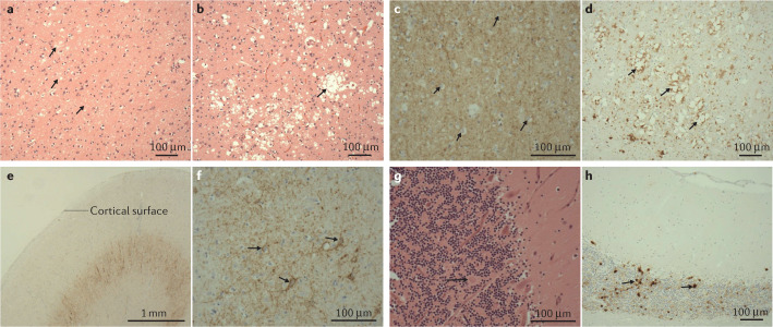
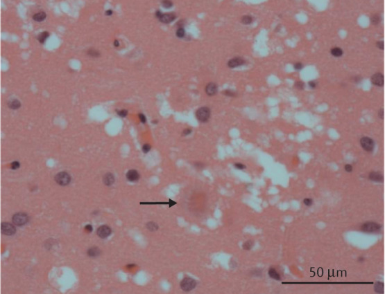
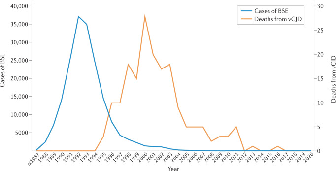
Creutzfeldt-Jakob disease (CJD) is a rapidly progressive, fatal and transmissible neurodegenerative disease associated with the accumulation of misfolded prion protein in the CNS. International CJD surveillance programmes have been active since the emergence, in the mid-1990s, of variant CJD (vCJD), a disease linked to bovine spongiform encephalopathy. Control measures have now successfully contained bovine spongiform encephalopathy and the incidence of vCJD has declined, leading to questions about the requirement for ongoing surveillance. However, several lines of evidence have raised concerns that further cases of vCJD could emerge as a result of prolonged incubation and/or secondary transmission. Emerging evidence from peripheral tissue distribution studies employing high-sensitivity assays suggests that all forms of human prion disease carry a theoretical risk of iatrogenic transmission. Finally, emerging diseases, such as chronic wasting disease and camel prion disease, pose further risks to public health. In this Review, we provide an up-to-date overview of the transmission of prion diseases in human populations and argue that CJD surveillance remains vital both from a public health perspective and to support essential research into disease pathophysiology, enhanced diagnostic tests and much-needed treatments.


Conflict of interest statement

The authors declare no competing interests.

Figures

Fig. 1
Fig. 1. The global burden of vCJD.
Graph shows the annual number of deaths from variant Creutzfeldt–Jakob disease (vCJD) in the 12 countries or territories for which data is available. Data obtained from the European Creutzfeldt–Jakob Disease Surveillance Network (EuroCJD).
Fig. 2
Fig. 2. Brain MRI in individuals with CJD.
Bilateral basal ganglia hyperintensities (arrowheads) are seen in sporadic Creutzfeldt–Jakob disease (CJD) (part a). Multifocal cortical ribboning can be seen in sporadic CJD, visible in the parietal (part b) and interhemispheric (part c) cortex in this individual. Bilateral pulvinar hyperintensities (arrows), known as the pulvinar sign, are seen in variant CJD (part d).
Fig. 3
Fig. 3. Histological features of sCJD.
Histologically, the characteristic feature of prion diseases is spongiform change. Sporadic, genetic and acquired forms of prion disease show different patterns of spongiform change and, in individuals with sporadic Creutzfeldt–Jakob disease (sCJD), PRNP codon 129 genotype and western blot typing of the abnormal prion protein (PrP) also influence the pattern of histological change. In this figure, we provide examples of the histology typically observed in the different forms sCJD. Frontal cortex section from an individual with the MM1 subtype of sCJD showing the fine cortical vacuolation (arrows) that is common in most prion disorders (H&E staining, x100 magnification) (part a). Frontal cortex section from an individual with the MM2 subtype of sCJD showing that fine vacuoles can coalesce to form coarse vacuoles (arrow), a feature particularly prominent in this subtype of sCJD (H&E staining, x100 magnification) (part b). Frontal cortex section from an individual with the MM1 subtype of sCJD showing a predominantly fine synaptic pattern of PrP antibody staining (brown) counterstained with haematoxylin to visualize cell nuclei (blue; x200 magnification) (part c). Frontal cortex section from an individual with the MM2 subtype of sCJD showing coarser PrP antibody staining (brown) that is accentuated around the coalesced vacuoles (arrows; x100 magnification) (part d). Frontal cortex section from an individual with the VV2 subtype of sCJD showing a linear pattern of PrP antibody staining in the deeper cortex (x20 magnification) (part e). A higher magnification image of the same sample shows that the staining in part e is peri-neuronal (frontal cortex section, x200 magnification) (part f). Cerebellar cortex section from an individual with the MV2 subtype of sCJD, showing kuru-like plaques (arrow; H&E staining, x400 magnification) (part g). These plaques (arrows) are easily visualized with PrP antibody staining (brown; cerebellum, x100 magnification) (part h).
Fig. 4
Fig. 4. Western blot typing in CJD.
Samples of brain tissue from individuals with Creutzfeldt–Jakob disease (CJD) can be treated with proteinase K before western blotting to detect protease-resistant fragments of misfolded prion protein (PrPres). PrPres is classified according to the molecular weight of the unglycosylated fragment, which is 21 kDa in type 1 PrPres and 19 kDa in type 2 PrPres. Type 2 is further classified into type 2A and 2B (variant CJD (vCJD)). Type 2B, present in both vCJD and bovine spongiform encephalopathy (BSE), is characterized by a predominant diglycosylated band (*). Examples are shown from individuals with sporadic CJD (sCJD) MM1, sCJD VV2, vCJD and from a case with BSE. MM, homozygous for methionine at codon 12 of the gene encoding prion protein; VV, homozygous for valine at codon 129 of the gene encoding prion protein.
Fig. 5
Fig. 5. Histological features of genetic forms of prion diseases.
Genetic forms of prion diseases show variable histological features and can resemble sporadic Creutzfeldt–Jakob disease; however, some show unique patterns of pathology. Image of a cerebellar section of an individual with the PRNP P102L-129M variant (the most common haplotype) of Gerstmann–Straussler–Scheinker disease showing multicentric plaques (arrows), which are most numerous in the molecular layer and granule cell layer (H&E staining, x200 magnification) (part a). A lower-magnification image from the same individual as in part a showing plaques (arrows) stained with prion protein antibody (brown; x40 magnification) (part b).
Fig. 6
Fig. 6. Histological features of vCJD.
Frontal cortex section from an individual with variant Creutzfeldt–Jakob disease (vCJD) showing a pathognomonic florid plaque (arrow), a fibrillar amyloid plaque surrounded by vacuolation (H&E staining, x400 magnification).
Fig. 7
Fig. 7. BSE and vCJD in the UK.
Graph shows the trajectory of the bovine spongiform encephalopathy (BSE) epizootic and the subsequent variant Creutzfeldt–Jakob disease (vCJD) epidemic in the UK. Data obtained from refs,.
Fig. 8
Fig. 8. A model CJD surveillance system.
A flowchart depicting a model system for comprehensive national Creutzfeldt–Jakob disease (CJD) surveillance, based on a combination of systems used in the authors’ nations, is shown. This system provides a detailed diagnostic assessment including the ascertainment of the CJD subtype, screening for important epidemiological risk factors, and evaluating for and mitigating risks of transmission to others. CSF, cerebrospinal fluid; PMCA, protein misfolding cyclic amplification; RT-QuIC, real-time quaking-induced conversion.

Comment in

  • CJD without borders.
    Cunha JEG, Nascimento JCF, Oliveira JRM. Cunha JEG, et al. Nat Rev Neurol. 2021 Nov;17(11):723. doi: 10.1038/s41582-021-00566-w. Nat Rev Neurol. 2021. PMID: 34561624 No abstract available.

References

    1. National CJD Research & Surveillance Unit. 28th Annual Report 2019. Creutzfeldt-Jakob Disease Surveillance in the UK https://www.cjd.ed.ac.uk/sites/default/files/Report28.pdf (2020).
    1. Uttley L, Carroll C, Wong R, Hilton DA, Stevenson M. Creutzfeldt-Jakob disease: a systematic review of global incidence, prevalence, infectivity, and incubation. Lancet Infect. Dis. 2020;20:e2–e10. doi: 10.1016/S1473-3099(19)30615-2. - DOI - PubMed
    1. Kovacs GG, et al. Genetic prion disease: the EUROCJD experience. Hum. Genet. 2005;118:166–174. doi: 10.1007/s00439-005-0020-1. - DOI - PubMed
    1. Masters CL, et al. Creutzfeldt-Jakob disease: patterns of worldwide occurrence and the significance of familial and sporadic clustering. Ann. Neurol. 1979;5:177–188. doi: 10.1002/ana.410050212. - DOI - PubMed
    1. Brown P, et al. Iatrogenic Creutzfeldt-Jakob disease, final assessment. Emerg. Infect. Dis. 2012;18:901–907. doi: 10.3201/eid1806.120116. - DOI - PMC - PubMed

Publication types