. 2021 Jul 1;162(7):bqab097.
doi: 10.1210/endocr/bqab097.

Ghrelin-induced Food Intake, but not GH Secretion, Requires the Expression of the GH Receptor in the Brain of Male Mice

Affiliations

Ghrelin-induced Food Intake, but not GH Secretion, Requires the Expression of the GH Receptor in the Brain of Male Mice

Frederick Wasinski et al. Endocrinology. .

Abstract

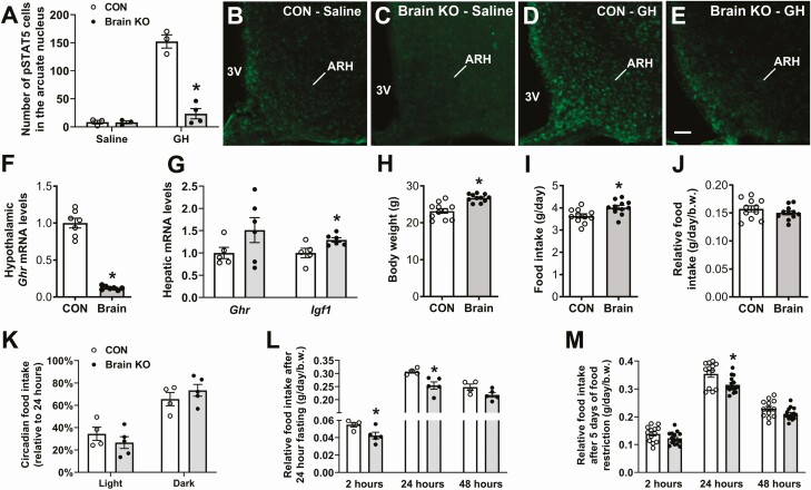
Ghrelin stimulates both GH secretion and food intake. The orexigenic action of ghrelin is mainly mediated by neurons that coexpress agouti-related protein (AgRP) and neuropeptide Y (NPY) in the arcuate nucleus of the hypothalamus (ARH). GH also stimulates food intake and, importantly, ARHAgRP/NPY neurons express GH receptor (GHR). Thus, ghrelin-induced GH secretion may contribute to the orexigenic effect of ghrelin. Here, we investigated the response to ghrelin in male mice carrying GHR ablation specifically in neurons (brain GHR knockout [KO] mice) or exclusively in ARHAgRP/NPY neurons (AgRP GHR KO mice). Although brain GHR KO mice showed normal ghrelin-induced increase in plasma GH levels, these mutants lacked the expected orexigenic response to ghrelin. Additionally, brain GHR KO mice displayed reduced hypothalamic levels of Npy and Ghsr mRNA and did not elicit ghrelin-induced c-Fos expression in the ARH. Furthermore, brain GHR KO mice exhibited a prominent reduction in AgRP fiber density in the ARH and paraventricular nucleus of the hypothalamus (PVH). In contrast, AgRP GHR KO mice showed no changes in the hypothalamic Npy and Ghsr mRNAs and conserved ghrelin-induced food intake and c-Fos expression in the ARH. AgRP GHR KO mice displayed a reduced AgRP fiber density (~16%) in the PVH, but this reduction was less than that observed in brain GHR KO mice (~61%). Our findings indicate that GHR signaling in the brain is required for the orexigenic effect of ghrelin, independently of GH action on ARHAgRP/NPY neurons.

Keywords: cytokines; energy balance; growth hormone receptor; hypothalamus.


Figures

Figure 1.
Figure 1.
Characterization of Brain GHR KO male mice. (A-E) STAT5 phosphorylation (pSTAT5) in the arcuate nucleus of the hypothalamus (ARH) of saline-injected control mice (B), saline-injected brain GHR KO mice (C), GH-injected control mice (D), and GH-injected brain GHR KO mice (E; n = 3-4/group). Abbreviation: 3V, third ventricle. Scale bar = 50 µm. (F) Ghr mRNA levels in the hypothalamus (n = 7/group). (G) Ghr and Igf1 mRNA levels in the liver (n = 5-6/group). (H) Body weight in 3-month-old male mice (n = 11/group). (I-J) Absolute and relative to body weight food intake (n = 11/group). (K) Percentage of food intake relative to 24 hours in the light and dark cycles (n = 4-5/group). (L) Food intake after a 24-hour fasting period (n = 4-5/group). (M) Refeeding response after 5 days of 60% food restriction (n = 13-15/group). *Significantly different compared with control mice (P < 0.05).
Figure 2.
Figure 2.
Brain GHR KO mice are unresponsive to ghrelin-induced food intake. (A) Area under the curve of plasma GH levels in control (n = 6) and brain GHR KO (n = 7) mice that received saline or ghrelin IP injections. (B) Cumulative food intake in control (n = 18) and brain GHR KO (n = 17) mice that received saline or ghrelin IP injections. (C-G). c-Fos expression in the ARH of control (n = 6) and brain GHR KO (n = 7) mice that received saline or ghrelin IP injections. Abbreviations: 3V, third ventricle; GHR, GH receptor; KO, knockout. Scale bar = 100 µm. *Significantly different compared with control mice (P < 0.05).
Figure 3.
Figure 3.
Reduced expression of NPY and ghrelin receptor in the hypothalamus of brain GHR KO mice. Relative mRNA levels in the hypothalamus of control (n = 8) and brain GHR KO (n = 7) mice. *Significantly different compared to control mice (P < 0.05). Abbreviations: GHR, GH receptor; KO, knockout; NPY, neuropeptide Y.
Figure 4.
Figure 4.
Reduced AgRP innervation in the hypothalamus of brain-specific GHR KO mice. (A-F) Immunostaining and quantification of AgRP fluorescent signal in the ARH and paraventricular nucleus of the hypothalamus (PVH) of control (n = 4) and brain GHR KO (n = 4) mice. (G-I) Immunostaining and quantification of POMC fluorescent signal in the PVH of control (n = 4) and brain GHR KO (n = 4) mice. Abbreviations: 3V, third ventricle: AgRP, agouti-related protein; GHR, GH receptor; KO, knockout. Scale bar = 100 µm. *Significantly different compared to control mice (P < 0.05).
Figure 5.
Figure 5.
Characterization of AgRP GHR KO male mice. (A-D) Colocalization of STAT5 phosphorylation (green) and AgRP neurons (magenta) in the ARH of GH-injected control and AgRP GHR KO mice (n = 3/group). Double-labeled cells appear in white. Note that AgRP fibers abundantly innervate the PVH. Scale bar = 50 µm. (E) Body weight in control (n = 11) and AgRP GHR KO (n = 8) male mice. (F) Food intake in control (n = 12) and AgRP GHR KO (n = 11) mice. (G) Food intake after a 24-hour fasting period (n = 3-4/group). (H) Refeeding response after 5 days of 60% food restriction (n = 15/group). *Significantly different compared with control mice (P < 0.05). Abbreviations: 3V, third ventricle; AgRP, agouti-related protein; ARH, arcuate nucleus of the hypothalamus; GHR, GH receptor; KO, knockout.
Figure 6.
Figure 6.
AgRP GHR KO mice exhibit reduced AgRP innervation in the hypothalamus. (A) Relative mRNA levels in the hypothalamus of control and AgRP GHR KO mice (n = 6/group). (B-G) Immunostaining and quantification of AgRP fluorescent signal in the ARH and PVH of control (n = 3) and AgRP GHR KO (n = 4) mice. Abbreviations: 3V, third ventricle; AgRP, agouti-related protein; GHR, GH receptor; KO, knockout. Scale bar = 100 µm. *Significantly different compared with control mice (P < 0.05).
Figure 7.
Figure 7.
AgRP GHR KO mice display normal orexigenic response to ghrelin. (A) Cumulative food intake in control (n = 19) and AgRP GHR KO (n = 18) mice that received saline or ghrelin IP injections. (B-D) c-Fos expression in the ARH of control and AgRP GHR KO mice that received ghrelin injection (n = 3/group). Abbreviations: 3V, third ventricle; AgRP, agouti-related protein; GHR, GH receptor; KO, knockout. Scale bar = 50 µm. *Significantly different compared with saline-injected mice (P < 0.05).

Similar articles

Cited by

References

    1. Kojima M, Hosoda H, Date Y, Nakazato M, Matsuo H, Kangawa K. Ghrelin is a growth-hormone-releasing acylated peptide from stomach. Nature. 1999;402(6762):656-660. - PubMed
    1. Müller TD, Nogueiras R, Andermann ML, et al. Ghrelin. Mol Metab. 2015;4(6):437-460. - PMC - PubMed
    1. Veldhuis JD, Reynolds GA, Iranmanesh A, Bowers CY. Twenty-four hour continuous ghrelin infusion augments physiologically pulsatile, nycthemeral, and entropic (feedback-regulated) modes of growth hormone secretion. J Clin Endocrinol Metab. 2008;93(9):3597-3603. - PMC - PubMed
    1. Arvat E, Di Vito L, Broglio F, et al. Preliminary evidence that Ghrelin, the natural GH secretagogue (GHS)-receptor ligand, strongly stimulates GH secretion in humans. J Endocrinol Invest. 2000;23(8):493-495. - PubMed
    1. Peino R, Baldelli R, Rodriguez-Garcia J, et al. Ghrelin-induced growth hormone secretion in humans. Eur J Endocrinol. 2000;143(6):R11-R14. - PubMed

Publication types

MeSH terms