. 2021 Aug;37(8):718-729.
doi: 10.1002/kjm2.12390. Epub 2021 May 11.

Astragaloside alleviates alcoholic fatty liver disease by suppressing oxidative stress

Affiliations

Astragaloside alleviates alcoholic fatty liver disease by suppressing oxidative stress

Zhi-Bin Jiang et al. Kaohsiung J Med Sci. 2021 Aug.

Abstract

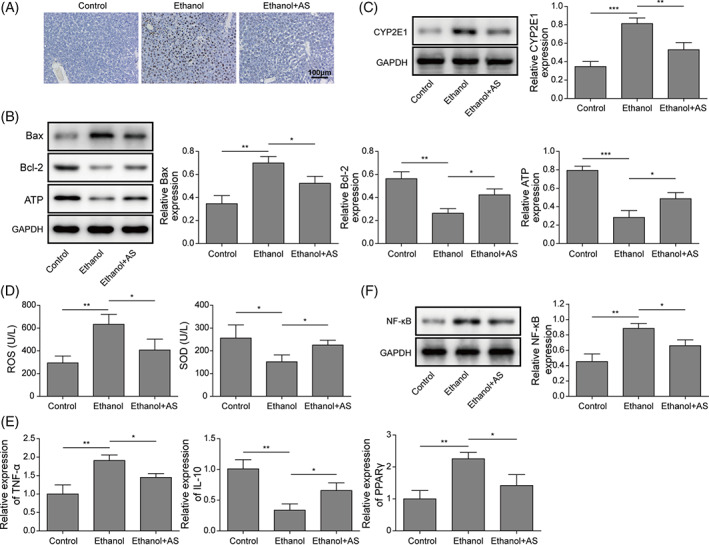
Alcoholic fatty liver disease (AFLD) is the most common liver disease and can progress to fatal liver cirrhosis and carcinoma, affecting millions of patients worldwide. The functions of astragaloside on the cardiovascular system have been elucidated. However, its role in AFLD is unclear. Ethanol-treated AML-12 cells were used as a cell model of alcoholic fatty liver. Real-time quantitative reverse transcription-PCR and Western blotting detected genes and proteins expressions. Reactive oxygen species (ROS), triglyceride, total cholesterol, low-density lipoprotein, albumin, ferritin, bilirubin, superoxide dismutase, aspartate aminotransferase (AST), and alanine aminotransferase (ALT) were examined using commercial kits. Lipid accumulation was assessed by Oil red O staining. MTT and flow cytometry measured cell viability and apoptosis. JC-1 was used to analyze mitochondrial membrane potential. A rat model of AFLD was established by treating rats with ethanol. Astragaloside suppressed ethanol-induced lipid accumulation, oxidative stress, and the production of AST and ALT in AML-12 cells. Ethanol induced TNF-α and reduced IL-10 expression, which were reversed by astragaloside. Ethanol promoted Bax expression and cytochrome C release and inhibited Bcl-2 and ATP expression. Astragaloside hampered these apoptosis effects in AML-12 cells. Impaired mitochondrial membrane potential was recovered by astragaloside. However, all these astragaloside-mediated beneficial effects were abolished by the ROS inducer pyocyanin. Ethanol-induced activation of NF-κB signaling was suppressed by astragaloside in vitro and in vivo, suggesting that astragaloside inhibited oxidative stress by suppressing the activation of NF-κB signaling, thus improving liver function and alleviating AFLD in rats. Our study elucidates the pharmacological mechanism of astragaloside and provides potential therapeutic strategies for AFLD.

Keywords: alcoholic fatty liver disease; astragaloside; lipid accumulation; oxidative stress.


Conflict of interest statement

The authors declare that there are no conflicts of interest.

Figures

FIGURE 1
FIGURE 1
Astragaloside attenuated alcohol‐induced lipid accumulation, inflammation, and reactive oxygen species (ROS) generation in AML‐12 cells. AML‐12 cells were treated with ethanol (0, 75, 100, 150, or 200 mM) and astragaloside (0, 50, 75, 100, or 200 μg/ml) for 48 h. (A) Cell viability was tested by MTT assay (n = 3). AML‐12 cells were treated with ethanol (200 mM) and astragaloside (200 μg/ml, ethanol + AS) or ethanol alone (ethanol) for 48 h. Untreated AML‐12 cells (control) were used as controls. (B) Lipid accumulation was detected by oil red O staining (n = 3). (C) Determination of TG, AST, and ALT levels in different groups (n = 3). (D) Analysis of ROS and superoxide dismutase (SOD) levels in different groups (n = 3). (E) RT‐qPCR was used to determine the relative expression of TNF‐α, IL‐10, and PPARγ in different groups (n = 3). (F) Western blotting detected the expression of NF‐κB (n = 3). GAPDH was used as a normalization control. The results were from at least three independent experiments. *p < 0.05, **p < 0.01, and ***p < 0.001
FIGURE 2
FIGURE 2
Astragaloside suppressed alcohol‐induced cell apoptosis. AML‐12 cells were treated with 200 mM ethanol alone or 200 mM ethanol and 200 μg/ml astragaloside for 48 h. Untreated AML‐12 cells were used as controls. (A) Mitochondrial membrane potential was tested by JC‐1 staining. Here, 50 μM CCCP was used to treat AML‐12 cells as a positive control, and the untreated group served as a negative control (n = 3). (B) Flow cytometry analysis of cell apoptosis (n = 3). (C) Western blot analysis of the expression of Bax, Bcl‐2, and cytoplasmic and mitochondrial Cyt‐C (n = 3). GAPDH and cox‐IV were used as normalization controls. The results were from at least three independent experiments. *p < 0.05, **p < 0.01, and ***p < 0.001
FIGURE 3
FIGURE 3
Pyocyanin reversed the protective effects of astragaloside on alcohol‐induced AML‐12. AML‐12 cells were untreated or treated with ethanol alone, ethanol and astragaloside, or ethanol and astragaloside in combination with 50 μM pyocyanin for 48 h. (A) Oil red O staining (n = 3). (B) Determination of TG, AST, and ALT levels (n = 3). (C) Determination of reactive oxygen species (ROS) and superoxide dismutase (SOD) levels (n = 3). (D) RT‐qPCR was used to analyze the expression of TNF‐α, IL‐10, and PPARγ (n = 3). (E) Western blot analysis of the expression of NF‐κB (n = 3). GAPDH was used as a normalization control. The results were from at least three independent experiments. *p < 0.05, **p < 0.01, and ***p < 0.001
FIGURE 4
FIGURE 4
Pyocyanin reversed astragaloside‐mediated inhibition of cell apoptosis. AML‐12 cells were untreated or treated with ethanol alone, ethanol and astragaloside, or ethanol and astragaloside in combination with pyocyanin for 48 h. (A) Mitochondrial membrane potential was detected by JC‐1 staining (n = 3). (B) Cell apoptosis was determined by Annexin V and propidium iodide staining (n = 3). (C) Western blotting was used to examine the expression of Bax, Bcl‐2, and cytoplasmic and mitochondrial Cyt‐C (n = 3). GAPDH and cox‐IV were used as normalization controls. The results were from at least three independent experiments. *p < 0.05, **p < 0.01, and ***p < 0.001
FIGURE 5
FIGURE 5
Astragaloside alleviated alcoholic fatty liver disease in vivo. An alcoholic fatty liver disease (AFLD) rat model was established with ethanol alone or ethanol and astragaloside treatment. (A) Hematoxylin and eosin (H&E) staining of rat liver tissues (n = 8 per group). (B) Oil red O staining of rat liver tissues (n = 8 per group). (C) Rat body weight monitoring (n = 8 per group). (D) Blood alcohol concentration (n = 8 per group). (E) The measurement of TG, TC, bilirubin, LDL, ALB, and ferritin in rats (n = 8 per group). The results were from at least three independent experiments. *p < 0.05, **p < 0.01, and ***p < 0.001
FIGURE 6
FIGURE 6
Astragaloside alleviated alcoholic fatty liver disease (AFLD) by suppressing NF‐κB signaling in rats. (A) TUNEL staining of rat liver tissues (n = 8 per group). (B) Western blot analysis of Bax, Bcl‐2, and ATP expression. (C) Western blot analysis of CYP2E1 expression (n = 8 per group). GAPDH was used as a normalization control. (D) Determination of reactive oxygen species (ROS) and superoxide dismutase (SOD) levels in rat liver tissues (n = 8 per group). (E) TNF‐α, IL‐10, and PPARγ expression was detected by RT‐qPCR (n = 8 per group). (F) Western blot analysis of the expression of NF‐κB (n = 8 per group). GAPDH was used as a normalization control. The results were from at least three independent experiments. *p < 0.05, **p < 0.01, and ***p < 0.001

Similar articles

Cited by

References

    1. Liu J. Ethanol and liver: recent insights into the mechanisms of ethanol‐induced fatty liver. World J Gastroenterol. 2014;20(40):14672–85. - PMC - PubMed
    1. Feldstein AE, Bailey SM. Emerging role of redox dysregulation in alcoholic and nonalcoholic fatty liver disease. Antioxid Redox Signal. 2011;15(2):421–4. - PMC - PubMed
    1. Rakha EA, Adamson L, Bell E, Neal K, Ryder SD, Kaye PV, et al. Portal inflammation is associated with advanced histological changes in alcoholic and non‐alcoholic fatty liver disease. J Clin Pathol. 2010;63(9):790–5. - PubMed
    1. Cederbaum AI, Lu Y, Wu D. Role of oxidative stress in alcohol‐induced liver injury. Arch Toxicol. 2009;83(6):519–48. - PubMed
    1. Meagher EA, Barry OP, Burke A, Lucey MR, Lawson JA, Rokach J, et al. Alcohol‐induced generation of lipid peroxidation products in humans. J Clin Invest. 1999;104(6):805–13. - PMC - PubMed

MeSH terms