. 2021 May 15;22(5):348-365.
doi: 10.1631/jzus.B2000441.

Protective effect of recombinant Lactobacillus plantarum against H2O2-induced oxidative stress in HUVEC cells

Affiliations

Protective effect of recombinant Lactobacillus plantarum against H2O2-induced oxidative stress in HUVEC cells

Guan Wang et al. J Zhejiang Univ Sci B. .

Abstract

This study probed the protective effect of recombinant Lactobacillus plantarum against hydrogen peroxide (H2O2)-induced oxidative stress in human umbilical vein endothelial cells (HUVECs). We constructed a new functional L. plantarum (NC8-pSIP409-alr-angiotensin-converting enzyme inhibitory peptide (ACEIP)) with a double-gene-labeled non-resistant screen as an expression vector. A 3-(4,5-dimethyl-2-thiazolyl)-2,5-diphenyl-2H-tetrazolium bromide (MTT) colorimetric assay was carried out to determine the cell viability of HUVEC cells following pretreatment with NC8-pSIP409-alr-ACEIP. Flow cytometry (FCM) was used to determine the apoptosis rate of HUVEC cells. Cysteinyl aspartate specific proteinase (caspase)-3/8/9 activity was also assayed and western blotting was used to determine protein expression of B-cell lymphoma 2 (Bcl-2), Bcl-2-associated X protein (Bax), inducible nitric oxide synthase (iNOS), nicotinamide adenine dinucleotide phosphate oxidase 2 (gp91phox), angiotensin II (AngII), and angiotensin-converting enzyme 2 (ACE2), as well as corresponding indicators of oxidative stress, such as reactive oxygen species (ROS), mitochondrial membrane potential (MMP), malondialdehyde (MDA), and superoxide dismutase (SOD). NC8-pSIP409-alr-ACEIP attenuated H2O2-induced cell death, as determined by the MTT assay. NC8-pSIP409-alr-ACEIP reduced apoptosis of HUVEC cells by FCM. In addition, compared to the positive control, the oxidative stress index of the H2O2-induced HUVEC (Hy-HUVEC), which was pretreated by NC8-pSIP409-alr-ACEIP, iNOS, gp91phox, MDA, and ROS, was decreased obviously; SOD expression level was increased; caspase-3 or -9 was decreased, but caspase-8 did not change; Bcl-2/Bax ratio was increased; permeability changes of mitochondria were inhibited; and loss of transmembrane potential was prevented. Expression of the hypertension-related protein (AngII protein) in HUVEC cells protected by NC8-pSIP409-alr-ACEIP decreased and expression of ACE2 protein increased. These plantarum results suggested that NC8-pSIP409-alr-ACEIP protects against H2O2-induced injury in HUVEC cells. The mechanism for this effect is related to enhancement of antioxidant capacity and apoptosis.

Keywords: Apoptosis; Human umbilical vein endothelial cell (HUVEC); Hydrogen peroxide (H 2 O 2); Oxidative stress.


Figures

Fig. 1
Fig. 1. Schematic diagram of NC8-pSIP409-alr-ACEIP construction and expression. (a) Schematic diagram of ACEIP recombinant expression vector. (b) Agarose gel electrophoresis of plasmid. Plasmid digested by restriction enzymes plaNcoI and BamHI (Lane 1) and undigested plasmid (Lane 2) were run on agarose gel and expected bands are shown (5808 bp for vector and 1988 bp for target gene). (c) Western blot determined that ACEIP fusion protein was successfully expressed in NC8-pSIP409-alr. (d) Growth curve of recombinant Lactobacillus plantarum NC8-pSIP409-alr-ACEIP. ACEIP: angiotensin-converting enzyme inhibitory peptide; OD600: optical density at 600 nm.
Fig. 2
Fig. 2. Effects of H2O2 on HUVEC cell growth. Cells were seeded at a density of 7000 cells/well and then treated with 100, 200, 300, 400, 500, 600, 700, and 800 μmol/L H2O2 for 1 h (a) or 3 h (b). The control group was treated with RPMI-1640. Cell viability was then determined by MTT assay. Data are presented as mean±standard deviation (SD) with three independent experiments. ** P<0.01, *** P<0.001, compared with the control.
Fig. 3
Fig. 3. Effects of NC8-alr (a) or NC8-pSIP409-alr-ACEIP (b) on HUVEC cell growth. Cells were seeded at a density of 7000 cells/well and then treated at bacterium/cell ratios of 20:1, 40:1, 80:1, 160:1, and 320:1 for 1 or 2 h. The control group was treated with RPMI-1640. Cell viability was then determined by MTT assay. Data are presented as mean±standard deviation (SD) with three independent experiments. ** P<0.01, compared with the control. ACEIP: angiotensin-converting enzyme inhibitory peptide.
Fig. 4
Fig. 4. Cell viabilities of Hy-HUVEC treated with NC8-alr (a) or NC8-pSIP409-alr-ACEIP (b) for 1 or 2 h determined by MTT assay. NC8-pSIP409-alr-ACEIP promotes proliferation of Hy-HUVEC cells. Cells were plated in the 96-well plates at 7000 cells/well. Next, the cells were treated with bacterium/Hy-HUVEC ratios of 20:1, 40:1, 80:1, 160:1, and 320:1 for 1 and 2 h, while the control group was treated with RPMI-1640. Data are presented as mean±standard deviation (SD) with three independent experiments. * P<0.05, ** P<0.01, *** P<0.001, compared with the positive H2O2 control group. ACEIP: angiotensin-converting enzyme inhibitory peptide; Hy-HUVEC: H2O2-induced human umbilical vein endothelial cell.
Fig. 5
Fig. 5. Flow cytometric analysis of apoptotic Hy-HUVEC cells induced by NC8-pSIP409-alr-ACEIP. Hy-HUVEC cells were treated with NC8-pSIP409-alr-ACEIP (a) or NC8-alr (b), which was added at bacterium/cell ratios of 20:1, 40:1, 80:1, 160:1, and 320:1 for 2 h. FITC: fluorescein isothiocyanate; PI: propidium iodide; ACEIP: angiotensin-converting enzyme inhibitory peptide.
Fig. 6
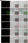
Fig. 6. Analysis of ROS levels in Hy-HUVEC cells with NC8-pSIP409-alr-ACEIP treatment. ROS generation in the control and Hy-HUVEC cells was assayed with DCFH-DA. Fluorescence intensity of the oxidation-insensitive probed DCF-DA in control and Hy-HUVEC cells was measured. Cells were examined under a fluorescence microscope. The large images and small images were observed under field of view of 5× and 20× magnifications (scale bar=100 μm), respectively. (a) Control group; (b) H2O2 positive control group; (c) NC8-alr group; (d) NC8-pSIP409-alr-ACEIP group; (e) NC8-alr+H2O2 group; (f) NC8-pSIP409-alr-ACEIP+H2O2 group. When ROS levels were increased in cells, the DCF signal was increased and the intensity of green fluorescence was enhanced. The intensity of the green fluorescence can be regarded as an indicator of ROS level. Hy-HUVEC: H2O2-induced human umbilical vein endothelial cell; ACEIP: angiotensin-converting enzyme inhibitory peptide; ROS: reactive oxygen species; DCFH-DA: 2',7'-dichlorofluorescein diacetate (no fluorescence); DCF-DA: 2',7'-dichlorofluorescein diacetate (with fluorescence) (Note: for interpretation of the references to color in this figure legend, the reader is referred to the web version of this article).
Fig. 7
Fig. 7. Effects of NC8-pSIP409-alr-ACEIP on MMP in Hy-HUVEC cells. Cells treated with NC8-pSIP409-alr-ACEIP (bacterium/cell ratio: 160:1) were incubated with JC-10, and their fluorescence intensity was measured. (a) Control group; (b) H2O2 positive control group; (c) NC8-alr group; (d) NC8-pSIP409-alr-ACEIP group; (e) NC8-alr+H2O2 group; (f) NC8-pSIP409-alr-ACEIP+H2O2 group. There was a significant difference in fluorescence between the H2O2-treated cells (b) and the NC8-pSIP409-alr-ACEIP-treated cells (f). Hy-HUVEC: H2O2-induced human umbilical vein endothelial cell; ACEIP: angiotensin-converting enzyme inhibitory peptide; MMP: mitochondrial membrane potential. Scale bar=100 μm (Note: for interpretation of the references to color in this figure legend, the reader is referred to the web version of this article).
Fig. 8
Fig. 8. Effects of NC8-pSIP409-alr-ACEIP on anti-oxidation-related indicators. (a) SOD activity in Hy-HUVEC cells. (b) MDA content of Hy-HUVEC cells. (c) Protein profiles of iNOS, gp91phox, and β-actin. β-Actin used as an internal control, and the induced cells were harvested by centrifugation at 1500 r/min for 3 min at 4 °C and suspended in 50 mmol/L PBS, followed by sonic disruption. The cell-free extract was analyzed on 12% SDS-PAGE and subjected to western blotting using iNOS rabbit polyclonal antibody, gp91phox rabbit polyclonal antibody, and β-actin mouse monoclonal antibody. (d) The ratio of iNOS protein to β-actin. (e) The ratio of gp91phox protein to β-actin. Results are expressed as mean±SEM (n=3). *** P<0.001, compared with the H2O2 positive control group, according to ANOVA. Hy-HUVEC: H2O2-induced human umbilical vein endothelial cell; ACEIP: angiotensin-converting enzyme inhibitory peptide; SOD: superoxide dismutase; MDA: malondialdehyde; iNOS: inducible nitric oxide synthase; gp91phox: nicotinamide adenine dinucleotide phosphate oxidase 2; SDS-PAGE: sodium dodecyl sulfate-polyacrylamide gel electrophoresis; SEM: standard error of the mean; ANOVA: analysis of variance.
Fig. 9
Fig. 9. Detection of HUVEC cell apoptosis-related protein expression and activity by NC8-pSIP409-alr-ACEIP. (a) Protein profiles of Bcl-2, Bax, and β-actin. β-Actin was used as an internal control, and the induced cells were harvested by centrifugation at 1500 r/min for 3 min at 4 °C and were suspended in 50 mmol/L PBS, followed by sonic disruption. The cell-free extract was analyzed on 12% SDS-PAGE and subjected to western blotting using Bal-2 rabbit polyclonal antibody, Bax rabbit polyclonal antibody, and β-actin mouse monoclonal antibody. (b) The ratio of Bcl-2 to β-actin. (c) The ratio of Bax to β-actin. (d) The ratio of Bcl-2 to Bax. (e‒g) Equal amounts of cell lysates were assayed for caspase-3 (e), -8 (f), and -9 (g) activity using DEVD-pNA, IETD-pNA, and LEHD-pNA as substrates, respectively. The concentrations of fluorescent products released were measured immediately at 405 nm using a microplate reader. Results represent the mean±standard deviation (SD) of triplicate assays. *** P<0.001, compared with the H2O2 positive control group, according to ANOVA. ACEIP: angiotensin-converting enzyme inhibitory peptide; Bcl-2: B-cell lymphoma 2; Bax: Bcl-2-associated X protein; SDS-PAGE: sodium dodecyl sulfate-polyacrylamide gel electrophoresis; Ac-DEVD-pNA: acetyl-Asp-Glu-Val-Asp-p-nitroanilide; Ac-IETD-pNA: acetyl-Ile-Glu-Thr-Asp-p-nitroanilide; Ac-LEHD-pNA: acetyl-Leu-Glu-His-Asp-p-nitroanilide; ANOVA: analysis of variance.
Fig. 10
Fig. 10. Effects of NC8-pSIP409-alr-ACEIP on AngII and ACE2 protein expression levels. (a) Protein profiles of AngII, ACE2, and β-actin. β-Actin was used as an internal control, and the induced cells were harvested by centrifugation at 1500 r/min for 3 min at 4 °C and were suspended in 50 mmol/L PBS, followed by sonic disruption. The cell-free extract was analyzed on 15% SDS-PAGE and subjected to western blotting using angiopoietin 2 rabbit polyclonal antibody, ACE2 rabbit polyclonal antibody, and β-actin mouse monoclonal antibody. (b) The ratio of AngII to β-actin. (c) The ratio of ACE2 to β-actin. Results represent the mean±standard deviation (SD) of triplicate assays. *** P<0.001, compared with the H2O2 positive control group according to ANOVA. ACEIP: angiotensin-converting enzyme inhibitory peptide; SDS-PAGE: sodium dodecylsulfate-polyacrylamide gel electrophoresis; ACE2: angiotensin-converting enzyme 2; ANOVA: analysis of variance.

Similar articles

Cited by

References

    1. Aldabbous L, Abdul-Salam V, McKinnon T, et al. , 2016. Neutrophil extracellular traps promote angiogenesis. Arterioscler Thromb Vasc Biol, 36(10): 2078-2087. 10.1161/ATVBAHA.116.307634 - DOI - PubMed
    1. Bali A, Singh N, Jaggi AS, 2014. Renin-angiotensin system in pain: existing in a double life? J Renin Angiotensin Aldosterone Syst, 15(4): 329-340. 10.1177/1470320313503694 - DOI - PubMed
    1. Białas AJ, Sitarek P, Miłkowska-Dymanowska J, et al. , 2016. The role of mitochondria and oxidative/antioxidative imbalance in pathobiology of chronic obstructive pulmonary disease. Oxid Med Cell Longev, 2016: 7808576. 10.1155/2016/7808576 - DOI - PMC - PubMed
    1. Bian YF, Yang HY, Yang ZM, et al. , 2011. Amlodipine treatment prevents angiotensin II-induced human umbilical vein endothelial cell apoptosis. Arch Med Res, 42(1): 22-27. 10.1016/j.arcmed.2011.01.012 - DOI - PubMed
    1. Camini FC, da Silva Caetano CC, Almeida LT, et al. , 2017. Oxidative stress in Mayaro virus infection. Virus Res, 236: 1-8. 10.1016/j.virusres.2017.04.017 - DOI - PubMed

MeSH terms