. 2021 Nov;53(9):1208-1219.
doi: 10.1002/lsm.23398. Epub 2021 May 11.

Low-Level Light Therapy Downregulates Scalp Inflammatory Biomarkers in Men With Androgenetic Alopecia and Boosts Minoxidil 2% to Bring a Sustainable Hair Regrowth Activity

Affiliations

Low-Level Light Therapy Downregulates Scalp Inflammatory Biomarkers in Men With Androgenetic Alopecia and Boosts Minoxidil 2% to Bring a Sustainable Hair Regrowth Activity

Yann F Mahe et al. Lasers Surg Med. 2021 Nov.

Abstract

Background and objectives: Low-level light therapies using visible to infrared light are known to activate several cellular functions, such as adenosine triphosphate and nitric oxide synthesis. However, few clinical observations report its biological consequences for skin and scalp homeostasis. Since scalp inflammation was recognized as a potential physiological obstacle to the efficacy of the reference hair regrowth drug Minoxidil in vivo and since perifollicular inflammation is the hallmark of about 50%-70% follicular units in androgenetic alopecia, we decided to investigate whether the anti-inflammatory activity of LLLT/GentleWaves® device were assigned to L'Oréal by Light BioScience L.L.C., Virginia Beach, VA (US) could enhance hair regrowth activity of Minoxidil.

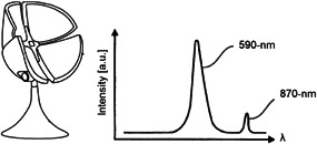
Study design/materials and methods: We conducted a first experimental clinical study on 64 men with androgenetic alopecia using LLLT/GentleWaves®, 590-nm predominant wavelength 70 seconds, specifically pulsed once per day, for 3 days, and we performed a whole-genome analysis of treated scalp biopsies. In a second clinical study, including 135 alopecic volunteers, we evaluated the hair regrowth activity in response to the upgraded LLLT/GentleWaves® device and Minoxidil.

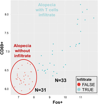
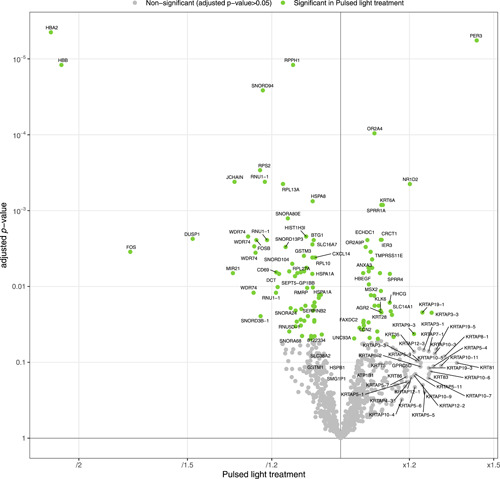
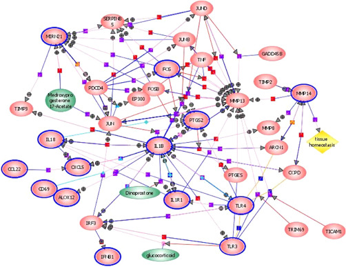
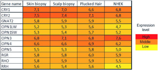
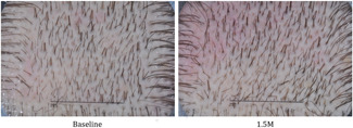
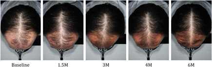
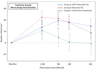
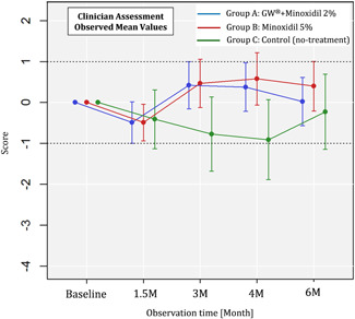
Results: In the first clinical study, whole-genome analysis of treated scalp biopsies showed downregulation of scalp inflammatory biomarkers, such as AP1/FOSB messenger RNA (mRNA) and mir21, together with the disappearance of CD69 mRNA, specific to scalp-infiltrating T cells of about 50% of the studied volunteers prior to the LLLT/GentleWaves® treatment. In the second clinical study, we observed that LLLT/GentleWaves® was able to boost the hair regrowth activity of a Minoxidil 2% lotion to the extent of the highest concentration (5%) in terms of efficacy, number of responders, and perceived performance.

Conclusions: Altogether, these observations suggest the potential benefit of LLLT/GentleWaves® as a noninvasive adjunctive technology for skin and scalp conditions, where a mild perifollicular inflammation is involved. Lasers Surg. Med. 2021. Copyright © 2021 Wiley Periodicals LLC.

Keywords: LLLT; Minoxidil; androgenetic alopecia; hair regrowth; inflammation; perifollicular fibrosis; whole genome.


Figures

Figure 1
Figure 1
Cartoon of the upgraded GentleWaves® device and its emission spectrum with a predominant peak at 590 nm (yellow‐orange) and a secondary peak at 870 nm (infrared) (cfr. EP2912509B1, EP2861203B1).
Figure 2
Figure 2
Diagram of co‐expression of inflammatory infiltrates (CD69+) and inflammatory status (FOS+) in the human scalp in vivo in 50% of the androgenetic alopecia men volunteers enrolled in the study (in cyan). Two groups and hence two stages of alopecia (i.e., inflammatory vs noninflammatory) can clearly be distinguished (red and cyan, respectively). Values represent the gene expression levels, as estimated by the Affymetrix arrays.
Figure 3
Figure 3
Detection of GentleWaves® significantly downregulated (left panel) and moderately upregulated (right panel) genes in vivo in the scalp from men with androgenetic alopecia based on transcriptional whole expression study. HBA2 and HBB (hemoglobin A2 and hemoglobin B), FOS, mir21, CD69+, and DUSP1 are the most downregulated genes, while keratins are slightly induced by GentleWaves®. Fold changes after pulsed light treatment (x axis) are expressed in log2 base, which means, for example, that Hba and Hbb are decreased more than 22.2 (i.e., divided by 4.4 = ×0.22); FOS is downregulated 21.7 (i.e., divided by 3.4 = ×0.29); CD69+ decreased by 21 (i.e., divided by 2 = ×0.5), etc. Genes in green were deemed significant by the statistical model.
Figure 4
Figure 4
Interactive map of inflammatory genes based on transcriptional whole expression study in 3.6‐mm Ø scalp biopsies and QRT‐PCR expression on cultured cells in vitro and in vivo: the not downregulated genes are shown in red ellipses, modulating molecules in green ellipses, and the genes downregulated in red ellipses circled in blue. The fold changes obtained from RT/PCR studies are as follows CCL22 (×0.45); IL1B (×0.19); IL1R1 (×0.36); 12Lipoxygénase (×0.38); MMP14 (×0.23); CXCL4 (×0.41); PTGD2S (×0.32); PTGS2 (×0.25); TLR3 (×0.16); TLR4 (×0.18); INFB1 (×0.47).
Figure 5
Figure 5
Comparative expression of photoreceptors in human skin and hair and normal human epidermal keratinocytes (NHEK). CRY1, cryptochrome circadian regulator 1; CRY2, cryptochrome circadian regulator 2; GNAT2, G protein subunit alpha transducin 2; OPN1LW, long‐wave‐sensitive opsin 1; OPN1SW, short‐wave‐sensitive opsin 1; OPN3, opsin 3; OPN4, opsin 4; OPN5, opsin 5; RGR, retinal G‐protein‐coupled receptor; RHO, rhodopsin; RRH, retinal pigment epithelium‐derived rhodopsin homolog.
Figure 6
Figure 6
Typical photos of the same subject of group A (GentleWaves® + Minoxidil 2%) obtained by the phototrichogram at baseline and at 1.5 months. Scale bar: 1 cm. In this example, a mean increase in hair density of +4.94% was observed in group A between baseline and 1.5 months (see also Table 5).
Figure 7
Figure 7
Typical photos were obtained by global photographs of the same subject of group A (GentleWaves® + Minoxidil 2%) at baseline, 1.5, 3, 4, and 6 months. These photos were used for self‐assessment by the subject and for evaluation by a clinician of scalp coverage. Note an increase in scalp coverage as observed by the subject and by the clinician from baseline up to 6 months. Also, note the increase in density of the central line that has been measured for the group as follow: +4.94% at 1.5 months, +7.33% at 3 months, +6.82% at 4 months, and +5.22% at 6 months (see also Table 5).
Figure 8
Figure 8
Total hair density (number of hair per cm2) using phototrichogram. Mean change from baseline (y axis) at different time points (x axis, baseline, 1.5 months, 3 months, 4 months, 6 months) for the three groups of volunteers: applying Minoxidil 2% twice daily in combination to GentleWaves® single daily exposure (group A, blue line), applying Minoxidil 5% alone twice daily (group B, red line), no treatment (group C, green line).
Figure 9
Figure 9
Clinicians’ single‐blind evaluation of scalp coverage. Mean values (y axis) at each observation time point (x axis, 1.5 months, 3 months, 4 months, 6 months) for each group. A 7‐point scale was used (−3: greatly decreased, −2: moderately decreased, −1: slightly, decreased, 0: no change, 1: slightly increased, 2: moderately increased, 3: greatly increased).
Figure 10
Figure 10
Perceived efficacy. Mean values of self‐assessment (y axis) at each observation time point (x axis, 1.5 months, 3 months, 4 months, 6 months) for each group. A 7‐point scale was used (−3: greatly decreased, −2: moderately decreased, −1: slightly decreased, 0: no change, 1: slightly increased, 2: moderately increased, 3: greatly increased).

Similar articles

Cited by

References

    1. Lattanand A, Johnson WC. Male pattern alopecia a histopathologic and histochemical study. J Cutan Pathol 1975;2(2):58–70. - PubMed
    1. Jawarosky C, Kligman A, Murphy G. Characterization of inflammatory infiltrates in male pattern alopecia: Implications for pathogenesis. Br J Dermatol 1992;127(3):239–246. - PubMed
    1. Whiting DA. Diagnostic and predictive value of horizontal sections of scalp biopsy specimens in male pattern androgenetic alopecia. J Am Acad Dermatol 1993;28(5):755–763. - PubMed
    1. Mahé YF, Buan B, Billoni N, et al. Pro‐inflammatory cytokine cascade in human plucked hair. Skin Pharmacol Physiol 1996;9(6):366–375. - PubMed
    1. Deloche C, De Lacharrière O, Misciali C, et al. Histological features of peripilar signs associated with androgenetic alopecia. Arch Dermatol Res 2004;295(10):422–428. - PubMed