. 2021 Jul;54(7):e13051.
doi: 10.1111/cpr.13051. Epub 2021 May 11.

Hypertrophic Preconditioning Attenuates Myocardial Ischaemia-Reperfusion Injury by Modulating SIRT3-SOD2-mROS-Dependent Autophagy

Affiliations

Hypertrophic Preconditioning Attenuates Myocardial Ischaemia-Reperfusion Injury by Modulating SIRT3-SOD2-mROS-Dependent Autophagy

Lei-Lei Ma et al. Cell Prolif. 2021 Jul.

Abstract

Background: Ischaemic preconditioning elicited by brief periods of coronary occlusion and reperfusion protects the heart from a subsequent prolonged ischaemic insult. Here, we test the hypothesis that short-term non-ischaemic stimulation of hypertrophy renders the heart resistant to subsequent ischaemic injury.

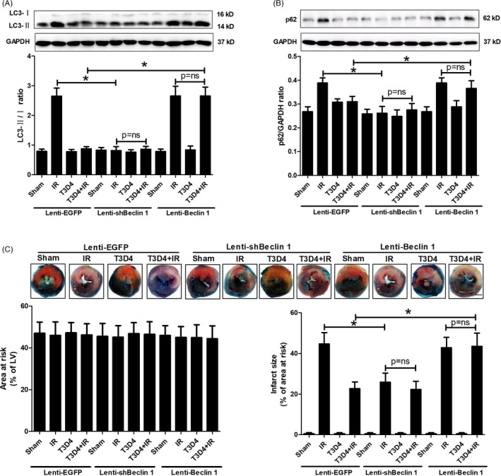
Methods and results: Transient transverse aortic constriction (TAC) was performed for 3 days in mice and then withdrawn for 4 days by aortic debanding, followed by subsequent exposure to myocardial ischaemia-reperfusion (I/R) injury. Following I/R injury, myocardial infarct size and apoptosis were significantly decreased, and cardiac dysfunction was markedly improved in the TAC preconditioning group compared with the control group. Mechanistically, TAC preconditioning markedly suppressed I/R-induced autophagy and preserved autophagic flux by deacetylating SOD2 via a SIRT3-dependent mechanism. Moreover, treatment with an adenovirus encoding SIRT3 partially mimicked the effects of hypertrophic preconditioning, whereas genetic ablation of SIRT3 in mice blocked the cardioprotective effects of hypertrophic preconditioning. Furthermore, in vivo lentiviral-mediated knockdown of Beclin 1 in the myocardium ameliorated the I/R-induced impairment of autophagic flux and was associated with a reduction in cell death, whereas treatment with a lentivirus encoding Beclin 1 abolished the cardioprotective effect of TAC preconditioning.

Conclusions: The present study identifies TAC preconditioning as a novel strategy for induction of an endogenous self-defensive and cardioprotective mechanism against cardiac injury. Specifically, TAC preconditioning reduced myocardial autophagic cell death in a SIRT3/SOD2 pathway-dependent manner.

Keywords: SIRT3; autophagy; hypertrophic preconditioning; ischaemia-reperfusion injury.


Conflict of interest statement

None.

Figures

FIGURE 1
FIGURE 1
Hypertrophic preconditioning decreased myocardial infarct size, inhibited cardiomyocyte apoptosis and improved cardiac function. A, Myocardial infarct size, as determined by Evans blue and TTC staining at 24 h after I/R; Scale bar = 1 mm. B, Cardiomyocyte apoptosis, as assessed by TUNEL staining (400×) and caspase‐3 activity measurement following in situ I/R; scale bar = 50 μm. C, Cardiac function (LVEF and LVFS), as measured by Doppler echocardiography at 24 h after I/R. n = 6‐8 per group, *P < .05
FIGURE 2
FIGURE 2
Hypertrophic preconditioning suppressed I/R‐induced autophagy. A, Myocardial SIRT3 protein expression and activity. B, Myocardial acetylated SOD2 levels and activity. COX4 was used as a loading control for mitochondrial proteins. C, Quantitative analysis of DHE and MitoSOX fluorescence intensity; scale bar = 50 μm. D, The expression of LC3B in I/R‐induced myocardial tissue, as detected by immunoblotting. E, Beclin 1. F, p62. GAPDH was used as a loading control. G, Representative autophagosome images indicated by the yellow arrows, and the number of autophagosomes was quantified by analysing 15 fields in each sample; scale bar = 500 nm. n = 5‐6 per group, *P < .05
FIGURE 3
FIGURE 3
SIRT3 deficiency reversed the cardioprotective effects of hypertrophic preconditioning. A, Myocardial infarct size, as determined by Evans blue and TTC staining at 24 h after I/R; Scale bar = 1 mm. B, Cardiomyocyte apoptosis, as assessed by TUNEL staining (400×) and caspase‐3 activity analysis following in situ I/R; scale bar = 50 μm. C, Cardiac function (LVEF and LVFS), as measured by Doppler echocardiography at 24 h after I/R. n = 6‐8 per group, *P < .05
FIGURE 4
FIGURE 4
Endogenous SIRT3 was required for the regulatory effects of hypertrophic preconditioning on the mROS‐autophagy pathway. A, The level of acetylated SOD2 in the I/R‐induced myocardium. B, SOD2 activity. C, Quantitative analysis of DHE and MitoSOX fluorescence intensity; scale bar = 50 μm. D, The expression of LC3B in I/R‐induced myocardial tissue, as detected by immunoblotting. E, Beclin 1. F, p62. G, Representative autophagosome photographs indicated by the yellow arrows, and the number of autophagosomes was quantified by analysing 15 fields in each sample; scale bar = 500 nm. n = 5‐6 per group, *P < .05
FIGURE 5
FIGURE 5
SIRT3 deacetylated SOD2 and modulated the mROS‐autophagy pathway. A, Myocardial SOD2 protein expression and activity. B, SIRT3 protein expression and activity. C, Quantitative analysis of DHE and MitoSOX fluorescence intensity; scale bar = 50 μm. D, Quantitative analysis of LC3B expression. E, Myocardial infarct size, as detected by Evans blue and TTC staining at 24 h after I/R; Scale bar = 1 mm. F, Cardiac function (LVEF and LVFS), as measured by Doppler echocardiography at 24 h after I/R. n = 6‐8 per group, *P < .05
FIGURE 6
FIGURE 6
mROS scavenging rescued I/R injury and was involved in the cardioprotective effect of hypertrophic preconditioning. A, Quantitative analysis of DHE and MitoSOX fluorescence intensity. B, Myocardial infarct size, as determined by Evans blue and TTC staining at 24 h after I/R; Scale bar = 1 mm. C, Cardiac function (LVEF and LVFS), as measured by Doppler echocardiography at 24 h after I/R. n = 6‐8 per group, *P < .05
FIGURE 7
FIGURE 7
Beclin 1 eliminated the protective effect of hypertrophic preconditioning against myocardial infarction. A, The expression of LC3B in I/R‐induced myocardial tissue, as detected by immunoblotting. B, p62. C, Myocardial infarct size, as determined by Evans blue and TTC staining at 24 h after I/R; Scale bar = 1 mm. n = 6‐8 per group, *P < .05
FIGURE 8
FIGURE 8
Chloroquine (CQ) attenuated hypertrophic preconditioning‐induced protection against myocardial infarction by regulating autophagic flux. A, Myocardial LC3B expression. B, p62. C, Myocardial infarct size; Scale bar = 1 mm. n = 6‐8 per group, *P < .05

Similar articles

Cited by

References

    1. Anderson JL, Morrow DA. Acute myocardial infarction. N Engl J Med. 2017;376(21):2053‐2064. - PubMed
    1. Cung T‐T, Morel O, Cayla G, et al. Cyclosporine before PCI in patients with acute myocardial infarction. N Engl J Med. 2015;373(11):1021‐1031. - PubMed
    1. Cabrera‐Fuentes HA, Aragones J, Bernhagen J, et al. From basic mechanisms to clinical applications in heart protection, new players in cardiovascular diseases and cardiac theranostics: meeting report from the third international symposium on "New frontiers in cardiovascular research". Basic Res Cardiol. 2016;111(6):69. - PMC - PubMed
    1. Heusch G, Rassaf T. Time to give up on cardioprotection? A critical appraisal of clinical studies on ischemic pre‐, post‐, and remote conditioning. Circ Res. 2016;119(5):676‐695. - PubMed
    1. Zhang L, Wang K, Lei Y, Li Q, Nice EC, Huang C. Redox signaling: potential arbitrator of autophagy and apoptosis in therapeutic response. Free Radic Biol Med. 2015;89:452‐465. - PubMed

MeSH terms

LinkOut - more resources