. 2021 Jun;10(11):3674-3688.
doi: 10.1002/cam4.3911. Epub 2021 May 11.

Ubiquitin-specific peptidase 53 inhibits the occurrence and development of clear cell renal cell carcinoma through NF-κB pathway inactivation

Affiliations

Ubiquitin-specific peptidase 53 inhibits the occurrence and development of clear cell renal cell carcinoma through NF-κB pathway inactivation

Dingwen Gui et al. Cancer Med. 2021 Jun.

Abstract

Background: Clear cell renal cell carcinoma (ccRCC) is one of the most prevalent malignant diseases in the urinary system with more than 140,000 related deaths annually. Ubiquitination-deubiquitination homeostasis is an important factor in ccRCC progression; ubiquitin-specific peptidase 53 (USP53) belongs to the family of deubiquitinating enzymes, but its functions are rarely reported.

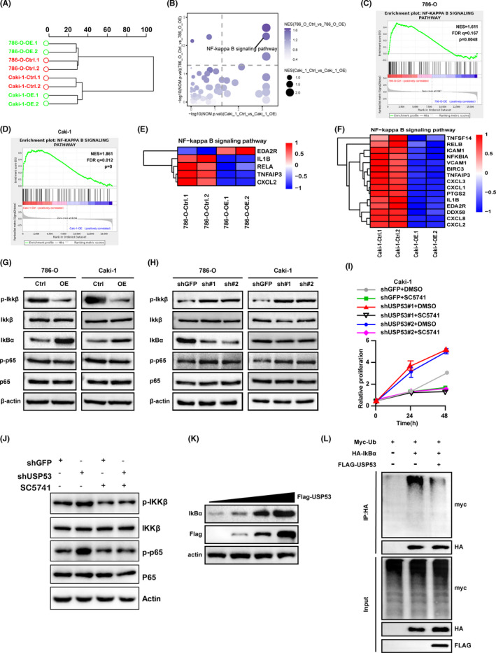
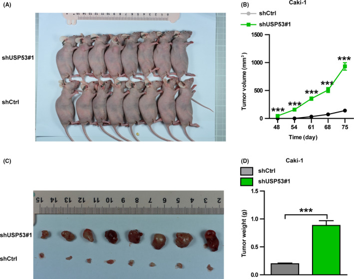
Methods: Databases obtained from GEO and TCGA were analyzed to reveal the role of USP53 in ccRCC. CCK-8/BrdU and EDU assays were used to detect the proliferation of ccRCC after USP53 overexpression or knockdown. A tumor xenograft experiment was used to verify the effect of the proliferation of ccRCC after USP53 knockdown. Transwell assays were used to detect the metastasis of ccRCC after USP53 overexpression or knockdown. RNA sequencing and western blot analysis were employed to detect the change in genes after USP53 overexpression and knockdown. Then we tested the effect of USP53 on IκBα protein stability through western blot analysis. Detect the effect of USP53 on IκBα ubiquitination in vitro by immunoprecipitation method.

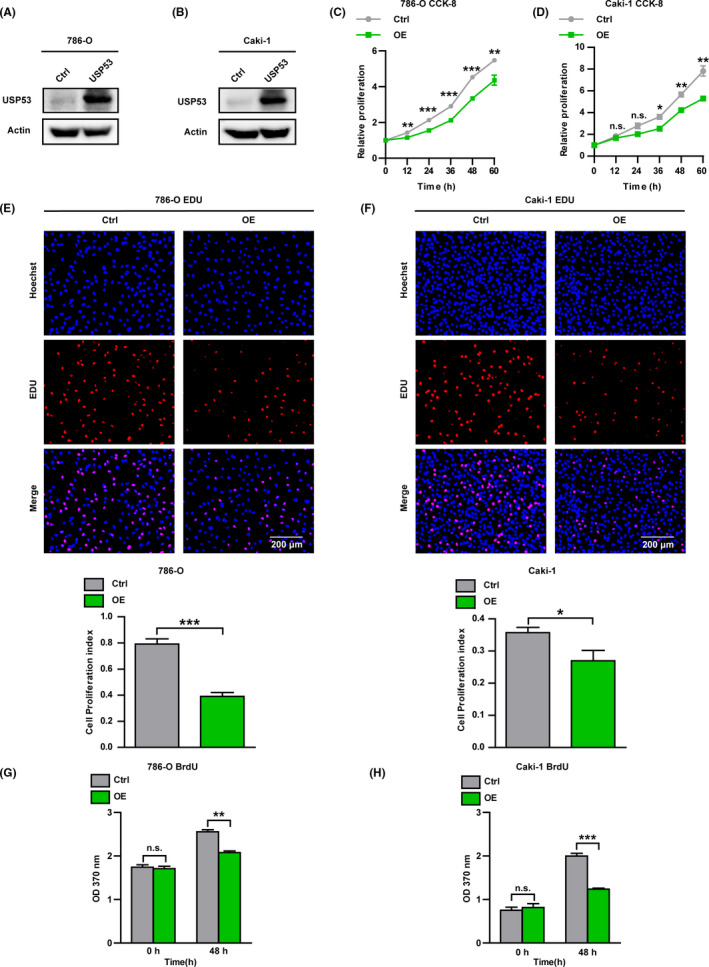
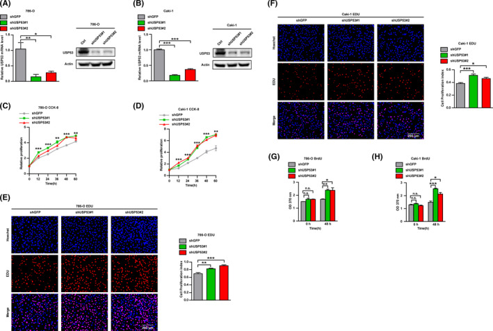
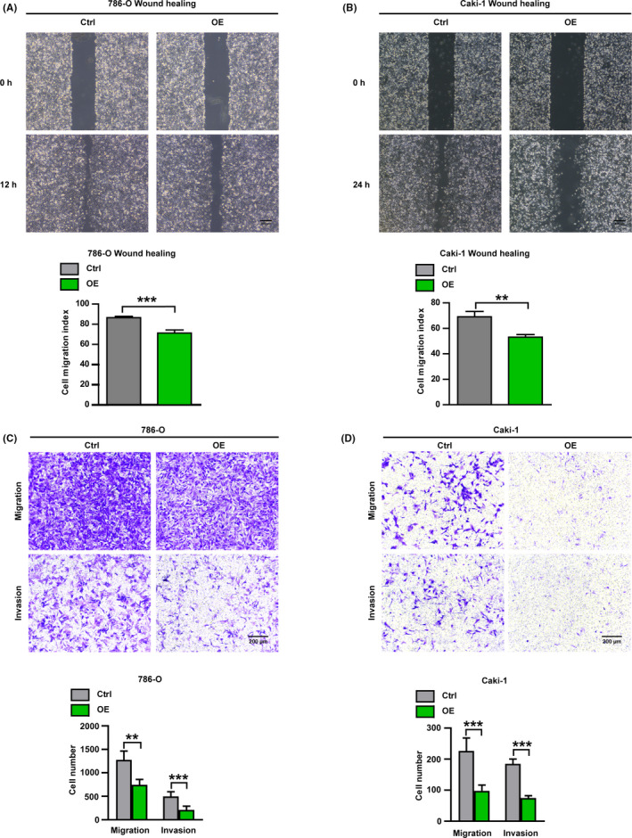
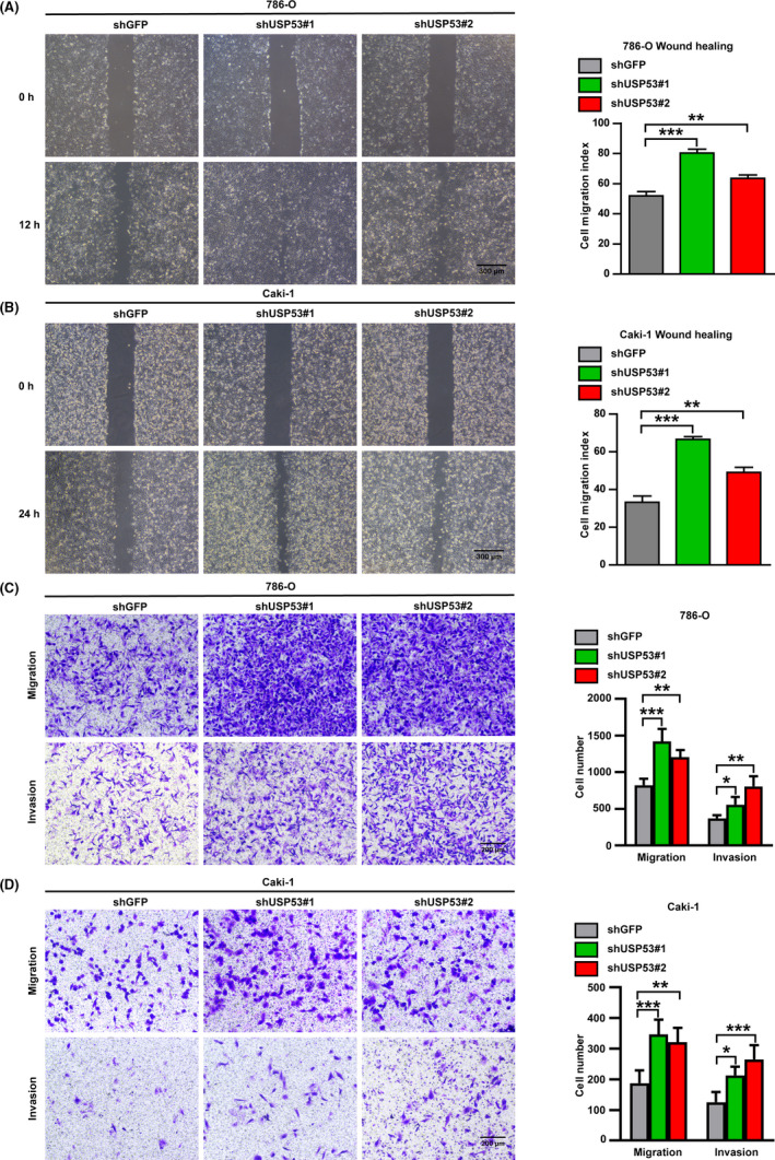
Results: USP53 expression was downregulated in ccRCC tissues and USP53 expression was significantly negatively correlated with the tumor progression and clinical prognosis. The ability of growth and metastasis of ccRCC was inhibited after USP53 overexpression. In addition, USP53 knockdown promoted ccRCC growth and metastasis. Moreover, USP53 knockdown promoted the ability of clone formation of ccRCC in vivo. NF-κB signaling pathway significantly enriched and downregulated in USP53 overexpressed cells, and genes in the NF-κB pathway (such as IL1B, CXCL1-3, RELA, RELB, etc.) were obviously downregulated in USP53 overexpressed cells. USP53 overexpression decreased the phosphorylation of IKKβ and P65 in both Caki-1 and 786-O cells, and the expression of IκBα was increased. Phosphorylation of IKKβ and P65 was increased in both Caki-1 and 786-O cells after USP53 knockdown. As the expression of USP53 increases, the protein expression of IκBα was also gradually increased and USP53 reduced the ubiquitination of IκBα.

Conclusion: In summary, our data indicate that USP53 inhibits the inactivation of the NF-κB pathway by reducing the ubiquitination of IκBα to further inhibit ccRCC proliferation and metastasis. These findings may help understand the pathogenesis of ccRCC and introduce new potential therapeutic targets for kidney cancer patients.

Keywords: NF-κB pathway; RNA sequencing; USP53; clear cell renal cell carcinoma.


Figures

FIGURE 1
FIGURE 1
Reduced expression of USP53 in renal cell carcinoma. (A) GES database (GES66271/GES76207/GES36895/GES26574) analysis showed compared with normal tissues; the expression of USP53 in ccRCC tissues was significantly reduced. (B) TCGA‐KIRC database analysis shows the expression of USP53 in ccRCC tissues was significantly reduced. (C) Low expression of USP53 is positively correlated with tumor grade. (D) Low expression of USP53 is positively correlated with poor prognosis. (E) Western blot assays show that the expression of USP53 in tumor cell lines (786‐O, Caki‐1, SK‐NEP‐1, SW‐13) is significantly reduced compared with normal cell lines (293T). (F) Immunohistochemical staining shoe that the expression of USP53 in tumor tissues was significantly lower than paracancerous tissues. (p < 0.0001, HR = 1.816, 59% CI: 1.347‐2.448)
FIGURE 2
FIGURE 2
Overexpression USP53 inhibits the growth and proliferation of ccRCC. (A, B) Immunoblotting assays were analyzed to determine the protein expression of USP53 in control and USP53 overexpression cells (786‐O‐USP53‐OE/Caki‐1‐USP53‐OE) or control cells (Ctrl). (C, D) Cell proliferation is impaired by overexpression in 786‐O and Caki‐1 cells, as assessed by CCK‐8 assay at the indicated time points. (E, F) EDU cell proliferation assay demonstrates the inhibitory effect of USP53 overexpression on both 786‐O and Caki‐1 cells. Cell proliferation index of 786‐O and Caki‐1 USP53 expression and control were statistically analyzed (bottom). (G, H) BrdU cell proliferation assays reflect 786‐O and Caki‐1 cells that overexpress the USP53 gene proliferation ability was significantly inhibited at 48 h after inoculation after USP53 overexpression. Cell numbers were counted by Image pro plus software and are shown at right (*p < 0.05, **< 0.01, ***p < 0.001. Data are shown as mean ± SEM)
FIGURE 3
FIGURE 3
Knockdown USP53 promotes the growth and proliferation of ccRCC. (A, B) mRNA levels of USP53 in Caki‐1 and 786‐O cells were analyzed by real‐time PCR after infection with Lentivirus targeting USP53 (USP53 shRNA) or negative controls (Ctrl). (C, D) Cell proliferation of the USP53 depleted or control cells was evaluated by CCK8 assays at the indicated time points. (E, F) EDU cell proliferation immunofluorescence assay were executed to determine the proliferation and viability of the USP53 depleted or control cells, Scale bar, 200 μm. Cell proliferation index of USP53 shRNA and control shRNA in 786‐O or Caki‐1 cells were statistically analyzed (right panel). (G, H) BrdU cell proliferation assay were executed to determine the growth and viability of the USP53 depleted or control cells. Representative images and quantification are shown (*p < 0.05, **p < 0.01, ***p < 0.001). Data are shown as mean ± SEM
FIGURE 4
FIGURE 4
Overexpression USP53 significantly inhibits migration and invasion of ccRCC. (A, B) Wound healing assays were used to evaluate the cell migration in USP53 overexpression or control cells. Representative images of USP53 overexpression cells or control cells in 786‐O and Caki‐1 cells in indicated time points were shown. Scale bar, 300 μm. Cell migration indexes were quantified at the bottom. (C, D) Cell migration and invasion assays were performed in Transwell assays with or without Matrigel, and USP53 overexpression significantly inhibits the cell migration and invasion. (C) Transwell assays in 786‐O cells with overexpressed USP53 or controls were determined at 10 h by taking images of the migrated cells, Scale bar, 200 μm. (D)Transwell assays in Caki‐1 cells with overexpressed USP53 or controls were determined at 24 h by taking images of the migrated cells, Scale bar, 200 μm. Representative photographs and the migration index are shown at right
FIGURE 5
FIGURE 5
Knockdown USP53 significantly promotes migration and invasion of ccRCC. (A, B) Cell wound healing ability was promoted at indicated time points after USP53 depleted compare with control through wound healing assay. In 786‐O cells (A) Caki‐1 cells (B). Scale bar 300 μm. Relative cell migratory distance of Caki‐1 USP53 overexpression and Control were quantified and statistically analyzed (right panel). (C, D) Transwell migration and invasion assays were performed in Transwell assays with or without Matrigel, and USP53 knockdown significantly inhibits the cell migration and invasion. (C) Representative Images of Transwell migration assay of USP53 depleted in 786‐O cells and Control cells for 10 h. Scale bar, 200 μm. (D) Representative images of Transwell invasion assay of USP53 depleted in Caki‐1 cells and Control cells for 24 h. Scale bar, 200 μm
FIGURE 6
FIGURE 6
NF‐κB pathway was changed in ccRCC cell lines after USP53 overexpression. (A) Hierarchical clustering analysis of RNA sequencing data revealed significant differences in gene expression between USP53 overexpressing cells and control cells. (B) GESA analysis showed that the NF‐κB pathway was changed significantly after USP53 overexpression in 786‐O and Caki‐1 cells. (C, D) Enrichment of an NF‐κB‐dependent gene expression signature in GSEA analysis of genes altered as described above. (E, F) Heat map representation of the differential expression of NF‐κB‐related genes between overexpressed and control cells. (G, H) Phosphorylation of IKKβ and p65 were estimated by Western blotting assay in USP53 overexpressed cells (G) or depletion cells (H). (I, J) Rescue experiments showed that USP53 inhibits the proliferation of Caki‐1 cell line by inhibiting the NF‐κB pathway. (K) The expression level of IκBα increased with the increase in USP53 expression in 293 cells; (L) USP53 can significantly reduce the ubiquitination of IκBα in 293 cells
FIGURE 7
FIGURE 7
Knockdown USP53 promotes the growth of renal cancer cells in vivo. (A) Representative images of subcutaneous tumor formation in nude mice derived from USP53‐depleted Caki‐1 cells and control cells 75 days after subcutaneous injection. (B) Tumor volumes were measured and statistically analyzed between Caki‐1 control and USP53‐depleted cells. (C) Nude mice were sacrificed and tumors were removed for photographing. (D) The isolated tumors were weighed. Representative images and quantification are shown (*p < 0.05, **p < 0.01, ***p < 0.001). Data are shown as mean ± SEM

Similar articles

Cited by

References

    1. Siegel RL, Miller KD, Jemal A. Cancer statistics, 2017. CA Cancer J Clin 2017;67(1):7–30. - PubMed
    1. Choueiri TK, Motzer RJ. Systemic therapy for metastatic renal‐cell carcinoma. N Engl J Med. 2017;376(4):354–366. - PubMed
    1. Capitanio U, Bensalah K, Bex A, et al. Epidemiology of renal cell carcinoma. Eur Urol. 2019;75(1):74–84. - PMC - PubMed
    1. Srigley JR, Delahunt B, Eble JN, et al. The International Society of Urological Pathology (ISUP) Vancouver classification of renal neoplasia. Am J Surg Pathol. 2013;37(10):1469–1489. - PubMed
    1. Nickerson ML, Jaeger E, Shi Y, et al. Improved identification of von Hippel‐Lindau gene alterations in clear cell renal tumors. Clin Cancer Res 2008;14(15):4726–4734. - PMC - PubMed

Publication types

MeSH terms