

Cell wall characteristics during sexual reproduction of Mougeotia sp. (Zygnematophyceae) revealed by electron microscopy, glycan microarrays and RAMAN spectroscopy
- PMID: 33974144
- PMCID: PMC8523461
- DOI: 10.1007/s00709-021-01659-5
Cell wall characteristics during sexual reproduction of Mougeotia sp. (Zygnematophyceae) revealed by electron microscopy, glycan microarrays and RAMAN spectroscopy
Abstract
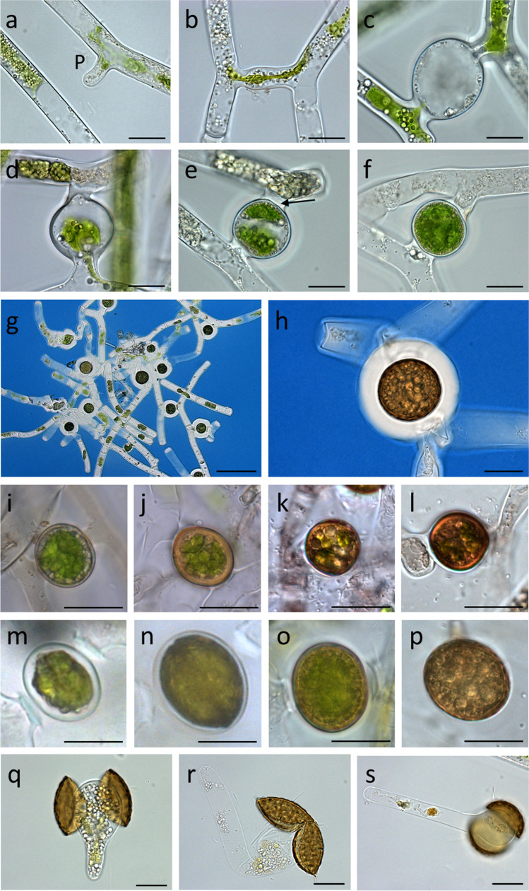
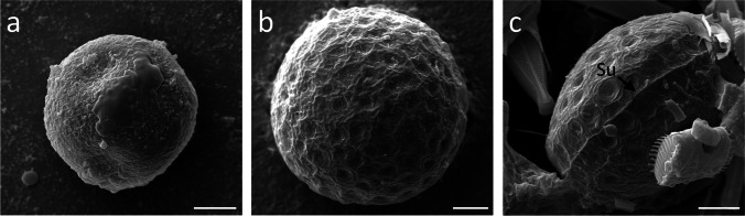
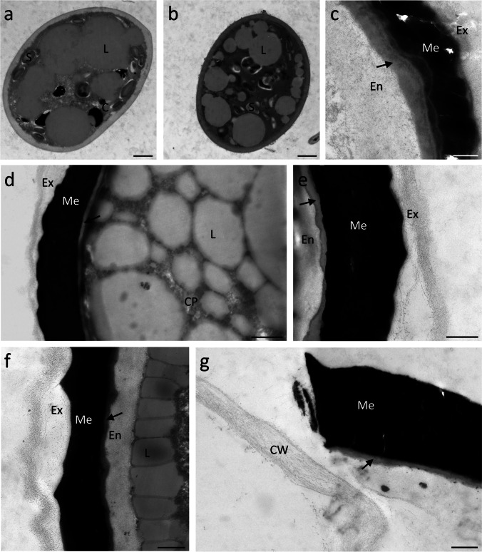
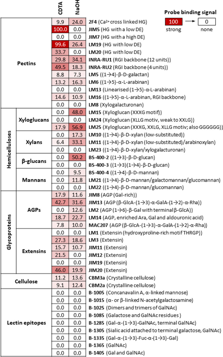
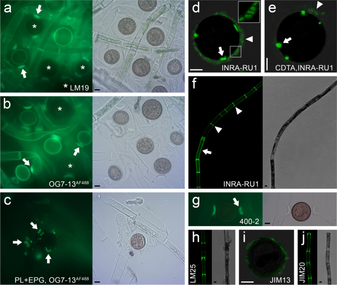
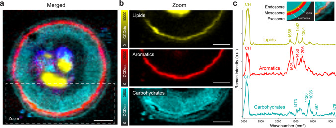
Mougeotia spp. collected from field samples were investigated for their conjugation morphology by light-, fluorescence-, scanning- and transmission electron microscopy. During a scalarifom conjugation, the extragametangial zygospores were initially surrounded by a thin cell wall that developed into a multi-layered zygospore wall. Maturing zygospores turned dark brown and were filled with storage compounds such as lipids and starch. While M. parvula had a smooth surface, M. disjuncta had a punctated surface structure and a prominent suture. The zygospore wall consisted of a polysaccharide rich endospore, followed by a thin layer with a lipid-like appaerance, a massive electron dense mesospore and a very thin exospore composed of polysaccharides. Glycan microarray analysis of zygospores of different developmental stages revealed the occurrence of pectins and hemicelluloses, mostly composed of homogalacturonan (HG), xyloglucans, xylans, arabino-galactan proteins and extensins. In situ localization by the probe OG7-13AF 488 labelled HG in young zygospore walls, vegetative filaments and most prominently in conjugation tubes and cross walls. Raman imaging showed the distribution of proteins, lipids, carbohydrates and aromatic components of the mature zygospore with a spatial resolution of ~ 250 nm. The carbohydrate nature of the endo- and exospore was confirmed and in-between an enrichment of lipids and aromatic components, probably algaenan or a sporopollenin-like material. Taken together, these results indicate that during zygospore formation, reorganizations of the cell walls occured, leading to a resistant and protective structure.
Keywords: Cell wall; Conjugation; Mougeotia; Sexual reproduction; Streptophyte; Zygospore.
© 2021. The Author(s).
Conflict of interest statement
The authors certify that there are no conflicts of interest/competing interests in the subject matter or materials discussed in this manuscript.
Figures
Similar articles
-
Zygospores of the green alga Spirogyra: new insights from structural and chemical imaging.Front Plant Sci. 2022 Dec 6;13:1080111. doi: 10.3389/fpls.2022.1080111. eCollection 2022. Front Plant Sci. 2022. PMID: 36561459 Free PMC article.
-
Induction of Conjugation and Zygospore Cell Wall Characteristics in the Alpine Spirogyra mirabilis (Zygnematophyceae, Charophyta): Advantage under Climate Change Scenarios?Plants (Basel). 2021 Aug 23;10(8):1740. doi: 10.3390/plants10081740. Plants (Basel). 2021. PMID: 34451785 Free PMC article.
-
Zygospore formation in Zygnematophyceae predates several land plant traits.Philos Trans R Soc Lond B Biol Sci. 2024 Nov 18;379(1914):20230356. doi: 10.1098/rstb.2023.0356. Epub 2024 Sep 30. Philos Trans R Soc Lond B Biol Sci. 2024. PMID: 39343014 Free PMC article. Review.
-
Zygospore development of Spirogyra (Charophyta) investigated by serial block-face scanning electron microscopy and 3D reconstructions.Front Plant Sci. 2024 Mar 14;15:1358974. doi: 10.3389/fpls.2024.1358974. eCollection 2024. Front Plant Sci. 2024. PMID: 38559764 Free PMC article.
-
Zygnematophycean algae: Possible models for cellular and evolutionary biology.Semin Cell Dev Biol. 2023 Jan 30;134:59-68. doi: 10.1016/j.semcdb.2022.03.042. Epub 2022 Apr 13. Semin Cell Dev Biol. 2023. PMID: 35430142 Review.
Cited by
-
Zygospores of the green alga Spirogyra: new insights from structural and chemical imaging.Front Plant Sci. 2022 Dec 6;13:1080111. doi: 10.3389/fpls.2022.1080111. eCollection 2022. Front Plant Sci. 2022. PMID: 36561459 Free PMC article.
-
Evolution of ethylene as an abiotic stress hormone in streptophytes.Environ Exp Bot. 2023 Oct;214:105456. doi: 10.1016/j.envexpbot.2023.105456. Environ Exp Bot. 2023. PMID: 37780400 Free PMC article. Review.
-
Temperature- and light stress adaptations in Zygnematophyceae: The challenges of a semi-terrestrial lifestyle.Front Plant Sci. 2022 Jul 19;13:945394. doi: 10.3389/fpls.2022.945394. eCollection 2022. Front Plant Sci. 2022. PMID: 35928713 Free PMC article. Review.
-
Search for evolutionary roots of land plant arabinogalactan-proteins in charophytes: presence of a rhamnogalactan-protein in Spirogyra pratensis (Zygnematophyceae).Plant J. 2022 Feb;109(3):568-584. doi: 10.1111/tpj.15577. Epub 2021 Nov 26. Plant J. 2022. PMID: 34767672 Free PMC article.
-
Induction of Conjugation and Zygospore Cell Wall Characteristics in the Alpine Spirogyra mirabilis (Zygnematophyceae, Charophyta): Advantage under Climate Change Scenarios?Plants (Basel). 2021 Aug 23;10(8):1740. doi: 10.3390/plants10081740. Plants (Basel). 2021. PMID: 34451785 Free PMC article.
References
-
- Allen MA (1958) The biology of a species complex in Spirogyra. Dissertation. Indiana University, Bloomington (USA)
-
- Blokker P. Structural analysis of resistant polymers in extant algae and ancient sediments. Geol Ultraiectina. 2000;193:1–145.
MeSH terms
Substances
Grants and funding
LinkOut - more resources
Full Text Sources
Other Literature Sources
Miscellaneous
