

Detection of liver cirrhosis in standard T2-weighted MRI using deep transfer learning
- PMID: 33974149
- PMCID: PMC8523404
- DOI: 10.1007/s00330-021-07858-1
Detection of liver cirrhosis in standard T2-weighted MRI using deep transfer learning
Abstract
Objectives: To investigate the diagnostic performance of deep transfer learning (DTL) to detect liver cirrhosis from clinical MRI.
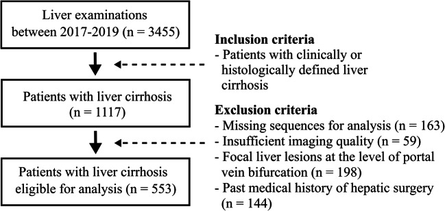
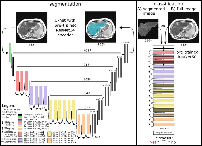
Methods: The dataset for this retrospective analysis consisted of 713 (343 female) patients who underwent liver MRI between 2017 and 2019. In total, 553 of these subjects had a confirmed diagnosis of liver cirrhosis, while the remainder had no history of liver disease. T2-weighted MRI slices at the level of the caudate lobe were manually exported for DTL analysis. Data were randomly split into training, validation, and test sets (70%/15%/15%). A ResNet50 convolutional neural network (CNN) pre-trained on the ImageNet archive was used for cirrhosis detection with and without upstream liver segmentation. Classification performance for detection of liver cirrhosis was compared to two radiologists with different levels of experience (4th-year resident, board-certified radiologist). Segmentation was performed using a U-Net architecture built on a pre-trained ResNet34 encoder. Differences in classification accuracy were assessed by the χ2-test.
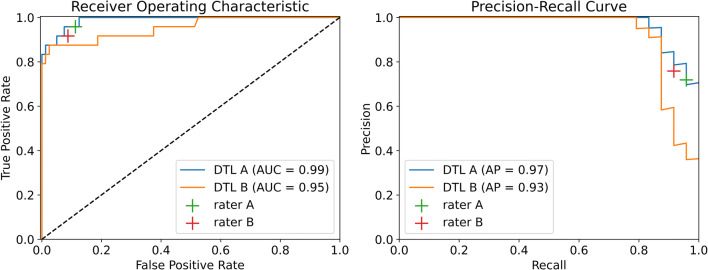
Results: Dice coefficients for automatic segmentation were above 0.98 for both validation and test data. The classification accuracy of liver cirrhosis on validation (vACC) and test (tACC) data for the DTL pipeline with upstream liver segmentation (vACC = 0.99, tACC = 0.96) was significantly higher compared to the resident (vACC = 0.88, p < 0.01; tACC = 0.91, p = 0.01) and to the board-certified radiologist (vACC = 0.96, p < 0.01; tACC = 0.90, p < 0.01).
Conclusion: This proof-of-principle study demonstrates the potential of DTL for detecting cirrhosis based on standard T2-weighted MRI. The presented method for image-based diagnosis of liver cirrhosis demonstrated expert-level classification accuracy.
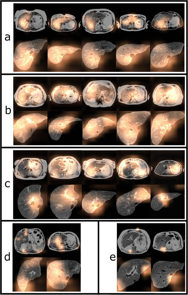
Key points: • A pipeline consisting of two convolutional neural networks (CNNs) pre-trained on an extensive natural image database (ImageNet archive) enables detection of liver cirrhosis on standard T2-weighted MRI. • High classification accuracy can be achieved even without altering the pre-trained parameters of the convolutional neural networks. • Other abdominal structures apart from the liver were relevant for detection when the network was trained on unsegmented images.
Keywords: Deep learning; Liver cirrhosis; Magnetic resonance imaging; Neural networks, computer.
© 2021. The Author(s).
Conflict of interest statement
The authors of this manuscript declare no relationships with any companies whose products or services may be related to the subject matter of the article.
Figures
References
MeSH terms
Grants and funding
LinkOut - more resources
Full Text Sources
Other Literature Sources
Medical
