. 2021 Jun 2;12(11):1873-1884.
doi: 10.1021/acschemneuro.0c00712. Epub 2021 May 11.

Signaling-Biased and Constitutively Active Dopamine D2 Receptor Variant

Affiliations

Signaling-Biased and Constitutively Active Dopamine D2 Receptor Variant

Dayana Rodriguez-Contreras et al. ACS Chem Neurosci. .

Abstract

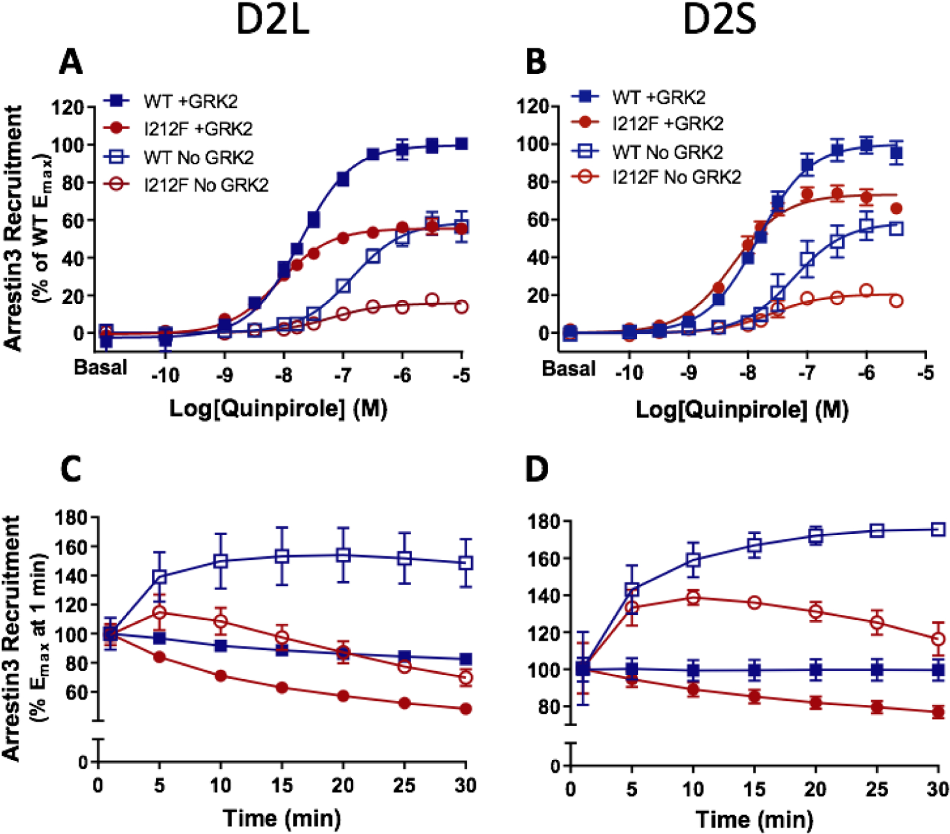
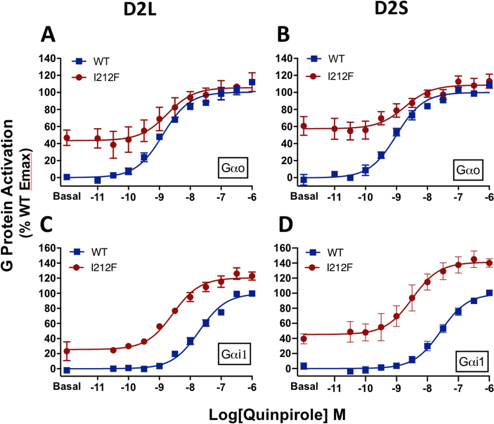
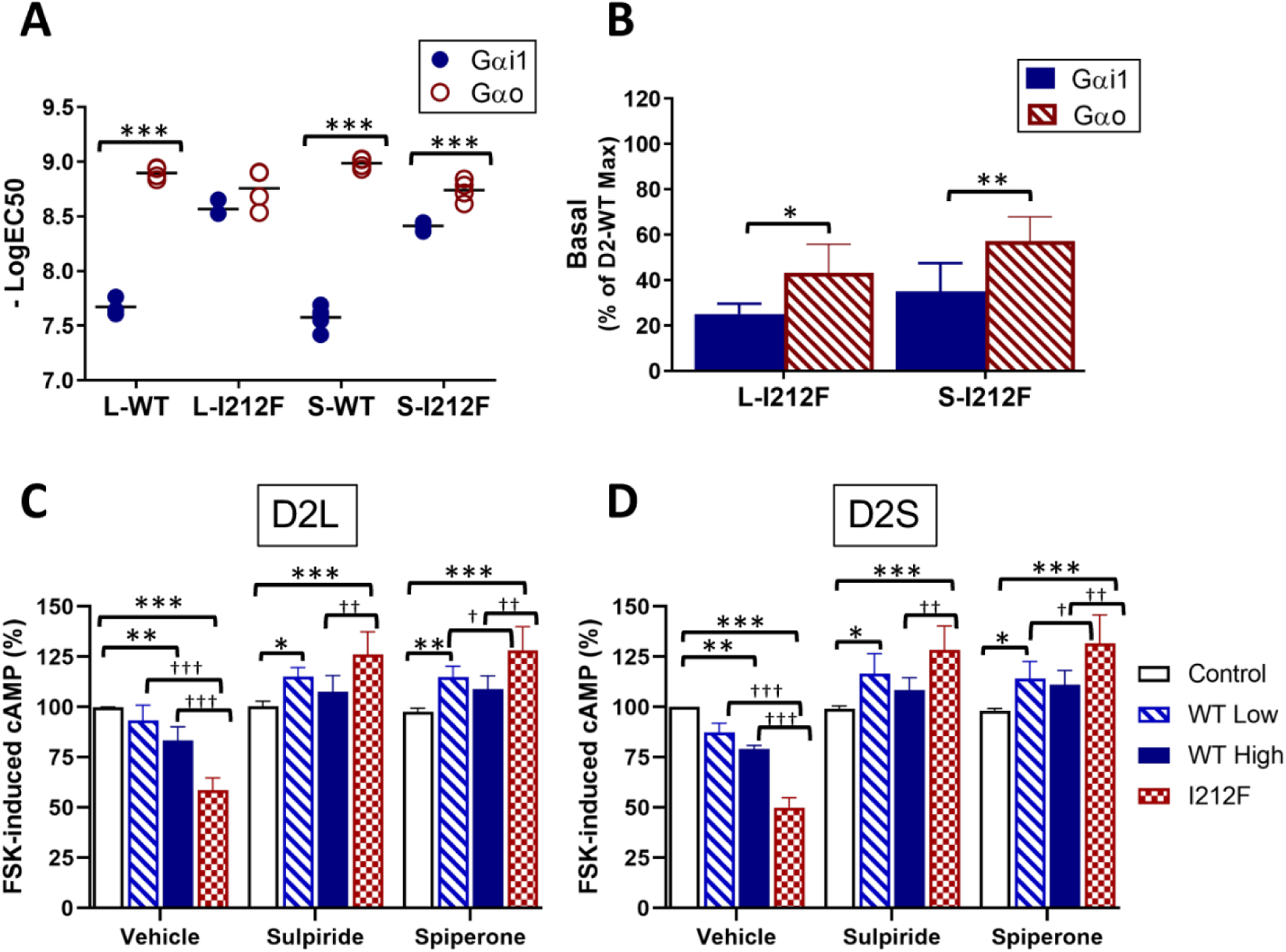
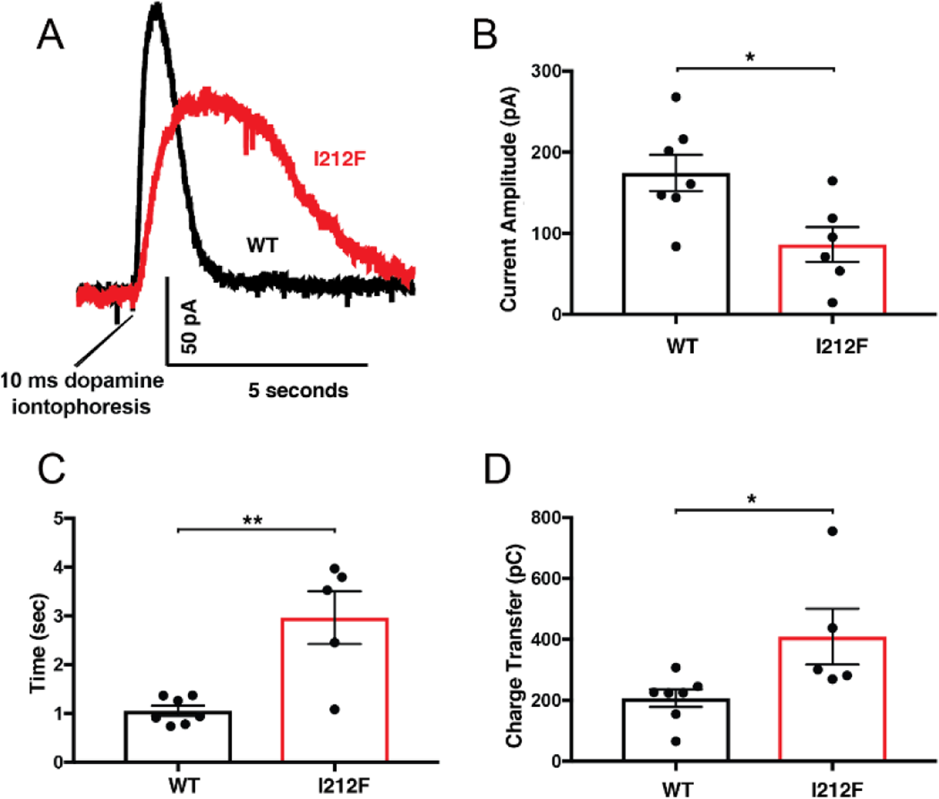
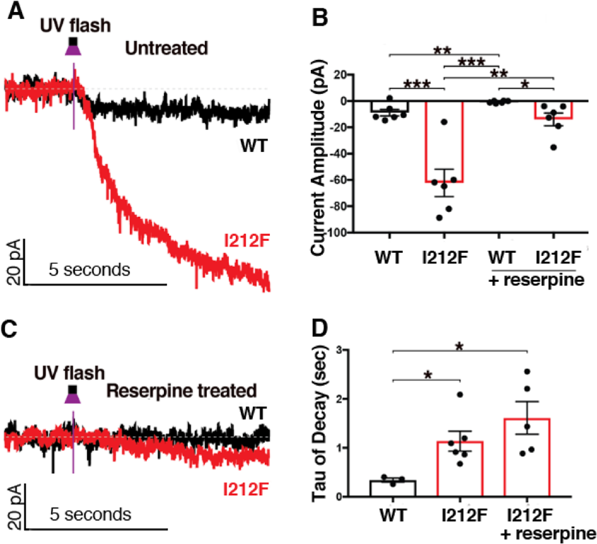
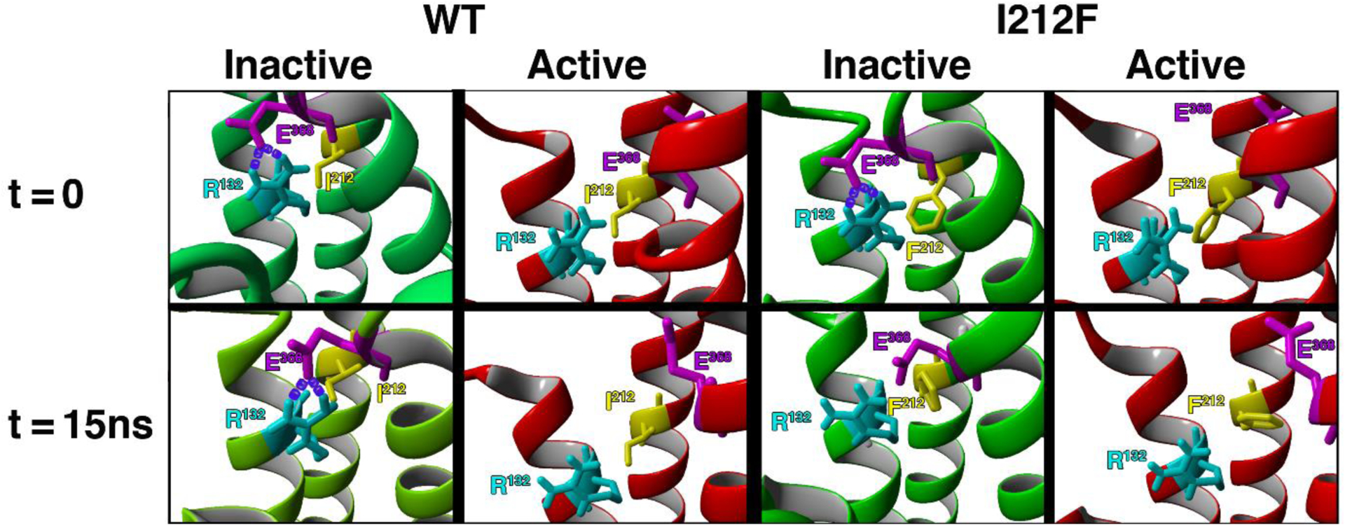
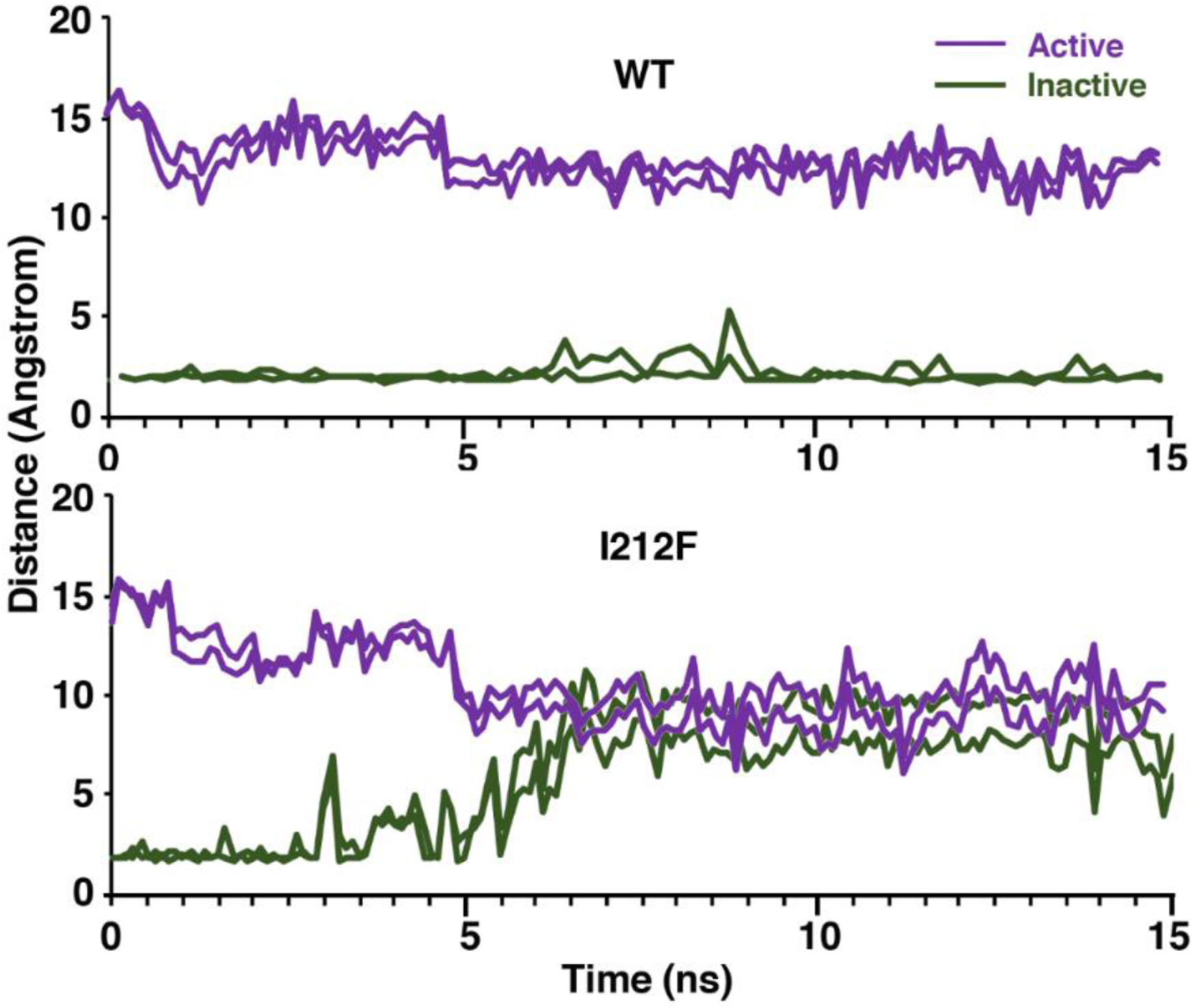
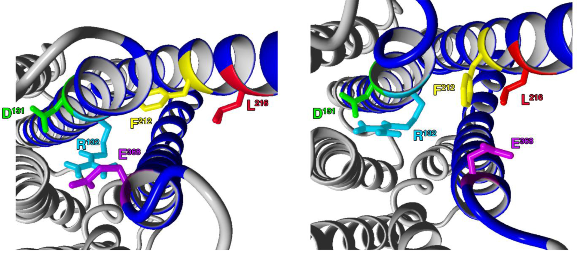
A dopamine D2 receptor mutation was recently identified in a family with a novel hyperkinetic movement disorder. Compared to the wild type D2 receptor, the novel allelic variant D2-I212F activates a Gαi1β1γ2 heterotrimer with higher potency and modestly enhanced basal activity in human embryonic kidney (HEK) 293 cells and has decreased capacity to recruit arrestin3. We now report that omitting overexpressed G protein-coupled receptor kinase-2 (GRK2) decreased the potency and efficacy of quinpirole for arrestin recruitment. The relative efficacy of quinpirole for arrestin recruitment to D2-I212F compared to D2-WT was considerably lower without overexpressed GRK2 than with added GRK2. D2-I212F exhibited higher basal activation of GαoA than Gαi1 but little or no increase in the potency of quinpirole relative to D2-WT. Other signs of D2-I212F constitutive activity for G protein-mediated signaling, in addition to basal activation of Gαi/o, were enhanced basal inhibition of forskolin-stimulated cyclic AMP accumulation that was reversed by the inverse agonists sulpiride and spiperone and a ∼4-fold increase in the apparent affinity of D2-I212F for quinpirole, determined from competition binding assays. In mouse midbrain slices, inhibition of tonic current by the inverse agonist sulpiride in dopamine neurons expressing D2-I212F was consistent with our hypothesis of enhanced constitutive activity and sensitivity to dopamine relative to D2-WT. Molecular dynamics simulations with D2 receptor models suggested that an ionic lock between the cytoplasmic ends of the third and sixth α-helices that constrains many G protein-coupled receptors in an inactive conformation spontaneously breaks in D2-I212F. Overall, these results confirm that D2-I212F is a constitutively active and signaling-biased D2 receptor mutant and also suggest that the effect of the likely pathogenic variant in a given brain region will depend on the nature of G protein and GRK expression.

Keywords: D2 receptor; Dopamine; G protein; allelic variant; arrestin; biased signaling; constitutive activity.


Conflict of interest statement

The authors declare no competing financial interest.

Figures

Figure 1.
Figure 1.
Dose-response curves for quinpirole-induced arrestin3 recruitment mediated by D2L/S-WT and D2L/S-I212F. Arrestin3 recruitment was measured in HEK293 cells co-transfected with GRK2 (+ GRK2) or nonspecific plasmid DNA (No GRK2). Values plotted are the means ± SD of 3–4 independent experiments performed in quadruplicate. A and B, quinpirole concentration-response curves measured at 10 min. Data from each independent experiment were normalized by subtracting the baseline and expressed as a percentage of maximum arrestin3 recruitment by D2-WT+GRK2. Data for + GRK2 are from the dataset described in van der Weijden et al. (8), where results were shown after 20 min of agonist stimulation. C and D, change in Emax values over 30 min, with each condition normalized to Emax for that condition at 1 min. Data for + GRK2 were previously described in van der Weijden et al. (8).
Figure 2.
Figure 2.
Concentration-response curves for Gαi/o protein activation mediated by D2-WT and D2-I212F in response to stimulation with quinpirole. Results are expressed as the percentage of maximum G protein activation by D2-WT, measured 10 min after adding coelenterazine h. A, Activation of GαoA by D2L-WT/I212F, B, Activation of GαoA by D2S-WT/I212F, C, Activation of Gαi1 by D2L-WT/I212F, and D, Data from van der Weijden et al. (8) for activation of Gαi1 by D2SWT/I212F. Values plotted represent means ± SD of three (panel C) or four (panels A, B, D) independent experiments performed in quadruplicate.
Figure 3.
Figure 3.
Increased constitutive activity of D2-I212F. A, B Concentration-response curves for Gαi/o protein activation mediated by D2-WT and D2-I212F were analyzed by nonlinear regression to determine quinpirole potency (A), expressed as the −LogEC50, and activation in the absence of quinpirole (B), expressed as the percentage of Emax for D2-WT. Data are from Table 2. Statistical differences were determined as described in Table 2 (*p<0.05, **p<0.01, ***p<0.001). C, D Cyclic AMP accumulation was measured in the presence of 10 μM forskolin (FSK) in HEK293 cells transfected with the cyclic AMP biosensor CAMYEL and either control plasmid DNA (control), a high (0.5 μg, WT High) or low (0.2 μg, WT Low) amount of D2L/S-WT DNA, or D2 L/S-I212F plasmid DNA (0.5 μg, I212F). Measurements were taken 10 min after addition of either vehicle, sulpiride (10 μM) or spiperone (1 μM), FSK (10 μM) and coelenterazine h. Results are expressed as a percentage of cyclic AMP accumulation by control cells treated with the inverse agonist vehicle. Values plotted are mean ± SD of four independent experiments performed in sextuplicate. Statistical differences were determined by 2-way ANOVA followed by Turkey’s post-hoc test (*p<0.05, **p<0.01, ***p<0.001 compared to the corresponding control condition; †p<0.05, ††p<0.01, †††p<0.001 compared to D2-WT).
Figure 4.
Figure 4.
Activation of GIRK currents by dopamine iontophoresis was assessed in mouse midbrain slices. AAV-DIO-D2S-WT or -D2S-I212F was used to restore D2 receptor expression in dopamine neurons of auto-D2-KO mice. A, representative outward currents in response to iontophoresis of dopamine (1 M) for 10 msec. Mean ± SEM is shown for (B) current amplitude, (C) peak half-width, and (D) charge transfer. The number of cells differs among panels for D2-I212F because kinetics in the lowest amplitude response in panel B could not be accurately resolved. Student’s t-test: *p < 0.05, **p < 0.01.
Figure 5.
Figure 5.
AAV-DIO-D2S-WT or -D2S-I212F was used to restore D2 expression in dopamine neurons of auto-D2-KO mice. CyHQ-sulpiride (5 μM) was circulated over a midbrain slice, and photolysis was by means of a 50 msec flash (365 nm) from a 6.5 mW LED light. The left panels depict representative traces for untreated slices (A) and slices incubated with reserpine to deplete endogenous dopamine (C). The right panels depict the mean ± SEM of the decreased current amplitude (B) and the rate of decay of the current after photorelease of sulpiride (D) in control slices and in slices pretreated with reserpine. It was not possible to calculate a decay rate for reserpine-treated slices from mice expressing D2-WT. For some conditions the number of cells differs between panels B and D because kinetics could not be accurately resolved in the lowest amplitude responses in panel B. Student’s t-test: *p < 0.05, **p < 0.01, ***p < 0.001.
Figure 6.
Figure 6.
The eight panels show TM3–6 ionic lock residues Arg132 (light blue) and Glu368 (magenta), as well as the variant residue Ile/Phe212 (yellow). Models are shown before (t=0) and after (t=15ns) MD simulations for 15 nsec. At t=0, the distances between OE1 of Glu368 and HH1 and HH2 of Arg132 are short enough to form salt bridges (purple) in both inactive models, but are too far apart in both active models. After 15 nsec MD simulations the WT model is essentially unchanged, whereas the presence of Phe212 separates the side chains of Arg132 and Glu368, preventing maintenance of the ionic lock in inactive D2-I212F. The cytoplasmic face is up.
Figure 7.
Figure 7.
The distances between the atoms that form the two bonds of the ionic lock are shown for all four models during 15 nsec MD simulations. Note that the distances are relatively stable for the active and inactive D2-WT models, whereas the distances increased 6–8 angstroms in the inactive D2-I212F model and decreased ~5 angstroms in the active model.
Figure 8.
Figure 8.
Residues involved in the disruption of the ionic lock are shown at t = 0.5 nsec (left panel) and 7.5 nsec (right panel) during MD simulation with the inactive D2-I212F model. Ballesteros-Weinstein numbering for the colored residues is Asp1313.49 (green), Arg1323.50 (light blue), Phe2125.61 (yellow), Leu2165.65 (red), and Glu3686.30 (magenta).

References

    1. Beaulieu JM; Sotnikova TD; Marion S; Lefkowitz RJ; Gainetdinov RR; Caron MG, An Akt/β-arrestin 2/PP2A signaling complex mediates dopaminergic neurotransmission and behavior. Cell 2005, 122 (2), 261–273. - PubMed
    1. Donthamsetti P; Gallo EF; Buck DC; Stahl EL; Zhu Y; Lane JR; Bohn LM; Neve KA; Kellendonk C; Javitch JA, Arrestin recruitment to dopamine D2 receptor mediates locomotion but not incentive motivation. Mol Psychiatry 2018, 25, 2086–2100. - PMC - PubMed
    1. Rose SJ; Pack TF; Peterson SM; Payne K; Borrelli E; Caron MG, Engineered D2R variants reveal the balanced and biased contributions of G-protein and β-arrestin to dopamine-dependent functions. Neuropsychopharmacology 2018, 43 (5), 1164–1173. - PMC - PubMed
    1. Cepeda C; Murphy KP; Parent M; Levine MS, The role of dopamine in Huntington’s disease. Prog. Brain Res 2014, 211, 235–254. - PMC - PubMed
    1. Moritz AE; Free RB; Sibley DR, Advances and challenges in the search for D2 and D3 dopamine receptor-selective compounds. Cell Signal 2018, 41, 75–81. - PMC - PubMed

Publication types

LinkOut - more resources