. 2021 May 11;17(5):e1009580.
doi: 10.1371/journal.ppat.1009580. eCollection 2021 May.

A Ran-binding protein facilitates nuclear import of human papillomavirus type 16

Affiliations

A Ran-binding protein facilitates nuclear import of human papillomavirus type 16

Kun-Yi Lai et al. PLoS Pathog. .

Abstract

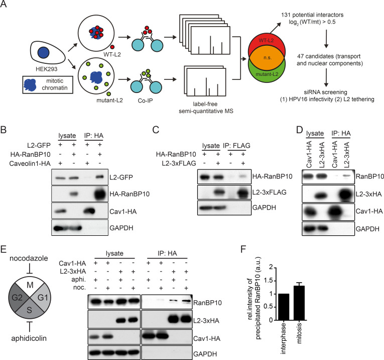
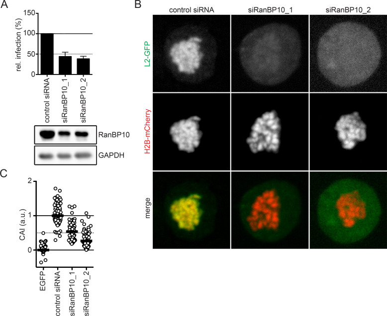
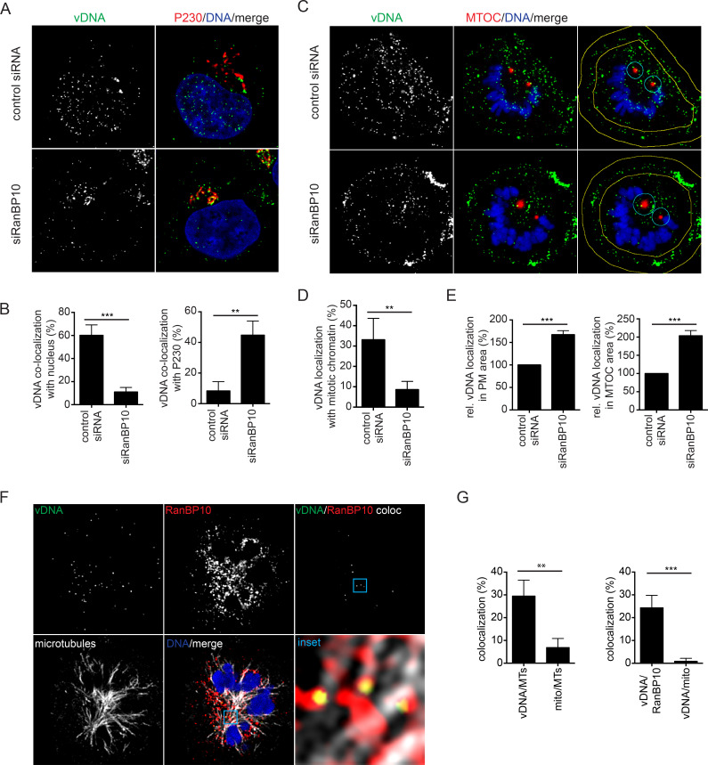
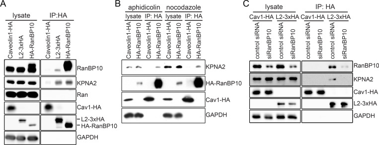
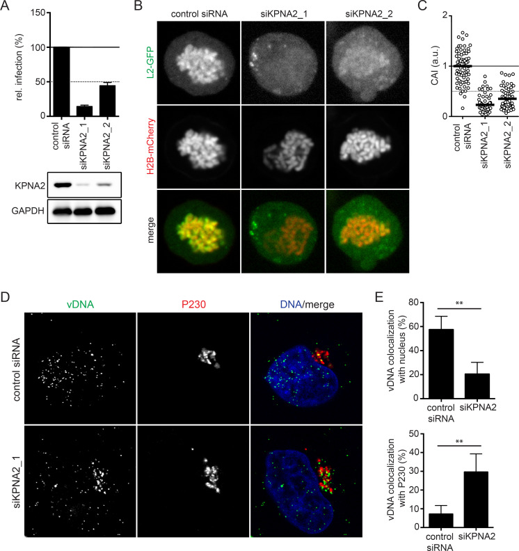
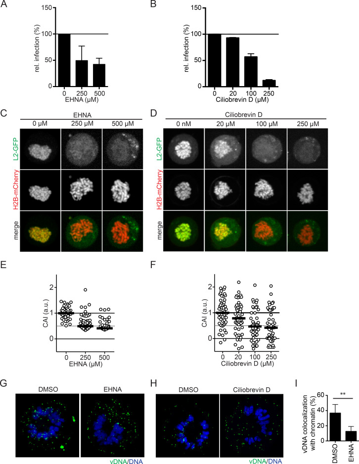
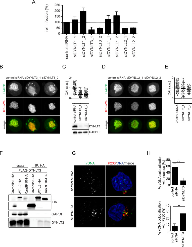
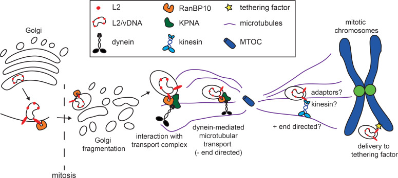
Human papillomaviruses (HPVs) utilize an atypical mode of nuclear import during cell entry. Residing in the Golgi apparatus until mitosis onset, a subviral complex composed of the minor capsid protein L2 and viral DNA (L2/vDNA) is imported into the nucleus after nuclear envelope breakdown by associating with mitotic chromatin. In this complex, L2 plays a crucial role in the interactions with cellular factors that enable delivery and ultimately tethering of the viral genome to mitotic chromatin. To date, the cellular proteins facilitating these steps remain unknown. Here, we addressed which cellular proteins may be required for this process. Using label-free mass spectrometry, biochemical assays, microscopy, and functional virological assays, we discovered that L2 engages a hitherto unknown protein complex of Ran-binding protein 10 (RanBP10), karyopherin alpha2 (KPNA2), and dynein light chain DYNLT3 to facilitate transport towards mitotic chromatin. Thus, our study not only identifies novel cellular interactors and mechanism that facilitate a poorly understood step in HPV entry, but also a novel cellular transport complex.


Conflict of interest statement

The authors have declared that no competing interests exist.

Figures

Fig 1
Fig 1. Identification of a novel L2 interaction partner during mitosis.
(A) Overview of the experimental strategy to identify mitotic L2 interactors using label-free semi-quantitative mass spectrometry. HEK293 cell lysates expressing wild-type L2-3xHA or RTR313EEE-mutant L2-3xHA were subjected to immunoprecipitation with an HA antibody. Cellular proteins co-precipitating with L2 were identified by label-free semi-quantitative mass spectrometry and analyzed with MaxQuant v1.5.3.12. 47 candidates were selected and preliminarily tested on their functional roles in HPV16 infectivity and L2 chromatin association. (B) Immunoprecipitation of HEK293 cell lysates co-expressing L2-GFP and HA-RanBP10. Caveolin 1-HA (Cav1-HA) was used as a negative control for the HA-tag pull-down. (C) Reverse pull-down for (B). Immunoprecipitation of HEK293 cell lysates co-expressing RanBP10 and L2-3xFLAG with a FLAG-tag antibody. (D)(E) Immunoprecipitation against HA in (D) unsynchronized or (E) synchronized HEK293 cells expressing L2-3xHA, followed by detection of endogenous RanBP10. Cells were synchronized for 16 hours with either aphidicolin (3 μM) to result in interphase-arrested cells or nocodazole (330 nM) to result in prometaphase-arrested cells. Caveolin 1-HA (Cav1-HA) was used as a negative control. (F) Quantification of RanBP10 immunoprecipitates from (E) of three independent experiments. Given are values relative to interphase levels ± SD.
Fig 2
Fig 2. A crucial role of RanBP10 for HPV16 infectivity and L2 tethering.
(A) RNAi of RanBP10 in HeLa cells was followed by HPV16-PsV infection for 48 hours. Infectivity was scored by flow cytometry based on the percentage of the cells expressing GFP. The infectivity was normalized to control siRNA transfected cells and depicted as relative (rel.) infection. The protein expression level of RanP10 upon siRNA knockdown was analyzed by Western blotting. (B) RNAi of RanBP10 in HeLa Kyoto_H2B-mCherry_L2-GFP cells was followed by arrest in prometaphase using nocodazole (330 nM) for 16 hours. Cells were fixed and images were acquired using a spinning disk confocal microscope. Depicted are single median slices. (C) Analysis of (B) quantifying the degree of chromosomal association as described in material and methods. Displayed is the chromosomal association index (CAI) relative to control siRNA-treated cells (1) and GFP expressing cells (0). In three independent experiments, at least 50 cells were analyzed. The median was indicated by a black bar.
Fig 3
Fig 3. RanBP10 facilitated nuclear delivery of incoming HPV16 viral DNA.
vDNA from incoming EdU-labelled HPV16 PsV was detected using EdU Click-iT chemistry, nuclei were stained by Hoechst-33258. All images were acquired by confocal microscopy. Images represent single medial slices. The degree of overlapping signals from at least three independent experiments was quantified using IMARIS. (A) RNAi of RanBP10 in HeLa cells was followed by infection with EdU-labelled HPV16. Cells were fixed at 20 h.p.i., and stained with an anti-P230 antibody and Hoechst-33258 to indicate the TGN and nucleus, respectively. (B) Quantification of co-localized vDNA signals with the nucleus or P230. At least 35 cells were analyzed in three independent experiments. The error bars indicate the SD. (C) Experiments as in (A) were performed in mitotically arrested cells. Yellow lines in the right column indicate margins for quantification of (E). (D) Quantification of co-localized vDNA on mitotic chromosomes. At least 35 cells were analyzed in three independent experiments. The error bars indicate the SD. (E) Quantification of vDNA localized to plasma membrane or MTOC area normalized to control siRNA-treated cells. (F) Co-localization of endogenous RanBP10 with incoming vDNA and MTs. HeLa cells were infected with EdU-labelled HPV16 and arrested in mitosis. Cells were stained for vDNA, endogenous RanBP10 and alpha-tubulin (MTs). The channel indicating co-localized signals of vDNA and RanBP10 (top right) was generated using IMARIS. (G) Quantification of co-localized vDNA with either MTs (left) or RanBP10 (right). Indicated is also coincidence overlap with mitochondria (mito), images not shown. At least 35 cells were analyzed in three independent experiments.
Fig 4
Fig 4. L2 and RanBP10 formed a complex with KPNA2.
Immunoprecipitation was used to determine potential interactions between L2, RanBP10 and KPNA2 in HEK293 and HeLa cells. (A) Caveolin1-HA, L2-3xHA or HA-RanBP10 were ectopically expressed in HEK293 cells, and immunoprecipitation against HA was performed on cell lysates. Caveolin1-HA was used as a negative control of immunoprecipitation. Endogenous RanBP10, KPNA2, and Ran were detected by Western blotting after immunoprecipitation. (B) aphidicolin (3 μM) or nocodazole (330 nM) were used to arrest HEK293 cells ectopically expressing caveolin1-HA, or HA-RanBP10 in interphase or mitosis, respectively. Cell lysates were subjected to immunoprecipitation against HA and endogenous KPNA2 was detected by Western blotting. (C) HeLa cells were transfected with RanBP10 siRNA, followed by ectopic L2-3xHA expression for 24 hours. Cell lysates were subjected to immunoprecipitation against HA. Caveolin1-HA was used as a negative control for HA-tag pull-down. Endogenous RanBP10 or KPNA2 were detected by Western blotting.
Fig 5
Fig 5. KPNA2 facilitated HPV16 infection and L2/vDNA nuclear import.
(A) RNAi of KPNA2 by two individual siRNAs was followed by HPV16-PsV infection. Infectivity was scored based on cells expressing GFP over total cells. The infectivity was normalized to control siRNA transfected cells as relative (rel.) infection. Error bars indicate the SD of three independent experiments. (B) A L2 chromosomal association assay after KPNA2 depletion in HeLa Kyoto_H2B-mCherry_L2-GFP cells was performed as in Fig 2B. (C) CAI of panel B. At least 50 cells were analyzed in three independent experiments, and the median was indicated by the black bar. (D) HPV16 vDNA localization upon RNAi of KPNA2. HeLa cells were depleted of KPNA2 by siRNA transfection, followed by EdU-labelled HPV16 infection for 20 hours. P230 and Hoechst-33258 were stained to indicate TGN and nucleus, respectively. Confocal images depict median slices. (E) Co-localization of vDNA with the nucleus or P230 was quantified using IMARIS. At least 40 cells were analyzed in three independent experiments. Error bars indicate the SD.
Fig 6
Fig 6. Dynein function was crucial for HPV16 infection and L2 tethering.
EHNA or ciliobrevin D was used to inhibit dynein-mediated transport. HeLa Kyoto cells were treated with EHNA (A) or ciliobrevin D (B) one hour prior to and during HPV16 infection. The infectivity was scored based on the percentage of the cells expressing GFP with flow cytometry. The infectivity was normalized to DMSO-treated cells as relative infection. (C)(D) HeLa Kyoto_H2B-mCherry_L2-GFP cells were treated with EHNA (C) or ciliobrevin D (D) and nocodazole (330 nM) for 16 hours. L2 chromosomal association assay was assessed as described in Fig 2B. Depicted are single confocal slices. (E) and (F): Quantification of (C) and (D), respectively. The CAI was normalized and analyzed as in Fig 2C. At least 40 cells were analyzed in three independent experiments, the median was indicated by the black bar. (G)(H) HeLa Kyoto cells were infected with EdU-labelled HPV16 and arrested in prometaphase as in Fig 3C. EHNA (G) or ciliobrevin D (H) was used to inhibit dynein activities for 2 hours prior to mitotic onset. Mitotic cells were fixed and vDNA was stained with EdU Click-iT chemistry. Host DNA was stained with Hoechst-33258 to indicate mitotic chromosomes. Depicted are single confocal slices. (I) Quantification of (G) of co-localized vDNA on mitotic chromosomes using IMARIS. More than 35 cells were analyzed in three independent experiments, error bars indicate the SD.
Fig 7
Fig 7. Dynein light chain DYNLT3 facilitates nuclear delivery of incoming HPV16.
(A) RNAi of various dynein light chains by each two individual siRNAs as indicated in HeLa cells was followed by HPV16-PsV infection. Infectivity was scored based on cells expressing GFP over total cells. The infectivity was normalized to control siRNA transfected cells as relative (rel.) infection. Error bars indicate the SD of three independent experiments. (B)(D) A L2 chromosomal association assay after DYNLT3 (B) or DYNLL2 (D) depletion in HeLa Kyoto_H2B-mCherry_L2-GFP cells was performed as in Fig 2B. (C)(E) CAI of panels (B)(D). At least 40 cells were analyzed in three independent experiments, and the median was indicated by the black bar. (F) Caveolin1-HA, L2-3xHA or HA-RanBP10 were expressed together with FLAG-DYNLT3 in HEK293 cells. Immunoprecipitation against HA was performed on cell lysates. Caveolin1-HA was used as a negative control of immunoprecipitation. HA, FLAG, and GAPDH were detected by Western Blotting after immunoprecipitation. (G) Knockdown of DYNLT3 in HeLa cells was followed by infection with EdU-labelled HPV16. Cells were fixed at 20 h.p.i., and stained with an anti-P230 antibody and Hoechst-33258 to indicate the TGN and nucleus, respectively. (H) Quantification of co-localized vDNA signals with nucleus or P230. At least 35 cells were analyzed in three independent experiments. The error bars indicate the SD.
Fig 8
Fig 8. Working model of RanBP10-mediated subcellular transport during the nuclear import of HPV16.
The L2 from incoming HPV16-PsV interacts with RanBP10, which interacts with cytosolic KPNA2 and thereby associates with MT-dependent motor proteins. The interaction forms a transport complex together with dynein motors that facilitates the subcellular trafficking of L2/vDNA complex. However, RanBP10 does not associate to mitotic chromosomes indicating a potential chromatin tethering factor still needs to be identified.

Similar articles

Cited by

References

    1. Modis Y, Trus BL, Harrison SC. Atomic model of the papillomavirus capsid. EMBO J. 2002;21: 4754–4762. 10.1093/emboj/cdf494 - DOI - PMC - PubMed
    1. Buck CB, Cheng N, Thompson CD, Lowy DR, Steven AC, Schiller JT, et al.. Arrangement of L2 within the Papillomavirus Capsid. J Virol. 2008;82: 5190–5197. 10.1128/JVI.02726-07 - DOI - PMC - PubMed
    1. Conway MJ, Meyers C. Replication and assembly of human papillomaviruses. J Dent Res. 2009;88: 307–317. 10.1177/0022034509333446 - DOI - PMC - PubMed
    1. Finnen RL, Erickson KD, Chen XS, Garcea RL. Interactions between Papillomavirus L1 and L2 Capsid Proteins. J Virol. 2003;77: 4818–4826. 10.1128/jvi.77.8.4818-4826.2003 - DOI - PMC - PubMed
    1. Zheng ZM, Baker CC. Papillomavirus genome structure, expression, and post-transcriptional regulation. Front Biosci. 2006;11: 2286–2302. 10.2741/1971 - DOI - PMC - PubMed

Publication types

MeSH terms