

Overview and recent developments in cell-based noninvasive prenatal testing
- PMID: 33974713
- PMCID: PMC9355411
- DOI: 10.1002/pd.5957
Overview and recent developments in cell-based noninvasive prenatal testing
Abstract
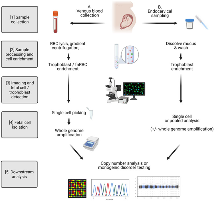
Investigators have long been interested in the natural phenomenon of fetal and placental cell trafficking into the maternal circulation. The scarcity of these circulating cells makes their detection and isolation technically challenging. However, as a DNA source of fetal origin not mixed with maternal DNA, they have the potential of considerable benefit over circulating cell-free DNA-based noninvasive prenatal genetic testing (NIPT). Endocervical trophoblasts, which are less rare but more challenging to recover are also being investigated as an approach for cell-based NIPT. We review published studies from around the world describing both forms of cell-based NIPT and highlight the different approaches' advantages and drawbacks. We also offer guidance for developing a sound cell-based NIPT protocol.
© 2021 John Wiley & Sons Ltd.
Conflict of interest statement
Conflict of interest
The authors declare no conflicts of interest.
Figures
Comment in
-
Non-invasive prenatal testing 10 years on.Prenat Diagn. 2021 Sep;41(10):1187-1189. doi: 10.1002/pd.6032. Prenat Diagn. 2021. PMID: 34418119 No abstract available.
References
-
- Salomon LJ, Sotiriadis A, Wulff CB, et al. Risk of miscarriage following amniocentesis or chorionic villus sampling: systematic review of literature and updated meta-analysis [Internet]. Vol. 54, Ultrasound in Obstetrics and Gynecology. John Wiley and Sons Ltd; 2019; p. 442–51. - PubMed
-
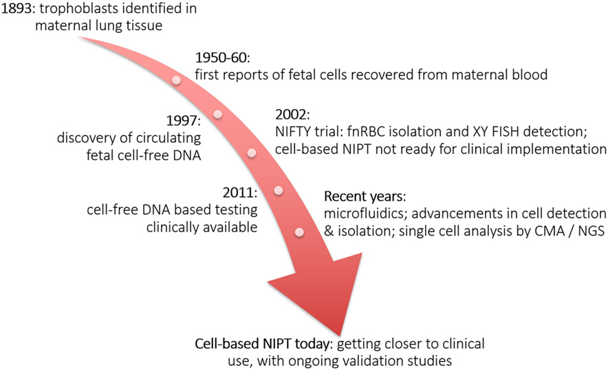
- Schmorl G Pathologisch-anatomische Untersuchungen über Puerperal-Eklampsie. Verlag FCW Vogel; 1893;
-
- Lapaire O, Holzgreve W, Oosterwijk JC, et al. Georg Schmorl on Trophoblasts in the Maternal Circulation. Placenta 2007;28(1):1–5. - PubMed
-
- Chown B Anaemia from bleeding of the fetus into the mother’s circulation. Lancet (London, England) 1954;266(6824):1213–5. - PubMed
-
- Zipursky A, Hull A, White FD, Israels LG. Foetal erythrocytes in the maternal circulation. Lancet (London, England) 1959;1(7070):451–2. - PubMed
Publication types
MeSH terms
Grants and funding
LinkOut - more resources
Full Text Sources
Other Literature Sources
